
Minced cartilage systems and methods

Active Publication Date: 2014-05-15
ALLOSOURCE
View PDF7 Cites 17 Cited by
  • Summary
  • Abstract
  • Description
  • Claims
  • Application Information

AI Technical Summary

Benefits of technology

The patent describes a method for manufacturing a cartilage composition by obtaining cartilage tissue from a human adult cadaveric donor, mincing the cartilage tissue into plural cartilage particles, and suspending them in a biocompatible carrier. The cartilage particles have a high percentage of viable chondrocytes and can be combined with a biological adhesive or demineralized bone. The method also involves washing the cartilage particles with a saline solution and using a cryopreservation medium for storage. The technical effect of this patent is to provide a reliable and effective method for manufacturing a cartilage composition with improved chondrocyte viability and a variety of biological adhesives for use in regenerative medicine and tissue engineering applications.

Problems solved by technology

Articular cartilage is not vascularized, and when damaged as a result of trauma or degenerative causes, this tissue has little or no capacity for in vivo self-repair.

Method used


Image

  • Minced cartilage systems and methods
  • Minced cartilage systems and methods
  • Minced cartilage systems and methods

Examples


example 1

Laser Cutting to Generate Minced Cartilage

[0102]Laser cutting techniques can provide a cost effective approach for the preparation of minced cartilage particles, with a decreased opportunity for tissue contamination during the mincing process. As described below, minced cartilage particles, tiles, mosaics, and the like as prepared by laser processing techniques showed cell viability results that were comparable to the cell viability results observed when using manual cutting techniques. By using a laser to prepare minced particles, cost, contamination, and processing time can be reduced. Further, it is possible to provide increased amounts of donor tissue product.

[0103]Tissue cutting experiments were performed using an Epilog Zing 30 Watt CO2 engraving laser on juvenile or adult cartilage slices. Table 1 shows the results of the tissue cutting experiments at varying speeds, powers, and frequencies.

TABLE 1Laser SettingsLaser SettingsSpeed(%)Power(%)Frequency(Hz)Result / outcome:A. Low ...

example 2

Characterization of Minced Articular Cartilage From Adult or Juvenile Donors

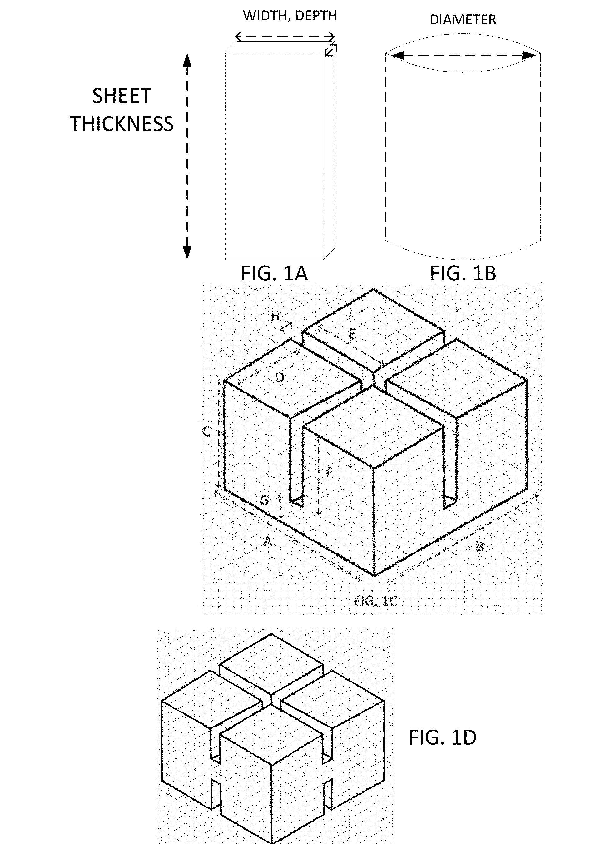
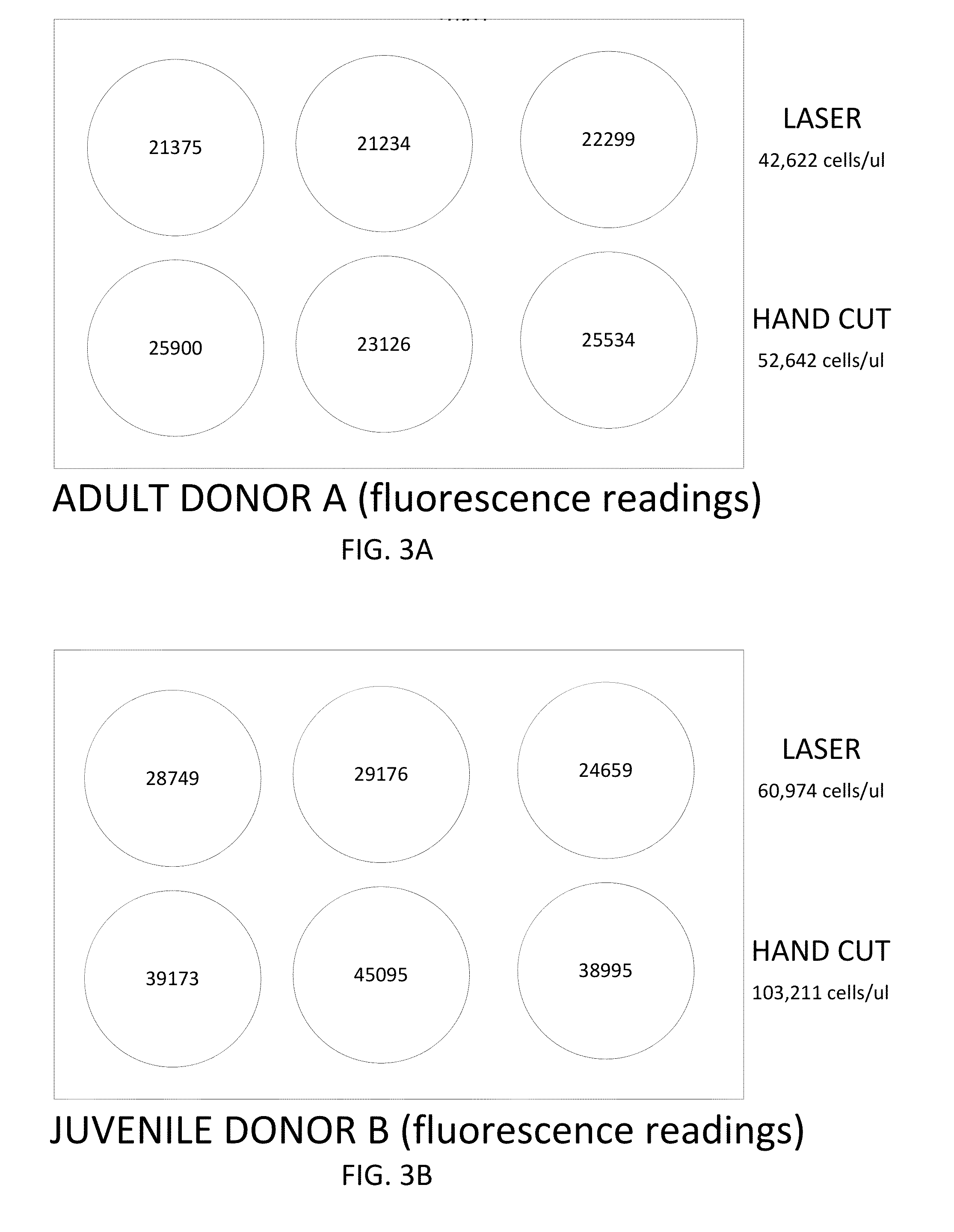
[0105]Fresh cadaveric adult and juvenile articular cartilage tissue samples were processed using either a laser cutting protocol or a hand cutting protocol. The adult donors were between fifteen and thirty six years of age, and the juvenile donors were between the ages of three months and 12 years. For the laser cutting method, the cartilage was shaved into thin slices (e.g., sheets having a thickness of 1-5 mm) using a scalpel, and the sliced sheets were minced into small particles (e.g., 1 mm, 2 mm, and / or 3 mm particles) using an Epilog Zing 30 Watt engraving laser. The laser cutting pattern was designed with a CorelDRAW® graphics software program. The cartilage was minced into square shaped particles, using energy levels and other laser parameters as described in Table 1. During the laser cutting procedure, the cartilage was maintained in a hydrated state. The minced particles were then washed with a pho...

example 3

12-Week Explant Study to Characterize Cartilage Samples

[0125]To further compare chondrocyte outgrowth and matrix production between adult and juvenile donors, a 12-week explant study was performed. Three research consented adult donors and two research consented juvenile donors were obtained. Samples were sliced by hand into 1 mm thick sheets and laser cut into 2 mm cubes. The samples were measured into 0.3 ml aliquots (5 samples per donor) and glued to a 12 well plate using TISSEEL (Baxter, Deerfield, Ill.) for a 12 week explant study to be performed. A 1:10 ratio of PrestoBlue® (Life Technologies, Carlsbad, Calif.) to media was used for weekly cell counting. Collagen type II immunohistochemistry was performed on samples after the 12 week time point, as well as sulfated glycosaminoglycans (sGAG) assay (Kamiya Biomedical Company, Seattle, Wash.), hydroxyproline assay (BioVison, Milpitas, Calif.), and DNA analysis with a Pico Green Assay (Invitrogen, Grand Island, N.Y.). All outcome ...


PUM

PropertyMeasurementUnit
Fractionaaaaaaaaaa
Fractionaaaaaaaaaa
Fractionaaaaaaaaaa

Abstract

Compositions comprising a plurality of cartilage particles from a human adult cadaveric donor, wherein the cartilage particles comprise viable chondrocytes, and a biocompatible carrier are provided. Methods of manufacturing cartilage compositions comprising a plurality of cartilage particles from a human adult cadaveric donor are also provided.

Description

CROSS-REFERENCES TO RELATED APPLICATIONS[0001]This application claims benefit of priority of U.S. Provisional Application No. 61 / 727,016, filed Nov. 15, 2012, the entire content of which is incorporated herein by reference.STATEMENT AS TO RIGHTS TO INVENTIONS MADE UNDER FEDERALLY SPONSORED RESEARCH AND DEVELOPMENT[0002]NOT APPLICABLEREFERENCE TO A “SEQUENCE LISTING,” A TABLE, OR A COMPUTER PROGRAM LISTING APPENDIX SUBMITTED ON A COMPACT DISK[0003]NOT APPLICABLEBACKGROUND OF THE INVENTION[0004]Cartilage tissue can be found throughout the human anatomy. The cells within cartilage tissue are called chondrocytes. These cells generate proteins, such as collagen, proteoglycan, and elastin, that are involved in the formation and maintenance of the cartilage. Hyaline cartilage is present on certain bone surfaces, where it is commonly referred to as articular cartilage. Articular cartilage contains significant amounts of collagen (about two-thirds of the dry weight of articular cartilage), a...

Claims


Application Information

Patent Timeline
IPC IPC(8): A61K35/12A61K35/32
CPCA61K35/35A61K35/32A61K47/20A61K47/42A61K47/36A61L27/3683A61L27/3645A61L27/3608A61L24/0005A61L2430/06
Inventor SHI, YALINGBARRETT, CAROLYNSTEVENS, PETER
Owner ALLOSOURCE
Learn More