Method for noninvasive prediction or diagnosis of inflammation and infection in amniotic fluid of patients with premature rupture of membranes

a technology of amniotic fluid and amniotic fluid, which is applied in the field of noninvasive prediction or diagnosis of inflammation and infection in amniotic fluid, can solve the problems of difficult to apply the cut-off value of il-6 in amniotic fluid collected by abdominal amniocentesis to cervical secretion, and the inability to predict inflammation or infection in vaginal fluid. it can be more stably performed on pregnant women

Active Publication Date: 2014-07-31
SEOUL NAT UNIV R&DB FOUND +1
View PDF0 Cites 1 Cited by
  • Summary
  • Abstract
  • Description
  • Claims
  • Application Information

AI Technical Summary

Benefits of technology

[0017]Compared to the conventional method for prediction or diagnosis of inflammation and infection in amniotic fluid by using invasively collected amniotic fluids, the method of the present invention can be performed more stably on pregnant women by using noninvasively collected amniotic fluid that is leaked through the cervix.

Problems solved by technology

However, collecting amniotic fluid for analysis requires abdominal amniocentesis, which is an invasive procedure.
In particular, as it is often difficult to perform amniocentesis when the volume of amniotic fluid is reduced in patients with premature rupture of membranes. therefore only ⅔ of patients undergo this procedure in US.
However, as there is a higher chance of infection since the vaginal fluid is exposed to outside, it is difficult to use vaginal fluid for prediction of inflammation or infection in amniotic fluid.
It has been confirmed that vaginal fluid can not have the predictability as much as the invasively collected amniotic fluid can.
However, as there is a significant difference between the absolute concentrations of two subject samples, it is difficult to apply the cut-off value of IL-6 in the amniotic fluid collected by abdominal amniocentesis to cervical secretion for analysis of inflammation or infection in amniotic fluid (see FIG. 1).
As the concentration of inflammatory cytokine in cervical secretion is extremely lower than those in amniotic fluid, it is difficult to directly use the concentration of cytokine in cervical fluid to predict the concentration of cytokine in amniotic fluid.
However, it is difficult to use MMP-8 in fluid leaked through the cervix as a. prediction marker for inflammation or infection in amniotic fluid, since in most cases, there was a significant increase in the concentration in amniotic fluid leaked through the cervix, even though the concentration in amniotic fluid collected by abdominal amniocentesis was low (See FIG. 10 and Table 8).
However, finding a noninvasive method for prediction of inflammation or infection in amniotic fluid was not easy, since there were significant differences in absolute values between inflammatory markers in vaginal secretion and those in amniotic fluid.
This caused the increased risk of contamination by bacteria in the vagina, making amniotic fluid from posterior fornix difficult to be used in prediction of infection / inflammation in the uterus.

Method used


Image

  • Method for noninvasive prediction or diagnosis of inflammation and infection in amniotic fluid of patients with premature rupture of membranes
  • Method for noninvasive prediction or diagnosis of inflammation and infection in amniotic fluid of patients with premature rupture of membranes
  • Method for noninvasive prediction or diagnosis of inflammation and infection in amniotic fluid of patients with premature rupture of membranes

Examples


example 1

Using IL-6 for Measuring Inflammation and / or Infection in Amniotic Fluid

[0087] Study Subject

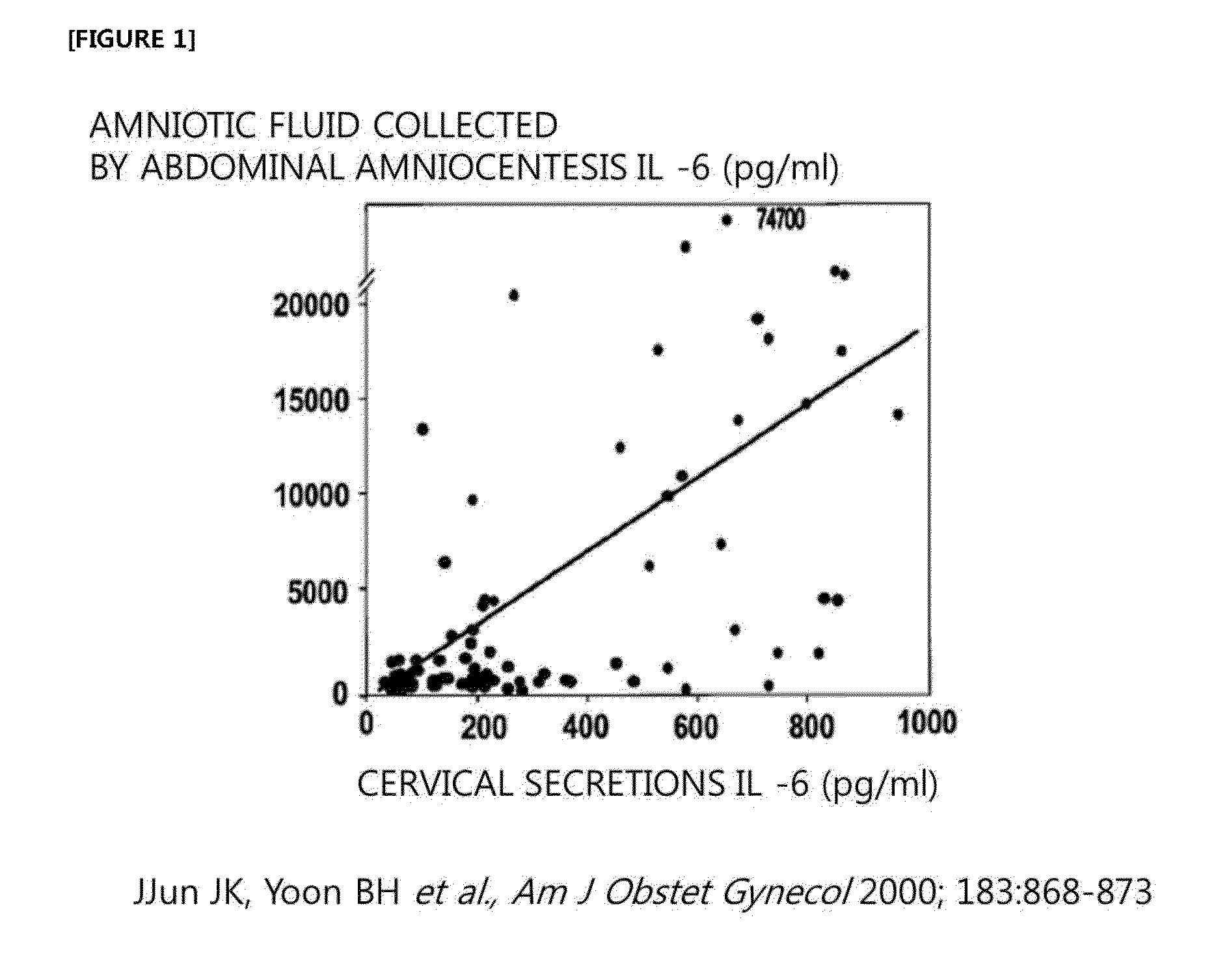
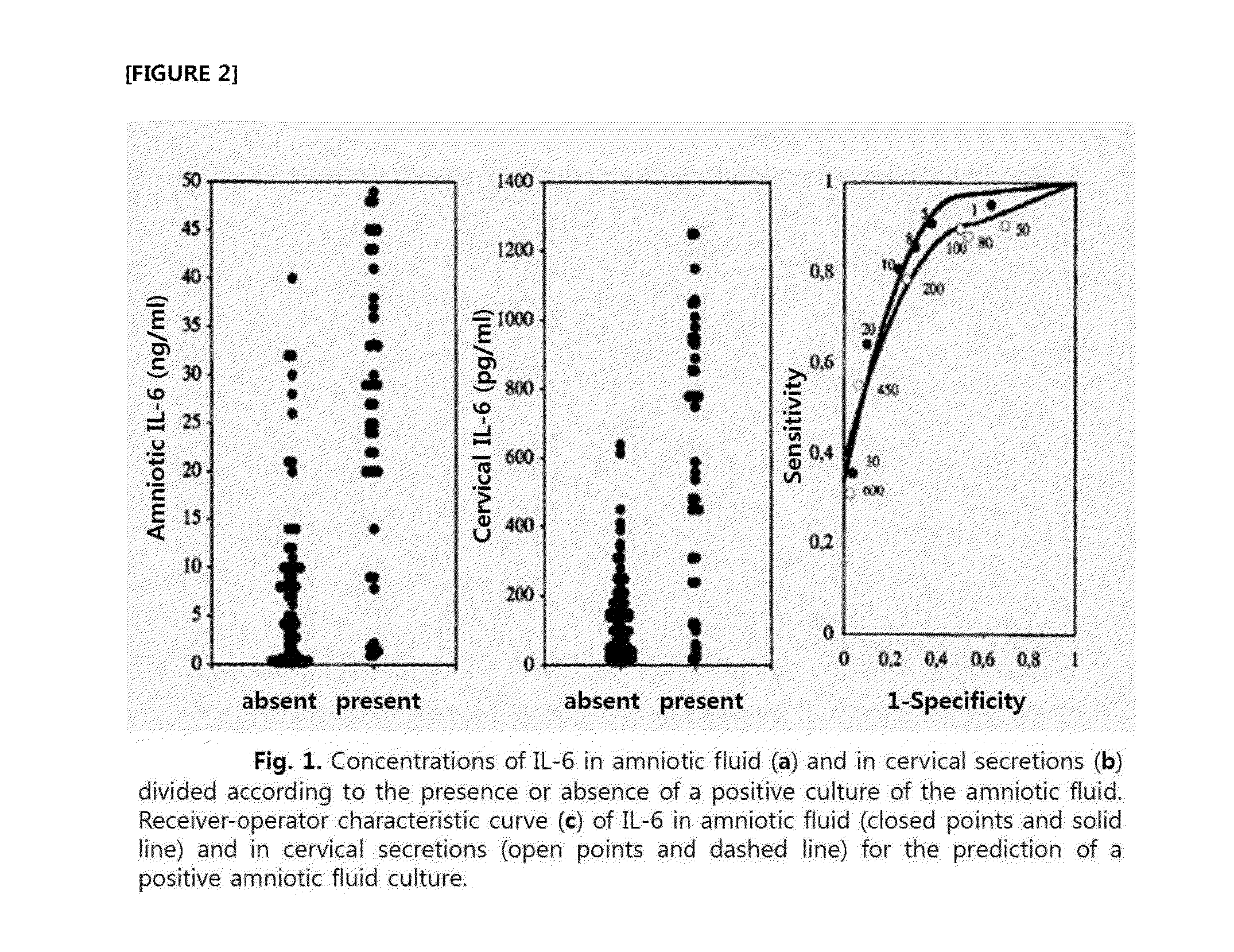
[0088]Amniotic fluid obtained by abdominal amniocentesis and amniotic fluid leaked from cervix into the vagina were collected from 18 pregnant mothers with premature rupture of membranes. The concentration of IL-6 was measured and compared by performing ELISA analysis on the above collected amniotic fluids using antibody against IL-6 (R&D system, Minneapolis, Minn.).

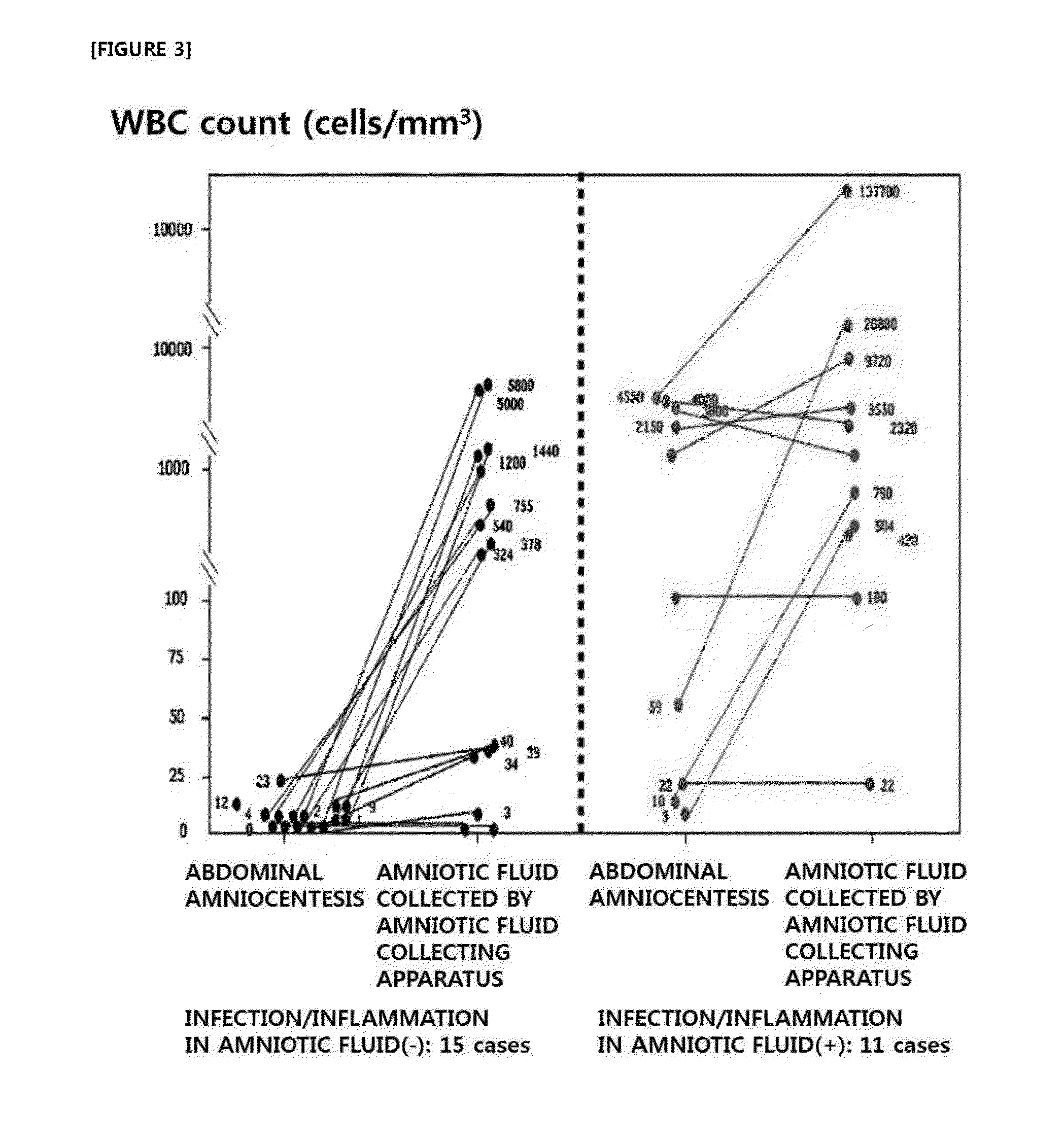
[0089] Definition of ┌Infection and Inflammation in Amniotic Fluid┘

[0090]‘Infection and inflammation in amniotic fluid’ is defined by culturing aerobic and anaerobic bacteria and genital mycoplasmas and by counting the number of white blood cells in the amniotic fluid collected by abdominal amniocentesis. In ‘infection and inflammation in amniotic fluid’, amniotic fluid infection is defined by the culture of aerobic bacteria, anaerobic bacteria and genital mycoplasma in the amniotic fluid, and amniotic fluid inflammation is define...

example 2

Using IL-8 for Measuring Inflammation and / or Infection in Amniotic Fluid

[0095] Analysis of Inflammation and / or Infection in Amniotic Fluid from Transabdominal Amniocentesis and Amniotic Fluid Leaked Through the Cervix into the Vagina

[0096]By using the test subjects from Example , the concentration of IL-8 was measured by ELISA analysis using 2 pg / ml of co-antibody-human IL-8 monoclonal anti body (Pierce endogen) and 0.05 ug / ml of biotinylated detecting antibody-anti-human IL-8 monoclonal antibody(pierce endogen). ‘Infection and inflammation in amniotic fluid’ was determined by the method of Example .

[0097]As a result, the concentration of IL-8 indicating inflammation and / or infection in amniotic fluid in patients with premature rupture of the membrane was defined as 6 ng / ml. Eight patients were confirmed as positive for ‘inflammation or infection’ in amniotic fluid collected by transabdominal amniocentesis. Out of these patients, 6 patients showed 6 ng / ml or higher concentration of ...

example 3

Using IL-1β for Measuring Inflammation and / or Infection in Amniotic Fluid

[0101] Analysis of Inflammation and / or Infection in Amniotic Fluid from Transabdominal Amniocentesis and Amniotic Fluid Leaked Through the Cervix into the Vagina

[0102]By using the test subjects from Example , the concentration of IL-1β was measured by ELISA analysis using Douset ELISA development system, human IL-1 beta / IF2 (R&D), and ‘infection and inflammation in amniotic fluid’ was determined by the method of Example .

[0103]As a result, the concentration of IL-1β indicating inflammation and / or infection in amniotic fluid in patients with premature rupture of membranes was defined as 200 pg / ml. Out of the eight patients who were confirmed as positive for ‘inflammation or infection’ in amniotic fluid collected by transabdominal amniocentesis, 7 patients showed 200 pg / ml or higher concentration of IL-1β in the amniotic fluid that was leaked from the cervix to vagina. Therefore, when the cut-off values of IL-1β ...


PUM

PropertyMeasurementUnit
concentrationaaaaaaaaaa

Abstract

Provided is a method for noninvasive prediction or diagnosis of inflammation and / or infection in amniotic fluid leaked through the cervix into the vagina to predict the concentration of inflammatory markers in the amniotic fluid inside uterus by measuring the concentration of inflammatory markers (various cytokines). Further provided is a method for prediction or diagnosis of inflammation and / or infection in amniotic fluid by measuring the concentration of markers (IL-6, IL-1β IL-8, MCP-1, GRO-α) in the amniotic fluid leaked through the cervix into the vagina in patients with premature rupture of membranes. The method can be performed more stably on pregnant women, as compared to the conventional method for prediction or diagnosis of inflammation and / or infection using invasively collected amniotic fluid.

Description

TECHNICAL FIELD[0001]The present invention relates to a method for noninvasive prediction or diagnosis of inflammation and infection in amniotic fluid.BACKGROUND ART[0002]Inflammation and / or infection in amniotic fluid is a risk factor for preterm birth and adverse neonatal outcome, which is present in about 50% of patients with preterm premature rupture of membranes. The prediction or diagnosis of infection / inflammation in amniotic fluid can be performed by microbial culture and amniotic fluid analysis, such as measuring inflammatory markers (cytokine concentration, matrix metal loproteinase enzyme concentration, white blood cell count, etc.). The amniotic fluid analysis method is the most sensitive and specific method for predicting the possibility of preterm birth and assessing the risk for neonatal complications. However, collecting amniotic fluid for analysis requires abdominal amniocentesis, which is an invasive procedure. In particular, as it is often difficult to perform amn...

Claims


Application Information

Patent Timeline
Patent Type & Authority Applications(United States)
IPC IPC(8): G01N33/543
CPCG01N33/54306G01N33/6893G01N2333/5412G01N2333/5421G01N2800/368G01N33/58G01N33/53G01N33/532
Inventor YOON, BO HYUNPARK, JOONG SHINJUN, JONG KWANPARK, CHAN WOOK
Owner SEOUL NAT UNIV R&DB FOUND
Why Eureka
Try Eureka