Peptides and compositions for treatment of joint damage
a technology of peptides and compositions, applied in the direction of fusion polypeptides, cell culture active agents, dna/rna fragmentation, etc., to achieve the effects of preventing arthritis, joint damage or joint injury, and improving pharmaceutical properties
- Summary
- Abstract
- Description
- Claims
- Application Information
AI Technical Summary
Benefits of technology
Problems solved by technology
Method used
Image
Examples
example 1
Protease-Resistant Angptl3 Peptide Constructs
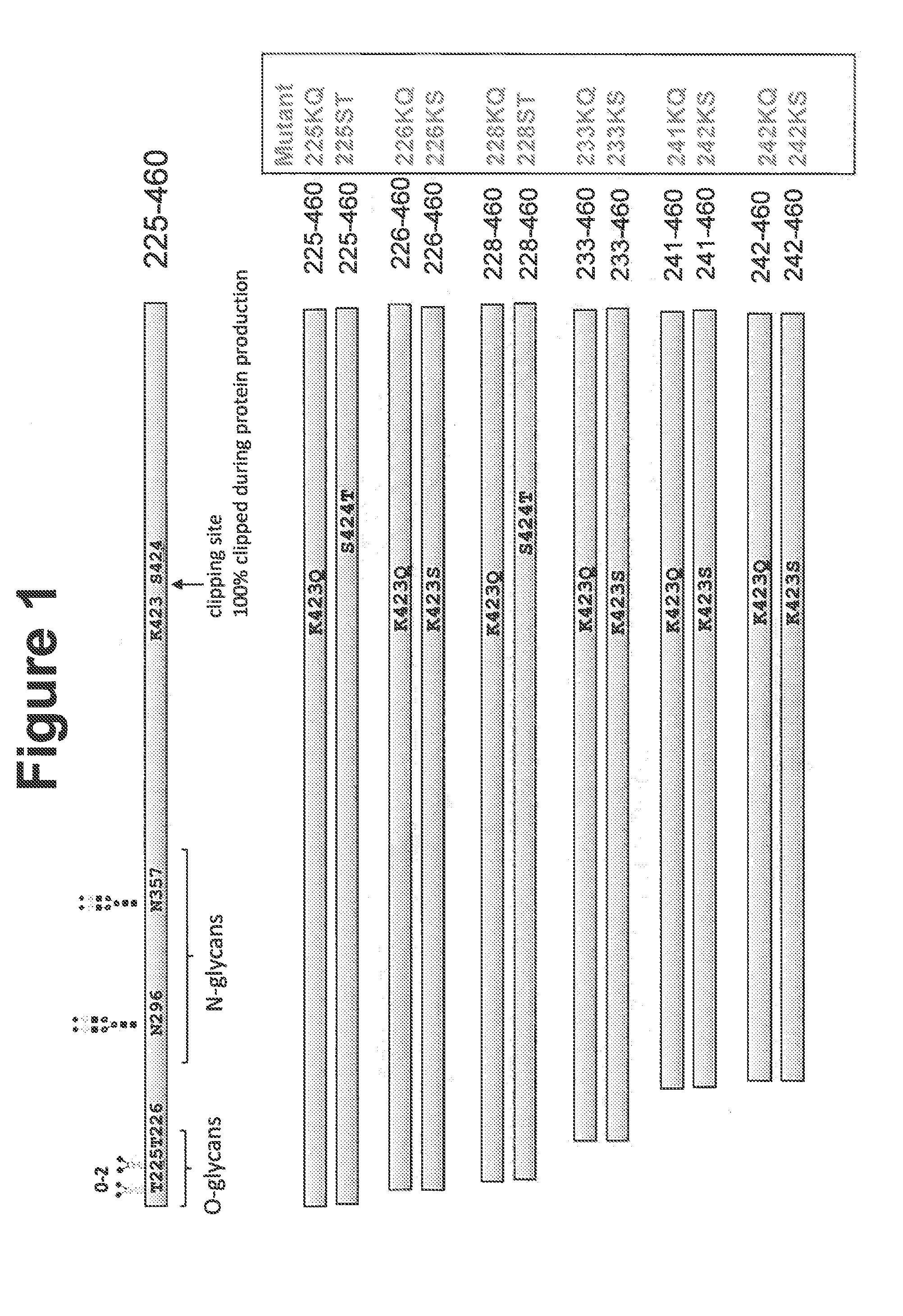
[0103]Various N-terminal truncation mutants were constructed to remove O-linked glycosylations and facilitate biophysical protein characterization. To identify protease-resistant peptides, amino acid substitutions were introduced into various positions of human Angptl3 peptide fragments corresponding to the C-terminal region of the peptide. FIG. 1 shows positions of mutations in the human Angptl3. Constructs were initially prepared with His tags. The mutant proteins were: 225-460 K423Q (225KQ), 225-460 S424T (225ST), 226-460 K423Q (226KQ), 226-460 K423S (226KS), 228-460 K423Q (228KQ), 228-460 S424T (228ST), 233-460 K423Q (233KQ), 233-460 K423S (233KS), 241-460 K423Q (241KQ), 241-460 K423S (241KS), 242-460 K423Q (242KQ), and 242-460 K423S (242KS).
[0104]His-tagged proteins were expressed in HEK Freestyle™ cells and purified by Ni-NTA column chromatography. Tag-less C-terminal constructs were also cloned, purified by previously described met...
example 2
Integrin Binding Assays
[0106]αVβ3 Integrin
[0107]Prepared peptides 225KQ, 228KQ, 233KQ, 241KQ and 242KQ were tested in vitro for binding to αVβ3 integrin. Briefly, Maxisorp plates were coated with 2 μg / ml Integrin αvβ3, and various concentrations of polypeptide construct (indicated) were added. Bound peptide was detected by the addition of Anti-ANGPTL3 mAb followed by horseradish peroxidase-conjugated Goat anti-Mouse IgG antibody. All tested peptides retained or improved integrin binding capacity. EC50 for each were determined from the binding data, and results are shown in TABLE 2.
[0108]α5β1 Integrin
[0109]Prepared peptides 225KQ, 228KQ, 233KQ, 241KQ and 242KQ were tested in vitro for binding to α5β1 integrin. Plates were coated with 2 μg / ml as described above but with Integrin α5β1, and various concentrations of polypeptide construct (indicated) were added, and detection carried out as described above. All tested peptides retained or improved integrin binding capacity. EC50 for each...
example 3
Functional Analysis of Constructs
[0110]Chondrogenesis.
[0111]Peptide constructs were evaluated in functional assays to assess chondrogenesis activity.
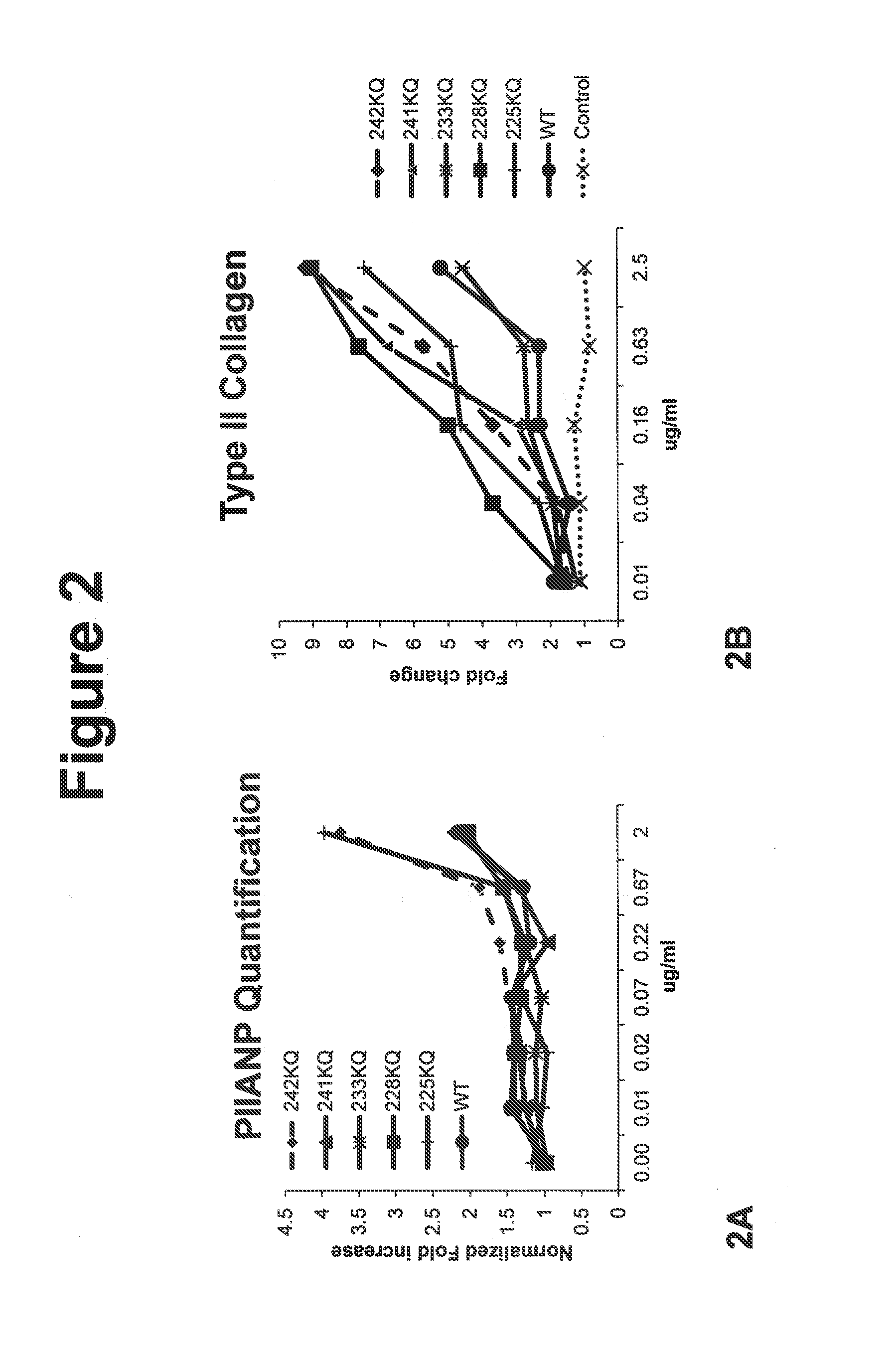
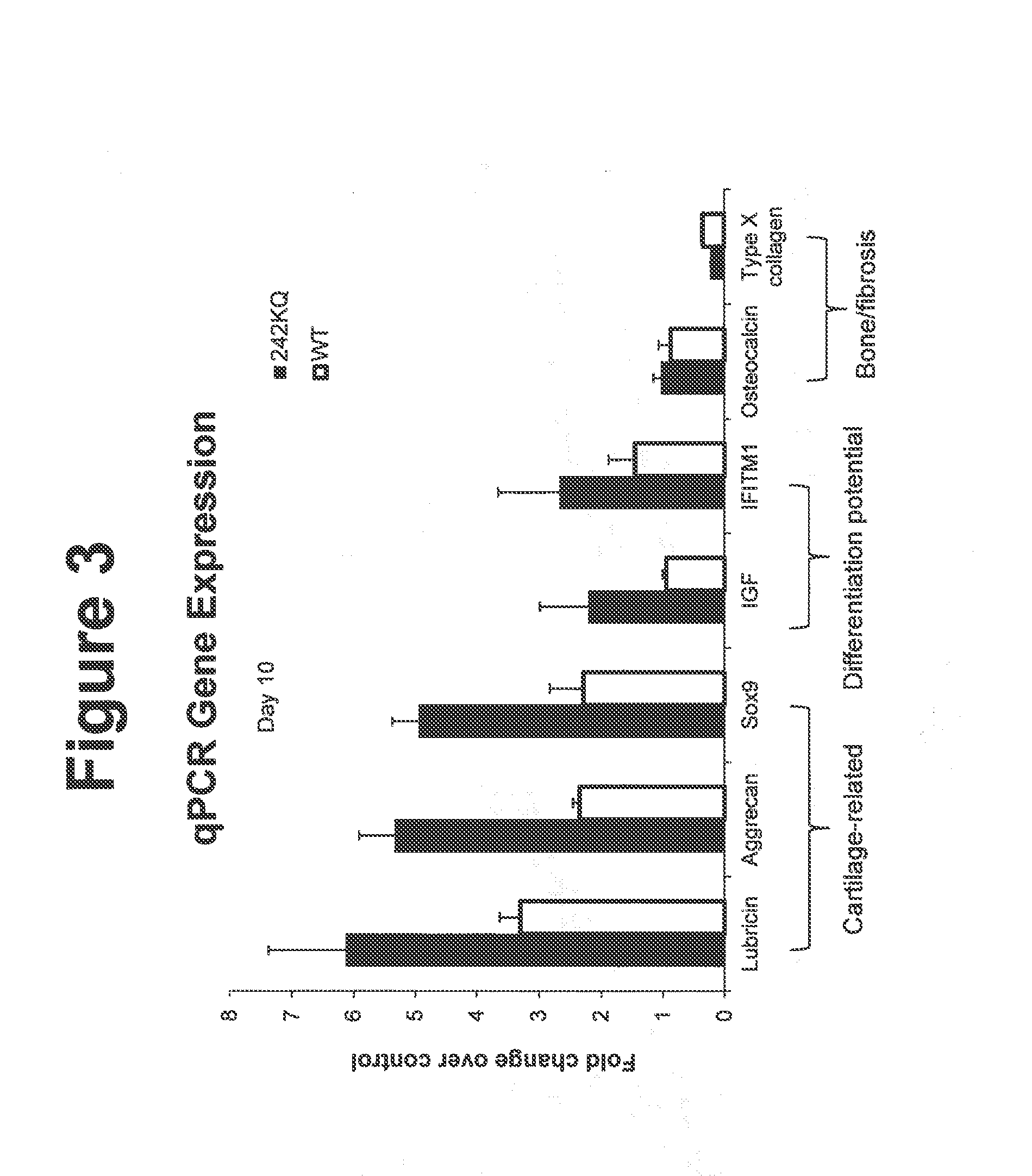
[0112]Cell-based 2D chondrogenesis was induced in vitro and assessed as described previously in Johnson, K., et al., (2012) Science 336, 717. Briefly, primary human bone marrow derived mesenchymal stem cells (hMSCs) were plated in growth media then subsequently changed to a chondrogenic stimulation media with and without constructs, and cultured for 7 or 14 days. Cells were then fixed with formaldehyde, washed and then stained using standard immuno-cytochemical techniques to detect primary cartilage proteins Pro-collagen Type 2A (PIIANP) and Type II collagen Immuno-fluorescence for each protein detected was quantified through high content imaging (Image Express Ultra) and as described previously. Results are exemplified in FIG. 2 for control (cells stimulated without construct, diluent alone), WT wild type C-terminal (225-460) ANGPTL3 o...
PUM
| Property | Measurement | Unit |
|---|---|---|
| pH | aaaaa | aaaaa |
| pH | aaaaa | aaaaa |
| pH | aaaaa | aaaaa |
Abstract
Description
Claims
Application Information
 - R&D
- Intellectual Property
- Life Sciences
- Materials
- Tech Scout
- Unparalleled Data Quality
- Higher Quality Content
- 60% Fewer Hallucinations
Browse by: Latest US Patents, China's latest patents, Technical Efficacy Thesaurus, Application Domain, Technology Topic, Popular Technical Reports.
© 2025 PatSnap. All rights reserved.Legal|Privacy policy|Modern Slavery Act Transparency Statement|Sitemap|About US| Contact US: help@patsnap.com


