
Instant, in-situ, nondestructive material differentiation apparatus and method

a non-destructive, material technology, applied in the field of material differentiation, can solve the problems of long turnaround time, lack of effectiveness and utility, and inability to clearly define or remov

Inactive Publication Date: 2014-12-04
UTAH VALLEY UNIVERSITY
View PDF5 Cites 19 Cited by
  • Summary
  • Abstract
  • Description
  • Claims
  • Application Information

AI Technical Summary

Benefits of technology

This patent describes a method for analyzing the structure of materials using ultrasonic waves. The technique involves placing two ultrasonic transducers facing each other on opposite sides of the material, and transmitting an ultrasonic beam through it. The resulting reflections are then received at the other transducer. By analyzing the structure of the power spectra and comparing it to model data, the method can provide information about the microstructure and properties of the material. The use of high-frequency ultrasonics and novel data collection methods make the technique sensitive to biological materials and enable nondestructive testing.

Problems solved by technology

Other tumors may have portions that extend into surrounding tissues with longer branches or fingers that are not well defined nor easily removed.
However, such processes have such a long turnaround time that they lack effectiveness and utility during the time of surgery.
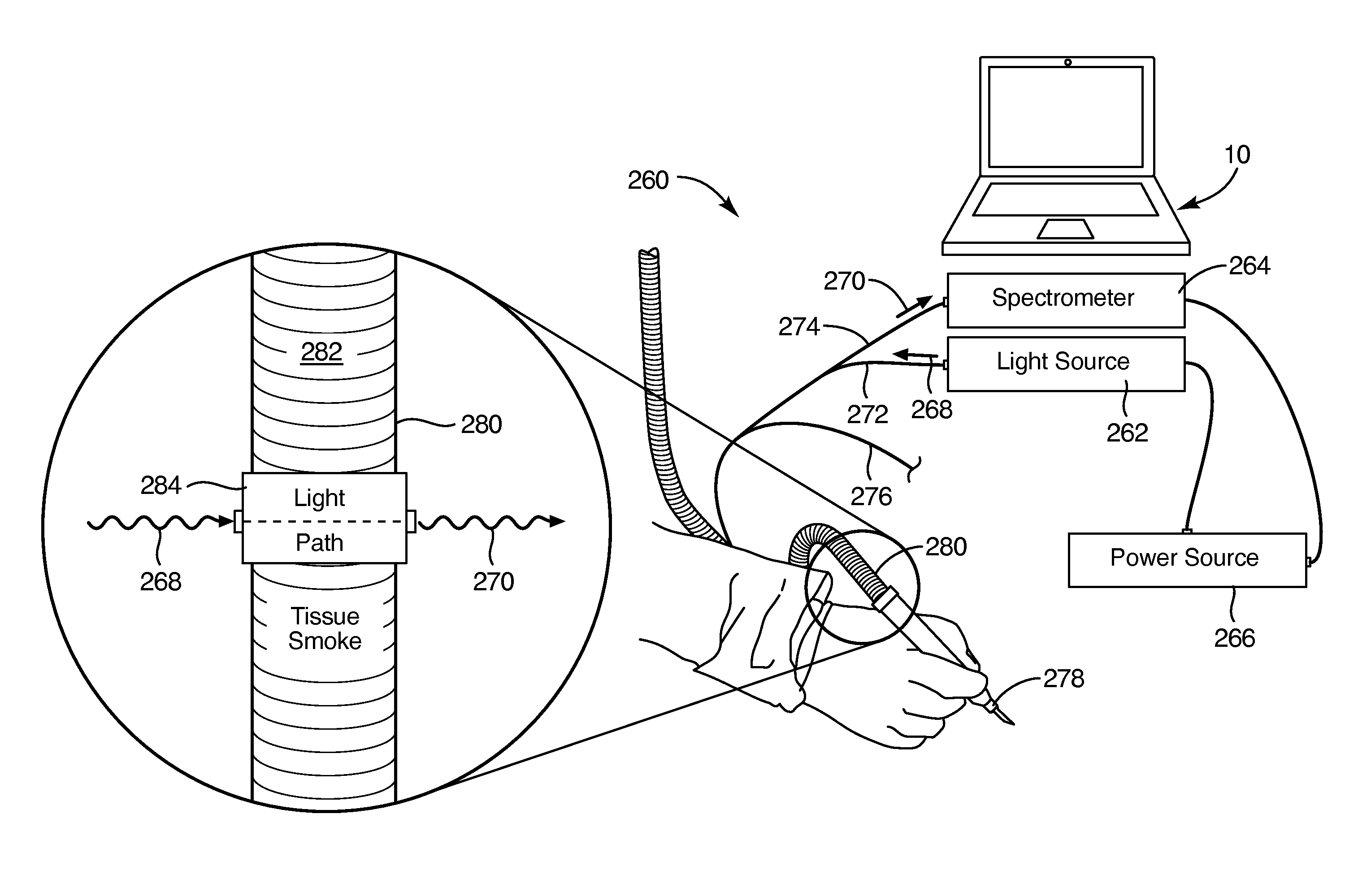
For example, in certain systems, electrical or laser cutters providing automatic cauterization of tissues produce smoke.
Such testing from data gathered from the smoke produced by cauterizing surgery still requires more time than is actually available during surgery, and lacks the desired simultaneity with the surgery.
The time of flight or passage within a conduit, sensitivity, aliquot size requirements, samplings, and light may be comparatively limited.
Moreover, more conventional methods of analysis including liquid chromatography and the like also are far removed from the time of surgery and therefore only have limited utility.
However, the analysis, whether by cytology, frozen section analysis, touch preparation cytology, or the like is still typically unable to identify close margins.
However, current techniques in conventional pathology may take days for accurate analysis of specimens.
Moreover, the specimens are necessarily excised, and thus involve destructive testing.

Method used


Image

  • Instant, in-situ, nondestructive material differentiation apparatus and method
  • Instant, in-situ, nondestructive material differentiation apparatus and method
  • Instant, in-situ, nondestructive material differentiation apparatus and method

Examples


Embodiment Construction

[0090]It will be readily understood that the components of the present invention, as generally described and illustrated in the drawings herein, could be arranged and designed in a wide variety of different configurations. Thus, the following more detailed description of the embodiments of the system and method of the present invention, as represented in the drawings, is not intended to limit the scope of the invention, as claimed, but is merely representative of various embodiments of the invention. The illustrated embodiments of the invention will be best understood by reference to the drawings, wherein like parts are designated by like numerals throughout.

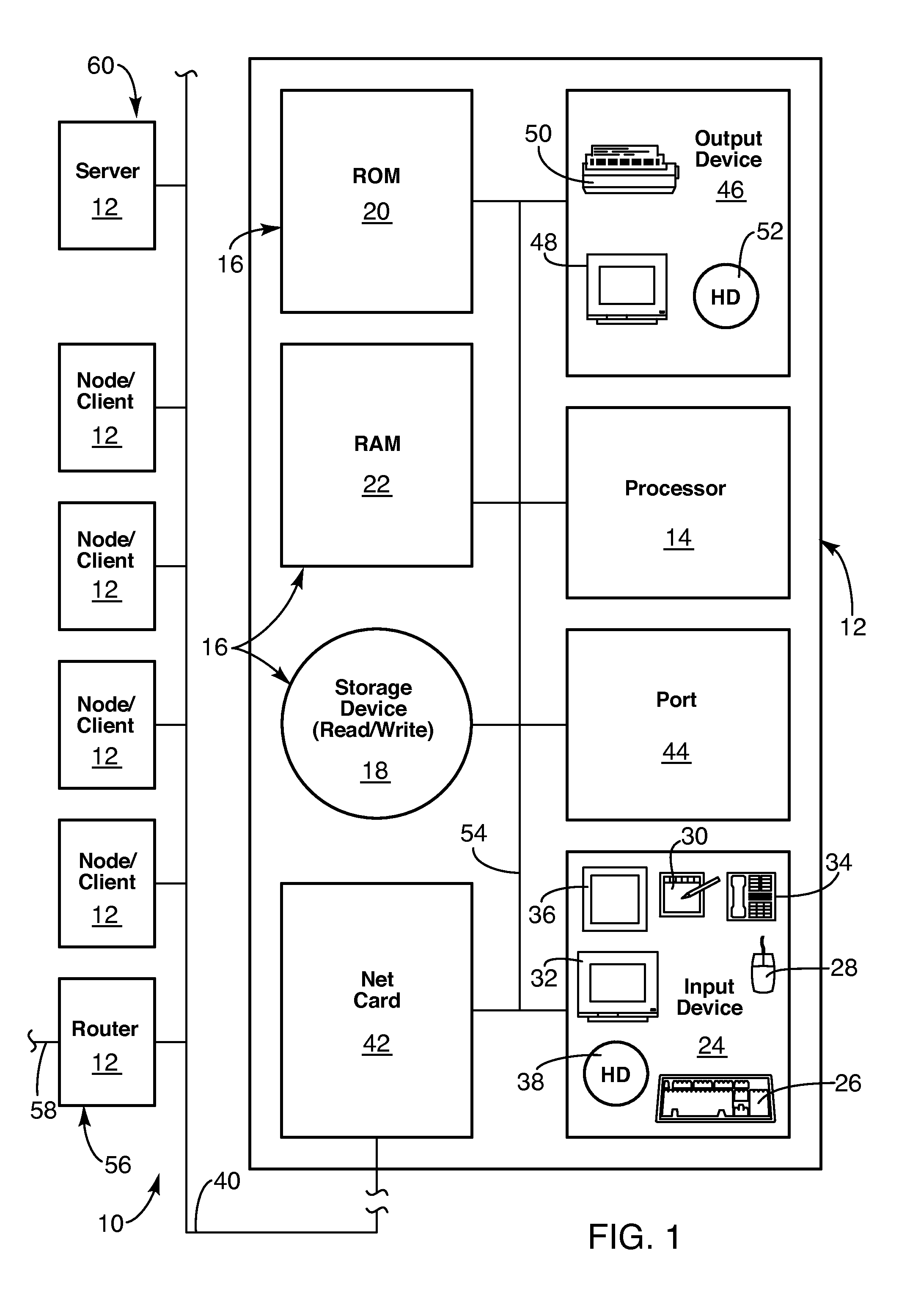
[0091]Referring to FIG. 1, an apparatus 10 or system 10 for implementing the present invention may include one or more nodes 12 (e.g., client 12, computer 12). Such nodes 12 may contain a processor 14 or CPU 14. The CPU 14 may be operably connected to a memory device 16. A memory device 16 may include one or more devices such as...


PUM


Abstract

Specular, ultrasonic, piezoelectric, detection devices provide real-time, analytical, edge finding in tissues during tumor surgery. Piezoelectric probe sensors at high frequencies (e.g., 10 to 100 MHz) characterize microstructure of cells and tissues. Through-transmission or specular reflection enables nondestructive testing in real time. Peak density analysis in power spectra, second-order spectrum analysis measuring the slope of the Fourier transform of the power spectrum, artificial intelligence pattern recognition, and modeling interpret the results. Model-based data analysis may compare experimental data with a computer simulation. Such comparisons may be based upon pattern classifications, including principal component analysis (PCA). Combining the above detection devices and analytical methods provides speed, accuracy, simplicity, and nondestructive mechanisms that militate for reliable, real-time diagnosis of tumor margins, tissue pathology, cell phenotypes, and molecular subtypes.

Description

RELATED APPLICATIONS[0001]This application claims the benefit of U.S. Provisional Patent Application Ser. No. 61 / 963,993 entitled ULTRASOUND MICROSENSOR FORCEPS FOR DETERMINING TISSUE PATHOLOGY, filed on Dec. 20, 2013; U.S. Provisional Patent Application Ser. No. 61 / 964,885 entitled HIGH-FREQUENCY ULTRASONIC SURFACE SENSOR FOR MICROSCOPIC ANALYSIS OF TISSUE PATHOLOGY, filed on Jan. 16, 2014; U.S. Provisional Patent Application Ser. No. 61 / 964,884 entitled DEVICE FOR ULTRASONIC AND OPTICAL PHENOTYPING OF BIOLOGICAL CELLS filed on Jan. 16, 2014; U.S. Provisional Patent Application Ser. No. 61 / 964,886 entitled ULTRASOUND MICRONEEDLE SENSOR FOR CHARACTERIZING BIOLOGICAL MATERIALS filed on Jan. 16, 2014; U.S. Provisional Patent Application Ser. No. 61 / 964,887 entitled ULTRASOUND MICROSENSOR SCALPEL FOR DETERMINING TISSUE PATHOLOGY, filed on Jan. 16, 2014; U.S. Provisional Patent Application Ser. No. 61 / 959,939 entitled OPTICAL METHOD FOR DETERMINING THE PATHOLOGY OF TISSUE AEROSOLS PRODU...

Claims


Application Information

Patent Timeline
IPC IPC(8): G01N29/44G01N33/483
CPCG01N33/4833G01N29/44A61B8/085
Inventor DOYLE, TIMOTHY EDWINTHOMPSON, LAUREL ANNLADANI, LEILA JANNESARI
Owner UTAH VALLEY UNIVERSITY
Learn More