Quantitative phantomless calibration of computed tomography scans

a computed tomography and phantomless technology, applied in tomography, instruments, applications, etc., can solve the problems of affecting the interpretation and clinical utility of quantitative assessment, affecting the accuracy of computed tomography, so as to improve the viewing and interpretation of ct scans.

Active Publication Date: 2014-12-25
O N DIAGNOSTICS
View PDF1 Cites 18 Cited by
  • Summary
  • Abstract
  • Description
  • Claims
  • Application Information

AI Technical Summary

Benefits of technology

[0010]Disclosed are systems and methods for phantomless calibration of a CT scan that do not require the use of an external calibration phantom and that account for machine-specific and patient-specific differences in X-ray attenuation characteristics within the body. The methods can be used to perform quantitative CT analysis on CT scans obtained without the use of an external calibration phantom, and, in some cases, can be used with contrast-enhanced CT scans. The methods can also be used to calibrate grayscale values in CT scans for the purposes of improving consistency in the appearance of the scan data.
[0012]Improved methods are presented for patient-specific, phantomless calibration of CT scans, to be used, for example, in measuring bone mineral density, bone mass, bone volume fraction, assessing bone or bone-implant strength, or quantifying mineral content, including amount of mineral in a blood vessel or in any soft tissue within the body, or measuring fat in the liver, in muscle, or in any other organs, or measuring the relative amount of grey and white matter in the brain, or in any application in which a patient-specific calibration of the attenuation data would be useful, including consistent display and qualitative viewing of CT scans. The methods do not require the use of an external calibration phantom; they are patient-specific in that the resulting calibration depends on measurements of grayscale attenuation data (referred to herein as the HU-values) in the patient's CT scan; they can be used retrospectively on previously acquired scans or prospectively on new scans; and in some applications they can also be used on scans containing intravenous contrast. Because the methods facilitate automation, they are easily implemented clinically and are highly repeatable.
[0022]For both the Effective-Energy and Equivalent-Density methods, one key improvement over the prior art is the use of air to provide a second reference calibration point. Air is easily sampled from a CT scan and represents a robust and repeatable measure for the purposes of phantomless calibration. The use of air to provide a calibration point results in the need for just one internal reference tissue for the overall phantomless calibration. Compared to the prior-art methods, this novel feature facilitates implementation and automation since only one tissue needs to be identified in the scan and processed; and this feature also leads to greater precision and repeatability of the overall phantomless calibration. Furthermore, the need for processing of just a single internal reference tissue enables the phantomless calibration method to be applied to anatomic sites and applications for which it would be difficult or impossible to process two internal reference tissues. One example of such includes analysis at the proximal femur hip region, for which visceral fat is present but little other tissue is consistently available except for muscle, which, as discussed, is inherently limited for phantomless calibration because it represents a mixture of actual muscle and fat. Another example is the tibial or femoral diaphysis, for which air and dense cortical bone can be used as reference tissues, since again, no other suitable tissues are available (again, excluding muscle since it is typically mixed with an unknown amount of fat). Another example is the analysis of contrast-enhanced images, for which blood and muscle cannot be used as reference tissues since they are highly perfused by the contrast agent, whereas a large proportion of the fat remains relatively non-perfused. Thus, the use of air to provide a second calibration point in the current invention not only facilitates the use of phantomless calibration, but also enables its application to situations that would otherwise not be amenable to phantomless calibration methods that are based on analysis of two or more internal reference tissues.
[0024]As depicted in FIG. 2, process 200 represents one general embodiment of the Effective-Energy method as applied to the measurement of a density of a tissue of interest in a patient's CT scan 101, many details of which are provided in subsequent paragraphs. In step 102, HU-values are measured from the scan for one internal reference tissue and for external air, the air serving as a second reference material. In general step 103, energy-specific values of attenuation are specified for said internal reference tissue and air. To do this, first, in sub-step 135, empirical relations are provided that relate certain scanner-acquisition information—for example, the scanner make and model, and the kVp value used in the scan acquisition—to effective energy of a scan. These relations are used to specify an effective energy of the patient's CT scan for their particular scanner-acquisition information. In sub-step 136, known values of attenuation as a function of effective energy are used to specify attenuation values as a function of effective energy for the internal reference tissue and air. In sub-step 130, information from sub-steps 135 and 136 is combined to provide energy-specific values of attenuation for the internal reference tissue and air for the estimated energy of the patient's CT scan. In general step 104, voxels in the tissue of interest in the scan are calibrated using the information provided by general step 103. To do this, in sub-step 140, a linear equation is developed between HU-value and attenuation, using the measured HU-values 102 and the energy-specific values of attenuation for the internal reference tissue and air 130. This equation can then be used for all voxels in the tissue of interest to map HU-values to attenuation values. In sub-step 155, a mixture-model for attenuation sharing is assumed for each voxel in the tissue of interest, in which the mass-attenuation of an individual voxel is assumed to be the mass-fraction-weighted sum of the mass-attenuations of each of the assumed components of the mixture (for which energy-specific values can also be obtained in sub-step 136). For a two-component mixture model, the only unknown is the mass-fraction of one of the components, which can be solved for, given the attenuation value for a voxel. In sub-step 150, these calculations are performed to provide as output a value of a measure of density for the tissue of interest based on the calculated value of mass-fraction. Any calibration results from sub-steps 140 or 150 can be saved on electronic media 105. This information can be used in any subsequent quantitative analysis of said tissue or scan or for any other purposes. This embodiment facilitates full automation since image processing of just a single internal reference tissue, for example, fat or blood or spinal cord, lends itself to automation, as does image processing of air, and no other user input is required.
[0028]A number of different approaches can be used in sub-step 140 to map HU-values into energy-specific attenuation values. In one such method, a linear mapping is obtained between the energy-specific attenuation values 130 and HU-values 102 for the internal reference tissues. If only two internal reference tissues are used (or one internal reference tissue and air are used) for such purposes, a straight line fit can be calculated analytically. If three or more internal reference tissues are used—which can reduce measurement error due to poor signal-to-noise ratios in the reference materials—statistical approaches can be used for such purposes, for example, least-squares best-fit linear regression analysis. Note that different internal reference tissues can be used in Steps 103 and 104. For example, in process 300 (FIG. 3), the internal reference tissues used to estimate the energy of the scan in sub-step 130 do not have to be the same as the internal reference tissues used to form the relation in sub-step 140; preferably, if HU-values are measured for multiple internal reference tissues in step 102, air is used in 130 but not used in 140. Alternatively, if three or more internal reference tissues are used, a non-linear equation can be fit to the data in sub-step 140, although the relation between HU-values and attenuation is usually assumed to be linear in most CT scanners.
[0039]A variety of clinical CT scans can be calibrated according to the current invention, with or without intravenous contrast, including but not limited to abdominal, spine (lumbar, thoracic, or cervical), pelvic, and lung CT scans, scans for CT colonography and CT enterography, or CT angiography or CT calcium-scoring scans of the heart or abdomen. Calibration of the scan or body part or tissue or organ can be used to provide a quantitative measure of a tissue of interest; it can also be used to enhance the viewing and interpretation of CT scans, including further image processing. For example, segmentation routines can exploit a consistent calibration to provide images of body parts in CT scans that are more consistent across different settings and CT machines. Gray-scale levels can be automatically set based on calibrated values of the attenuation. For example, the output of step 140 in FIG. 1 can be used to convert HU-values in the original scan to true attenuation values or to corrected HU-values. Once the HU-values have been calibrated or corrected in a consistent manner in this way, fixed threshold values can be defined to optimize viewing of CT scans for specific clinical applications and those thresholds can lead to more consistent viewing across different scanners and patients and over time.

Problems solved by technology

Without such calibration, for example, the same patient would yield different CT-numbers if imaged on different CT machines or with different scanner settings, confounding the interpretation and clinical utility of the quantitative assessment.
However, the need for an external calibration phantom, which must be placed under the patient during scanning, adds expense and complexity to the clinical imaging and is therefore not widely used.
Another limitation of using an external calibration phantom is that the outcome measures of calibrated density of a tissue are always expressed in terms of the density or concentration of the materials used in the actual phantom; such measures of density are referred to as “equivalent-density” measures.
This makes clinical interpretation difficult, which hampers widespread clinical adoption of the quantitative CT technology.
However, while this method can account for between-machine differences in calibration characteristics for any particular CT machines that are pre-calibrated, it does not account for any patient-specific X-ray attenuation characteristics and thus does not provide a patient-specific calibration; nor it is applicable to CT machines that have not been pre-calibrated or to pre-calibrated CT machines with hardware components that deteriorate over time.
However, since muscle is mixed with fat—and since the ratio of pure fat to pure muscle tissue varies both across patients and within the body—it is difficult to separate out the fat and muscle in a repeatable and reliable fashion.
As noted by Pickhardt (2011), these limitations present barriers to clinical use.
However, this technique is limited since it also requires the use of an external calibration phantom either before or during imaging the patient.
This contrast agent alters the appearance of the blood and (highly perfused) muscle in the CT scan, so these tissues cannot be used as internal reference tissues.

Method used


Image

  • Quantitative phantomless calibration of computed tomography scans
  • Quantitative phantomless calibration of computed tomography scans
  • Quantitative phantomless calibration of computed tomography scans

Examples


Embodiment Construction

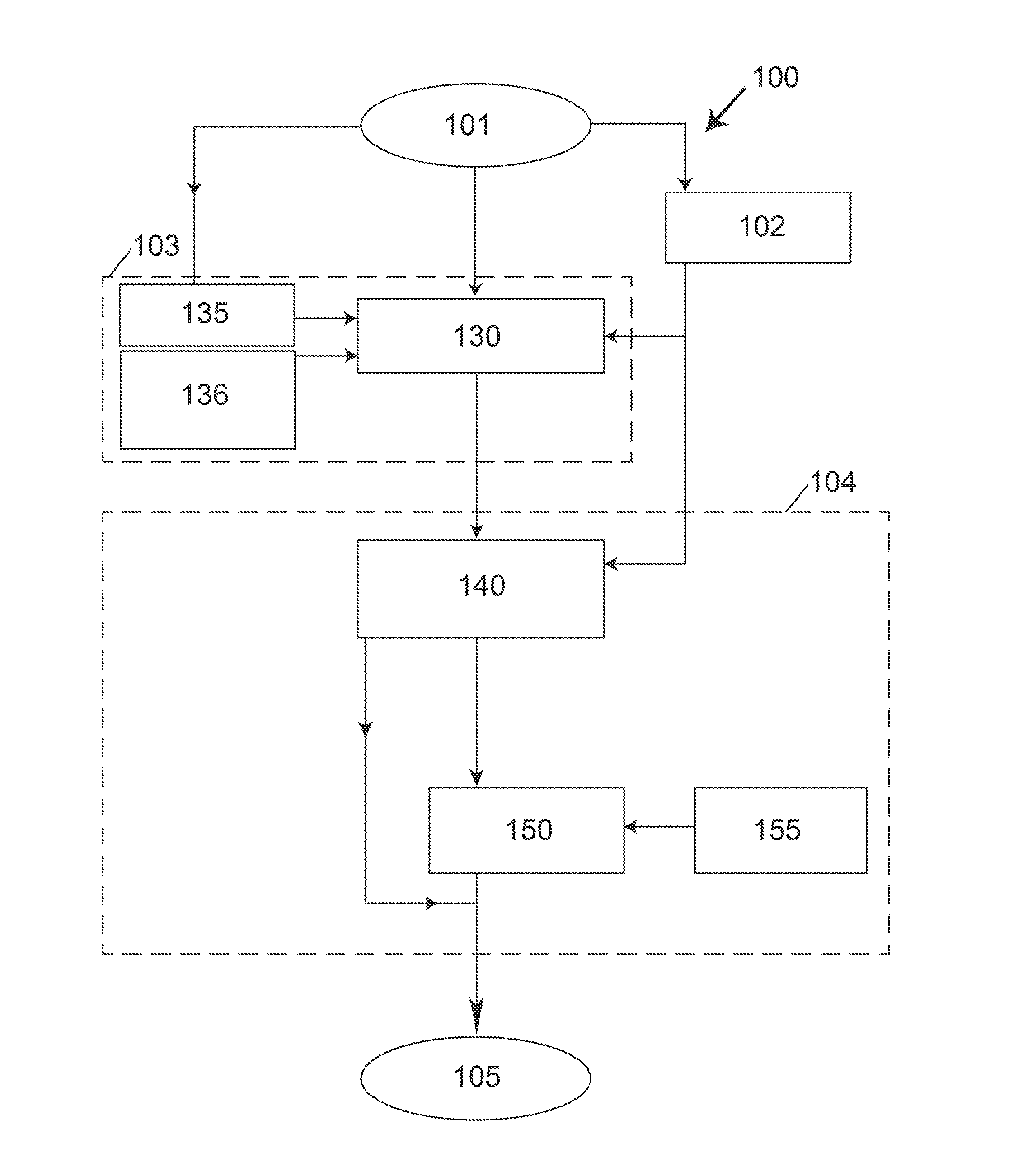
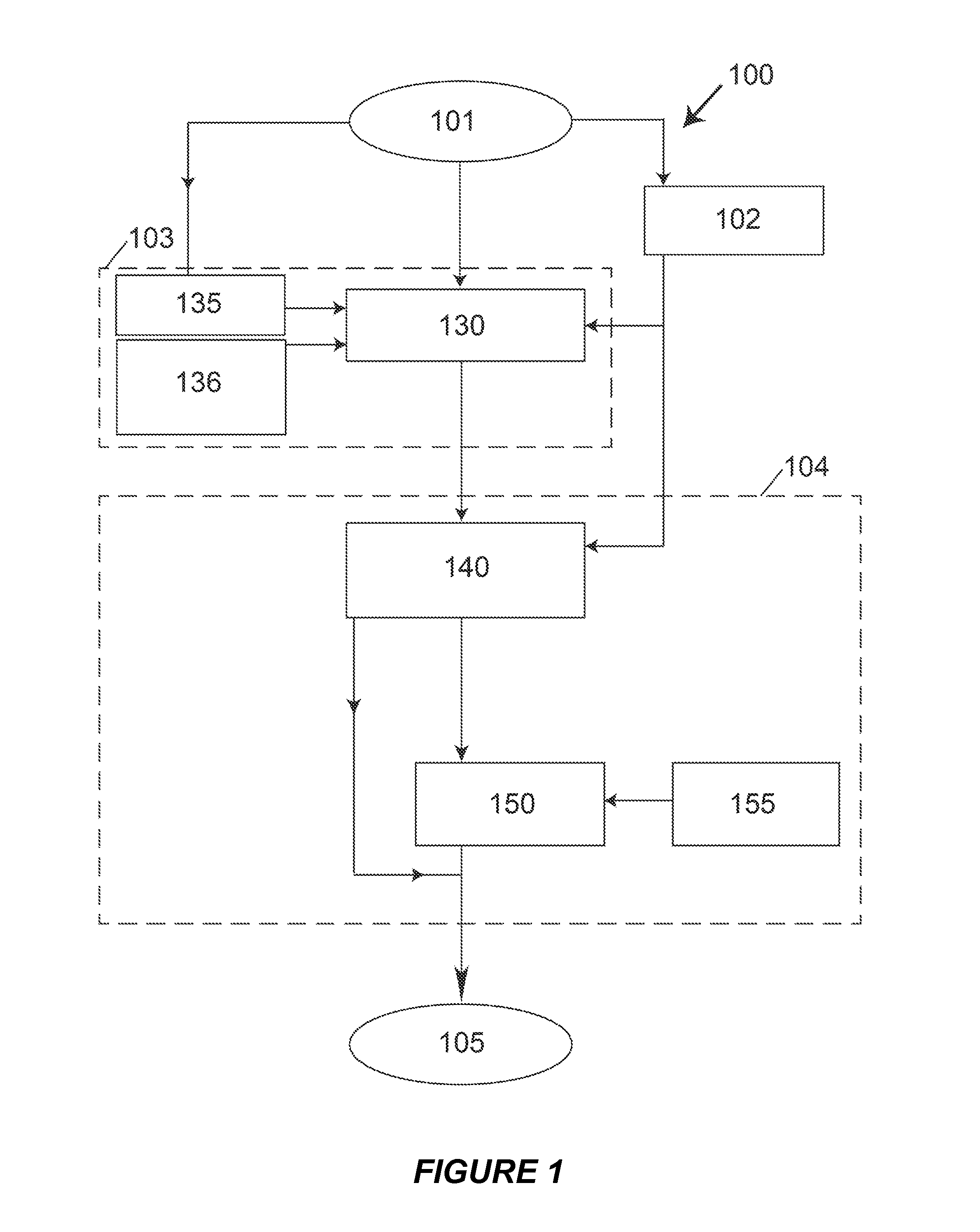
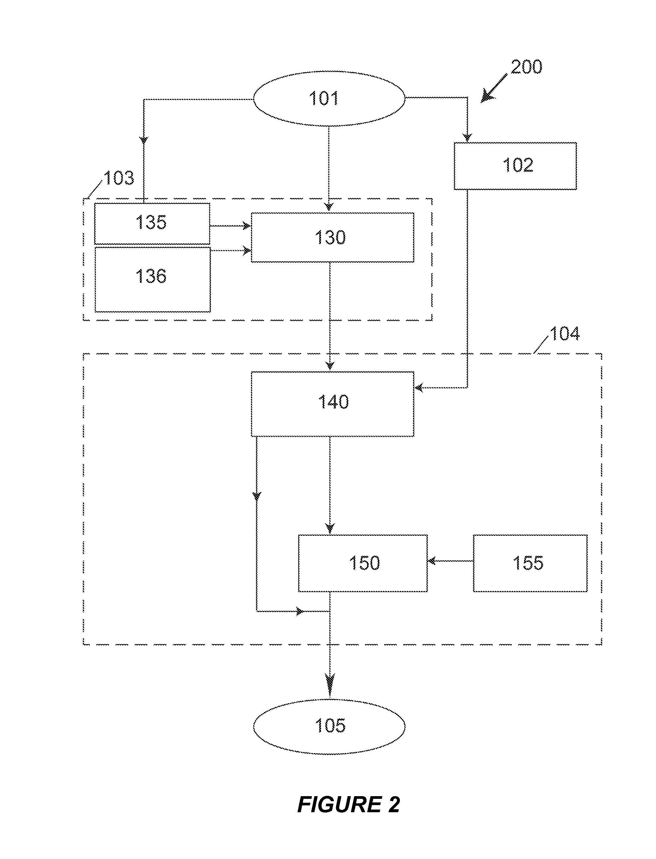
[0047]Embodiments of the present invention provide a system and method for phantomless quantitative CT using the Effective-Energy and Equivalent-Density methods. The following description is presented to enable one of ordinary skill in the art to make and use the invention and is provided in the context of a patent application and its requirements.

[0048]Various modifications to the preferred embodiments and the generic principles and features described herein will be readily apparent to those skilled in the art. Thus, the present invention is not intended to be limited to the embodiments shown but is to be accorded the widest scope consistent with the principles and features described herein.

DEFINITIONS

[0049]The following definitions apply to some of the aspects described with respect to some embodiments of the invention. These definitions may likewise be expanded upon herein.

[0050]As used herein, the term “or” is generally intended to mean “and / or” unless otherwise indicated.

[0051]...


PUM


Abstract

An apparatus, method, and computer program product for calibrating a CT scan without the use of an external calibration phantom, to enable quantitative assessment of internal body tissues and organs and additionally for any application that would benefit from a calibration of the scan attenuation data, such as viewing CT images in a consistent fashion. Embodiments are described with applications to quantitative assessment of bone density in the spine and hip, mineral content in blood vessels, hepatic-fat content in the liver, and gray-to-white matter ratio in the brain. The primary advantages of the method are that it does not require the use of an external calibration phantom, it is robust across different CT machines and scanner settings, and it is also highly precise, lending itself to a high degree of automation.

Description

CROSS REFERENCE TO RELATED APPLICATIONS[0001]This application claims benefit of U.S. Patent Application No. 61 / 838,159, the content of which is hereby expressly incorporated in its entirety for all purposes.FIELD OF THE INVENTION[0002]The present invention relates generally to computed tomography (CT), and more specifically, but not exclusively, to a phantomless calibration of CT scans, without the need for any external calibration phantom, for any application that would benefit from a quantitative calibration of the scan attenuation data, such quantitatively assessing internal body tissues and organs and consistently viewing CT images.BACKGROUND OF THE INVENTION[0003]The subject matter discussed in the background section should not be assumed to be prior art merely as a result of its mention in the background section. Similarly, a problem mentioned in the background section or associated with the subject matter of the background section should not be assumed to have been previously...

Claims


Application Information

Patent Timeline
Patent Type & Authority Applications(United States)
IPC IPC(8): A61B6/00
CPCA61B6/582A61B6/032A61B6/505A61B6/5211G16H50/20
Inventor KOPPERDAHL, DAVID L.LEE, DAVID CHOENKEAVENY, TONY M.
Owner O N DIAGNOSTICS
Why Eureka
Try Eureka