Method of analyzing multi-sequence MRI data for analysing brain abnormalities in a subject

a brain abnormality and multi-sequence technology, applied in the field of medical imaging, can solve the problems of difficult manual delineation and time-consuming, automated segmentation of mri, and little to no interpretability of parameters

Active Publication Date: 2015-02-12
THE HENRY M JACKSON FOUND FOR THE ADVANCEMENT OF MILITARY MEDICINE INC +2
View PDF5 Cites 29 Cited by
  • Summary
  • Abstract
  • Description
  • Claims
  • Application Information

AI Technical Summary

Benefits of technology

[0017]In accordance with another aspect of the present invention a sequence of the method includes isolating probable abnormalities from each modality, each of the blurred images including in a logistic regression model having estimates ofvoxel-specific probability of the voxel being part of the abnormality in an interaction between the images and the blurred images, removing said abnormality from the image, reblurring said image, and providing a refined description of regional patterns including spatial inhomogeneity and inhomogeneity and underlying anatomy of said abnormalities. The method also includes using said more refined blurred images and said normalized data and the interactions between the images and the blurred images, refitting logistic regression model, refining estimates of said voxel-specific probability of said abnormality, directly interpreting said probabilities, and yielding binary segmentation maps. The abnormality can take the form of a brain lesion and the method can further include utilizing a single cross-sectional imaging study and identifying how much said lesion load said subject has and where in the brain said lesions exist. The abnormality can be associated with multiple sclerosis. Additionally, the method can include resampling said multi-sequence MRI data over time, reestimating coefficients and performance using partial ROC methods, and obtaining a robust analysis of the image data.

Problems solved by technology

Manual delineation is challenging and time consuming, as three-dimensional information from several MRI modalities must be integrated.
One difficulty in automated segmentation of MRI is due to variable imaging acquisition parameters.
These parameters are not informed by the data and therefore must be tuned empirically, often with little to no interpretability of the parameter.
A second difficulty in intensity-based segmentation is that MRI data are acquired in arbitrary units; units can vary widely between and within imaging centers.
Many of the segmentation methods use intensity-normalized images, but these methods do not demonstrate robustness of the normalization procedure to changes in imaging acquisition parameters and imaging centers.
A third difficulty is intensity inhomogeneity, the slow spatial intensity variations of the same tissue within an MRI volume.
Inhomogeneity can significantly reduce the accuracy of image segmentation, and therefore some form of spatial normalization is necessary for accurate lesion segmentation.

Method used


Image

  • Method of analyzing multi-sequence MRI data for analysing brain abnormalities in a subject
  • Method of analyzing multi-sequence MRI data for analysing brain abnormalities in a subject
  • Method of analyzing multi-sequence MRI data for analysing brain abnormalities in a subject

Examples


example

[0050]An exemplary implementation of the present invention is described herein, in order to further illustrate the present invention. The exemplary implementation is included merely as an example and is not meant to be considered limiting. Any implementation of the present invention on any suitable subject known to or conceivable by one of skill in the art could also be used, and is considered within the scope of this application.

2. Materials and Methods

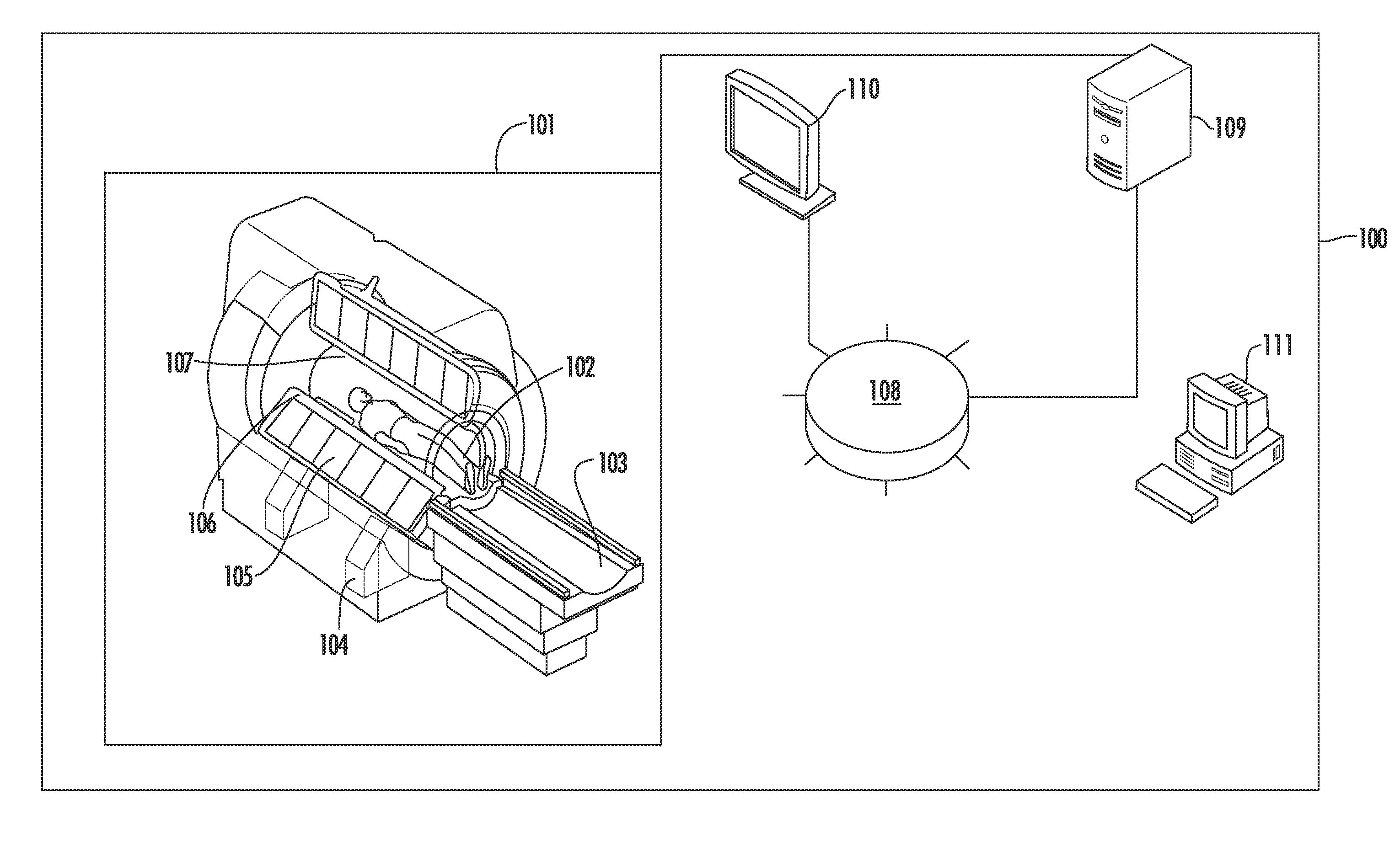
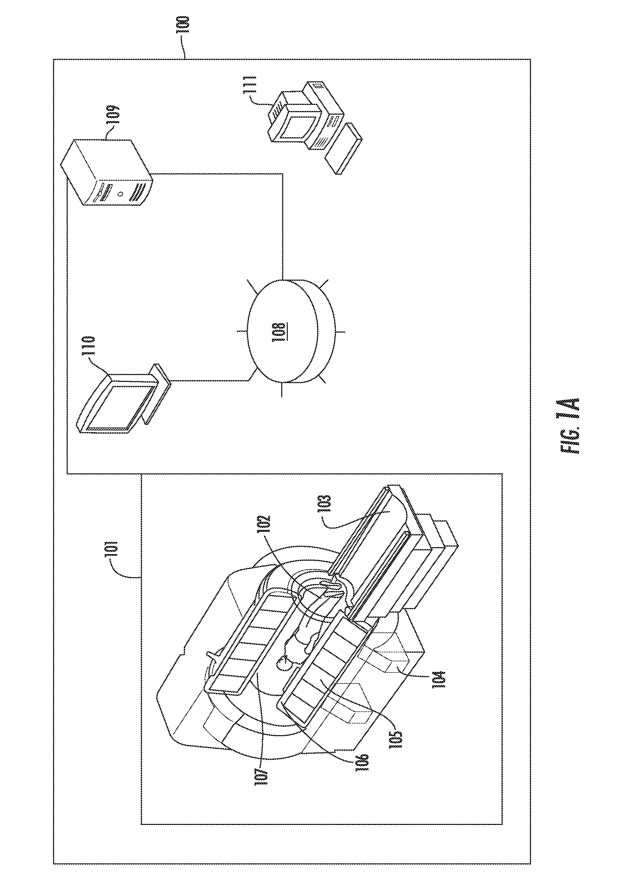
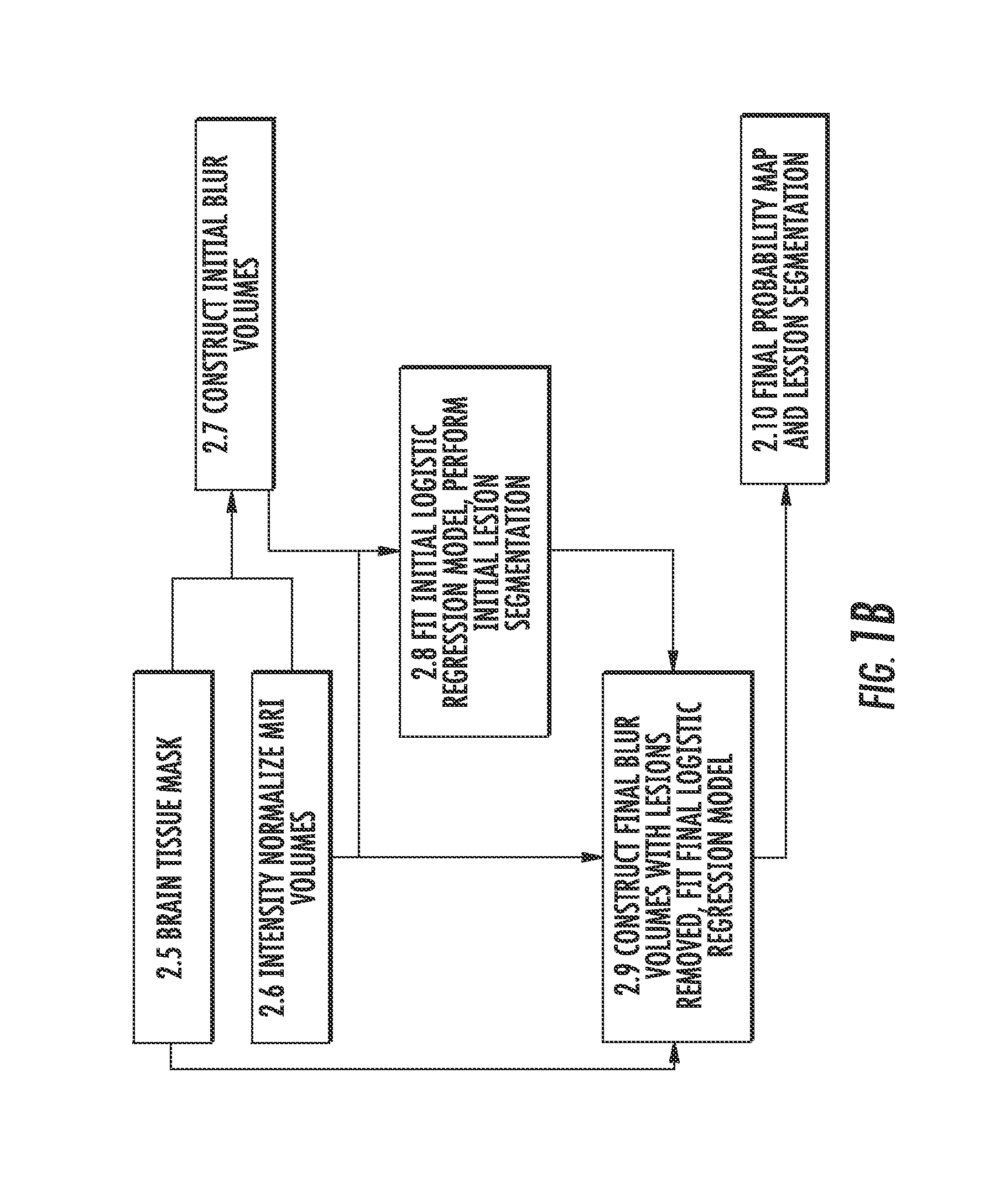
[0051]OASIS is a method inspired by Subtraction Based Inference for Modeling and Estimation (SuBLIME), an automated method for the longitudinal segmentation of incident and enlarging MS lesions described in International Application No. PCT / US2012 / 067997, and incorporated herein, by reference. Before the OASIS logistic regression model is fit, a brain tissue mask is created, all MRI volumes are intensity normalized, and BLUR volumes are created to capture local spatial information and adjust for remaining field inhomogeneities. The O...


PUM


Abstract

The present invention, referred to as Oasis is Automated Statistical Inference for Segmentation (OASIS), is a fully automated and robust statistical method for cross-sectional MS lesion segmentation. Using intensity information from multiple modalities of MRI, a logistic regression model assigns voxel-level probabilities of lesion presence. The OASIS model produces interpretable results in the form of regression coefficients that can be applied to imaging studies quickly and easily. OASIS uses intensity-normalized brain MRI volumes, enabling the model to be robust to changes in scanner and acquisition sequence. OASIS also adjusts for intensity inhomogeneities that preprocessing bias field correction procedures do not remove, using BLUR volumes. This allows for more accurate segmentation of brain areas that are highly distorted by inhomogeneities, such as the cerebellum. One of the most practical properties of OASIS is that the method is fully transparent, easy to implement, and simple to modify for new data sets.

Description

CROSS REFERENCE TO RELATED APPLICATIONS[0001]This application claims the benefit of U.S. Provisional Patent Application No. 61 / 613,569 filed on Mar. 21, 2012, which is incorporated by reference, herein, in its entirety.GOVERNMENT SUPPORT[0002]This invention was made with government support under NINDS RO1 NS070906 awarded by the National Institutes of Health, as well as R01NS060910 from the Intramural Research Program of the National Institute of Neurological Disorders and Stroke (NINDS) at the National Institutes of Health. The government has certain rights in the invention.FIELD OF THE INVENTION[0003]The present invention relates generally to medical imaging. More particularly the present invention relates to methods of analyzing brain abnormalities in a subject.BACKGROUND OF THE INVENTION[0004]Multiple sclerosis (MS) is an inflammatory disease of the brain and spinal cord characterized by demyelinating lesions that are most easily identified, at least on magnetic resonance imagin...

Claims


Application Information

Patent Timeline
Patent Type & Authority Applications(United States)
IPC IPC(8): A61B5/00G01R33/36G06T7/00G01R33/385
CPCA61B5/4064G01R33/385G01R33/3614G06T7/0012G06T2207/30096G06T7/0087G06T2207/20076G06T2207/10088G06T2207/30016G06T7/0081A61B5/055G01R33/5608
Inventor CRAINICEANU, CIPRIANGOLDSMITH, ARTHUR JEFFREYPHAM, DZUNGREICH, DANIEL S.SHIEE, NAVIDSHINOHARA, RUSSELL T.SWEENEY, ELIZABETH M.
Owner THE HENRY M JACKSON FOUND FOR THE ADVANCEMENT OF MILITARY MEDICINE INC
Why Eureka
Try Eureka