Electrostretched polymer microfibers for microvasculature development

a microfiber and electrode technology, applied in general culture methods, biochemical equipment and processes, bioreactors/fermenters, etc., can solve the problems of inability to analyze the organization of different ecm components in detail in microvasculature studies, and the difficulty of creating clinically relevant functional microvascular conduits (i.e. arterioles and venules)

Inactive Publication Date: 2015-04-30
THE JOHN HOPKINS UNIV SCHOOL OF MEDICINE
View PDF5 Cites 4 Cited by
  • Summary
  • Abstract
  • Description
  • Claims
  • Application Information

AI Technical Summary

Benefits of technology

[0012]The present invention is a three-dimensional (3D) fibrin microfiber scaffold for a novel in vitro model of the microvasculature that recapitulates endothelial cell alignment and ECM deposition. The microfiber scaffold allows the sequential co-culture of endothelial cells and other cells, such as perivascular cells. This model establishes that the fiber curvature affects the circumferential deposition of ECM from endothelial cells independently of cellular organization. The invention further presents a multicellular microvascular structure with an organized endothelium and multicellular perivascular tunica media. The present invention provides unique opportunities to assess microvasculature development and regeneration.

Problems solved by technology

In tissue engineering there is currently a limit on the size of tissues that can be constructed in vitro due to the diffusion limit of nutrients into developing organs.
However, studies on microvasculature have not analyzed the organization of different ECM components in detail (Movat H Z, and Fernando N V P, Experimental and Molecular Pathology, 1963; 2: 549-563; Hibbs R G, et al., American Heart Journal, 1958; 56: 662-670; Fernando N V, and Movat H Z, Experimental and Molecular Pathology, 1964; 3: 1-9).
The creation of clinically relevant functional microvascular conduits (i.e. arterioles and venules) remains a challenge.
Nonetheless, these systems have limited control over topographical cues presented by the ECM and impart a barrier for the high-resolution, dynamic, and detailed study of vascular organization as well as specific cell-ECM and multi-cellular interactions.
Furthermore, the organization of other ECM components, such as fibronectin and laminin, remains widely uninvestigated.

Method used


Image

  • Electrostretched polymer microfibers for microvasculature development
  • Electrostretched polymer microfibers for microvasculature development
  • Electrostretched polymer microfibers for microvasculature development

Examples


example 1

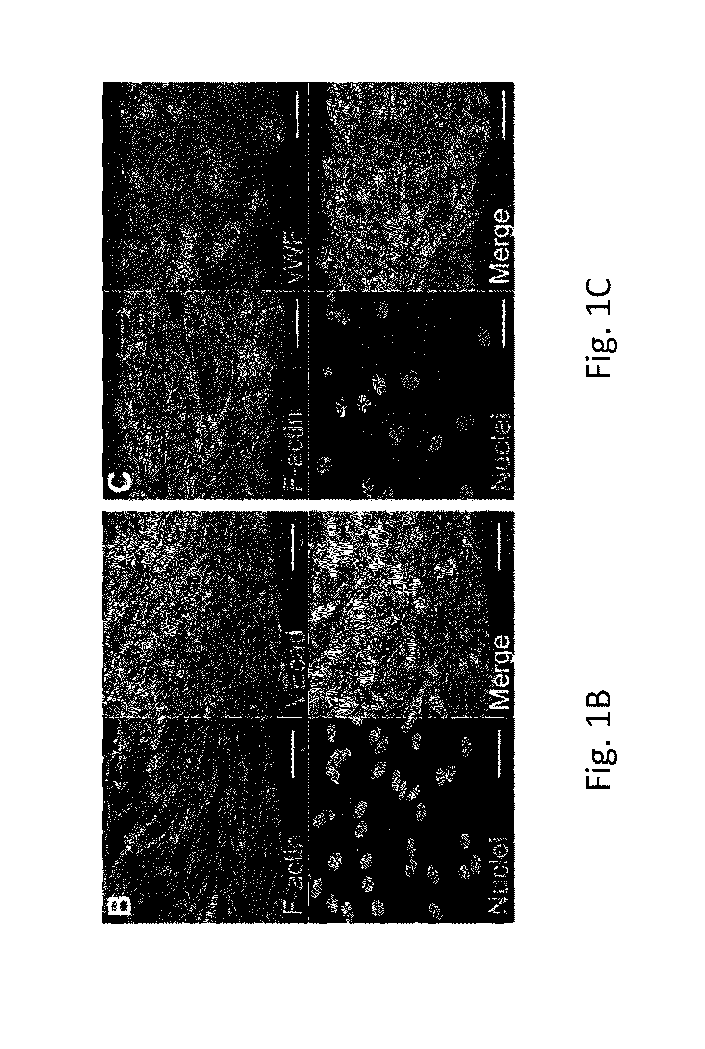
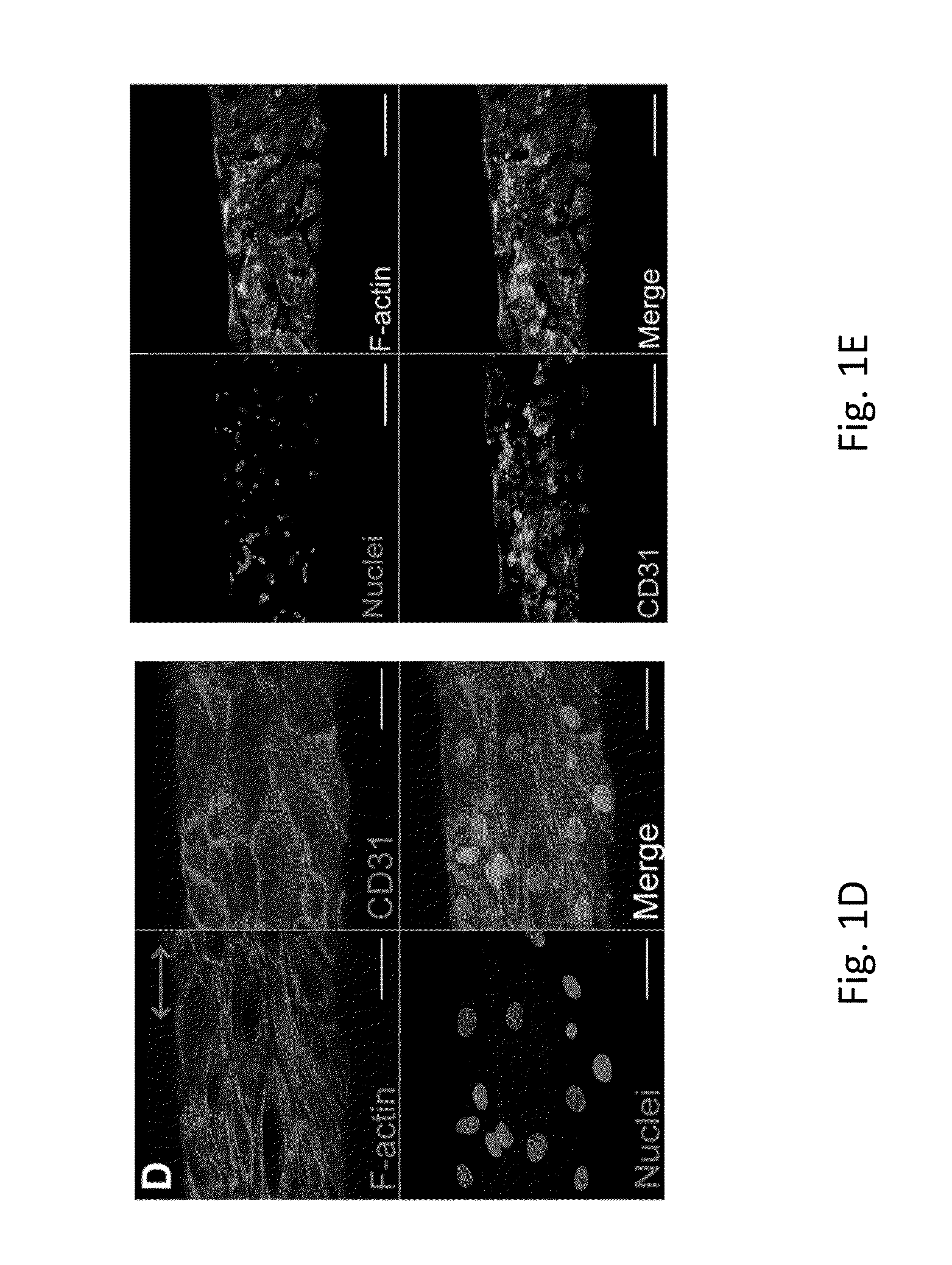
ECFC Attachment and Alignment on Fibrin Microfibers

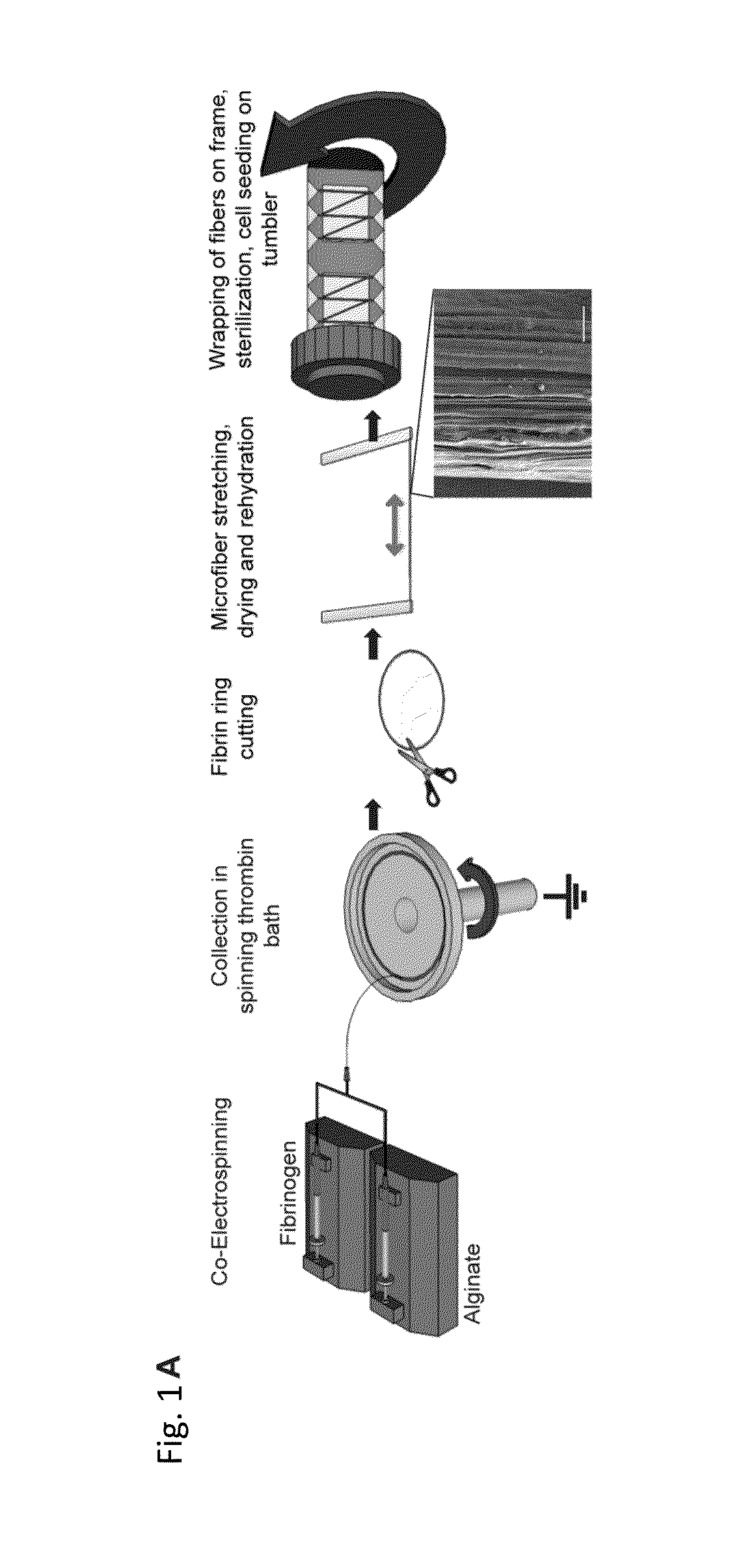
[0080]A new approach to create aligned hydrogel microfibers using an electrostretching process of polymer materials is disclosed. Unique characteristics of the electrostretched hydrogel microfibers are the internal and topographical alignment of the fibrous structure, generated as a result of both electrical field and mechanical shear-induced polymer chain alignment as described above. Furthermore, the diameter of a microfiber is controlled and uniform as a result of the bundling and processing of the individual nanofibers that compose the hydrogel microfibers. Fibrin gels have been extensively used to study microvasculature assembly (Dickinson L E, et al., Soft Matter, 2010; 6: 5109-5119; Bayless, K J, and Davis, G E, Biochemical and Biophysical Research Communications, 2003; 312: 903-913; Davis G E, and Bayless K J, Microcirculation, 2003; 10: 27-44; Bayless K J, et al., RGD-Dependent American Journal of Pathology, 2000; 156: 1673...

example 2

ECM Deposition from ECFCs on Fibrin Microfibers

[0088]While the importance of ECM deposition in vascular development has been recognized, few studies have looked at ECM production by the endothelium. ECFCs deposit collagen IV, fibronectin and laminin in an organized web-like structure when cultured on Petri dishes (Kusuma S, et al., FASEB J, 2012; 26: 4925-4936). To establish a reliable in vitro model of microvasculature, we characterized the ECM protein deposition by the endothelium on hydrogel microfibers.

[0089]ECFCs were seeded on fibrin hydrogel microfibers as described in Example 1.

[0090]ECFC seeded on fibrin microfibers deposited laminin, collagen IV, and fibronectin after one day in culture (FIG. 2A). On Day 5, ECFCs completely covered the fibrin microfiber, and abundant ECM deposition was observed (FIG. 2B). In contrast to what was observed previously on Petri-dishes (Kusuma S, et al., FASEB J, 2012; 26: 4925-4936), the ECM proteins deposited by ECFCs on hydrogel microfibers ...

example 3

ECM Deposition from ECFCs on Fibrin Sheets and PES Fibers

[0091]It was previously demonstrated that line-grating topography influences EC adhesion, alignment, and elongation (Ranjan A, and Webster T, Nanotechnology, 2009; 20: 305102; Liliensiek S, et al., Biomaterials, 2010; 31: 5418-5426; Bettinger C J, et al., Adv Mater, 2008; 20: 99-103; Lu J, et al., Acta Biomater, 2008; 4: 192-201). To probe if the aligned nanotopography on fibrin microfibers is responsible for ECFC alignment and coordinated deposition of laminin, collagen IV, and fibronectin, we first examined their deposition on flat (2D) fibrin sheets with similar aligned nanotopography (FIG. 3E) as the fibrin hydrogel microfibers by varying the dimensionality and cylindrical shape of the scaffold.

[0092]Preparation of 2D Fibrin Nanofiber Sheets

[0093]Fibrin-alginate hydrogel nanofibers were prepared according to the same electrostretching method as described in Example 1 above until the collection step in a rotating bath. The ...


PUM

PropertyMeasurementUnit
diameteraaaaaaaaaa
diameteraaaaaaaaaa
diameteraaaaaaaaaa

Abstract

An in vitro model system that guides the development of microvasculature, recapitulating the detailed organization of both its cellular and a-cellular components is established. Use of electrostretched fibrin microfibers enables both endothelial layer organization and co-culture of supporting perivascular (mural) cells such as vascular smooth muscle cells and pericytes. The fiber curvature affects the circumferential deposition of endothelial-produced ECM independently of cellular organization and induces deposition of higher quantities of vascular ECM proteins. Further, a luminal multicellular microvascular structure is disclosed.

Description

CROSS REFERENCE TO RELATED APPLICATION[0001]This claims benefit of U.S. Provisional Application Ser. No. 61 / 897,955, filed Oct. 31, 2013, the entire contents of which is incorporated by reference herein.[0002]This invention was made with U.S. Government support under government grants R01HL107938 and U54CA143868 awarded by the National Institutes of Health, and grant number DMR-0748340 awarded by the National Science Foundation. The government has certain rights in this invention.FIELD OF INVENTION[0003]This technology is a microvascular structure developed in vitro using a novel polymer microfiber with tunable diameter and internal and external longitudinal alignment.BACKGROUND[0004]The development of artificial microvascular vessels is of critical importance in the fields of cardiovascular diseases, cancer growth, and tissue engineering of vascularized organs. In tissue engineering there is currently a limit on the size of tissues that can be constructed in vitro due to the diffus...

Claims


Application Information

Patent Timeline
Patent Type & Authority Applications(United States)
IPC IPC(8): C12N5/00
CPCC12N5/0068C12N2535/00C12N2533/80C12N2533/50C12N2533/74C12M25/14C12M21/08C12N5/069C12N5/0691C12N2513/00C12N2533/30C12N2533/56
Inventor GERECHT, SHARONZHANG, SHUMINGBARRETO ORTIZ, SEBASTIAN F.MAO, HAI-QUAN
Owner THE JOHN HOPKINS UNIV SCHOOL OF MEDICINE
Why Eureka
Try Eureka