
Method And Apparatus For The Detection Of Neural Tissue

Inactive Publication Date: 2015-07-30
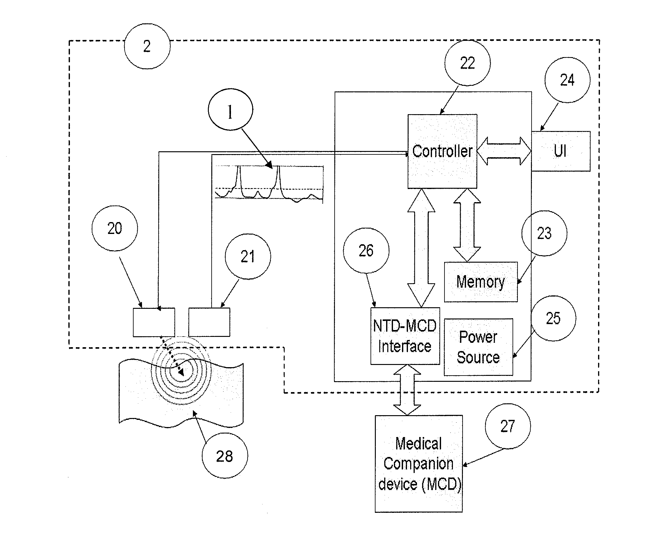
SZTRUBEL GENEVIEVE +1
View PDF5 Cites 88 Cited by
  • Summary
  • Abstract
  • Description
  • Claims
  • Application Information

AI Technical Summary

Benefits of technology

The invention is a method and apparatus for detecting and stimulating neural tissue during surgery. The apparatus can be used with a surgical robot or an intravascular catheter and can also be disposable for hygienic reasons. The method involves using impulses to stimulate targeted tissue and measuring the response of the tissue to detect the presence of neural tissue. The detected neural tissue can then be spared or denervated, as desired. The apparatus can also create a tridimensional mapping of the neural tissue and monitor the progress of a denervation procedure in real-time. The invention is fully automated and can influence the functioning of the surgical robot or intravascular catheter. The apparatus can also detect the nature of the tissue and take real-time actions in response to the detection.

Problems solved by technology

Often, carrying out a surgical procedure entails damaging neural tissue in the operated area, for example, during the removal of a tumor.
The surgical team who carries out the operation faces the challenge of recognizing neural tissue and avoiding harming it in the course of tumor resection.
Since the distinction between the neural tissue and tissue to be removed is generally carried out by visual inspection of the medical team, there is a significant risk of harming neural tissue.
In the event of such damage, post operatory consequences like, for instance, erectile dysfunction would be caused.
Even in the field of elective surgery such as plastic surgery, iatrogenic visual impairment may occur as a result of neural tissue damage.
Even if the neural tissue is successfully identified the spatial density of the embedded nerve network and its proximity to the tissues which are in the area subject to the surgery, make it technically difficult to avoid neural tissue harming.
The first three methods are based on neuromuscular activity and therefore are clearly restricted to motor nerves as their measurement principle relies on the recorded electrical, mechanical or magnetic activity of a connected muscle.
The last three methods are only indirectly related to the phrenic nerve stimulation, meaning the measured signal is also influenced by several uncontrolled parameters that make it hard to extract the specific contribution of the phrenic nerve stimulation to the overall recorded signal.
Moreover, no solution is proposed for sensory or central nerves for which no practically accessible position is usually known a priori for placing the detecting means.
For the desired purpose of denervation, however, this approach has two main inconveniences.
First, the positions at which the energy is released are not necessarily close to a targeted nerve since the actual nerve's position is unknown.
This may result in insufficient denervation and poor therapeutic effect.
Moreover, since Ardians' Simplicity system is not capable of distinguishing between renal nerve and the renal artery itself, some of the energy releases may hit undesired locations on the renal artery.
Secondly, the amount of energy delivered at each position is constant and does not account for the actual progression of the denervation process.
This lack of feedback may result in unnecessarily large energy release, possibly damaging the artery wall, or, alternatively, in insufficient energy release, resulting in poor denervation.
As we have already mentioned before, neural tissue is not easy to detect, among other reasons, because of its size, shape and its being embedded with other tissues.

Method used


Image

  • Method And Apparatus For The Detection Of Neural Tissue
  • Method And Apparatus For The Detection Of Neural Tissue
  • Method And Apparatus For The Detection Of Neural Tissue

Examples


third exemplary embodiment

[0135]A third embodiment comprises a variation of the first or the second embodiment.

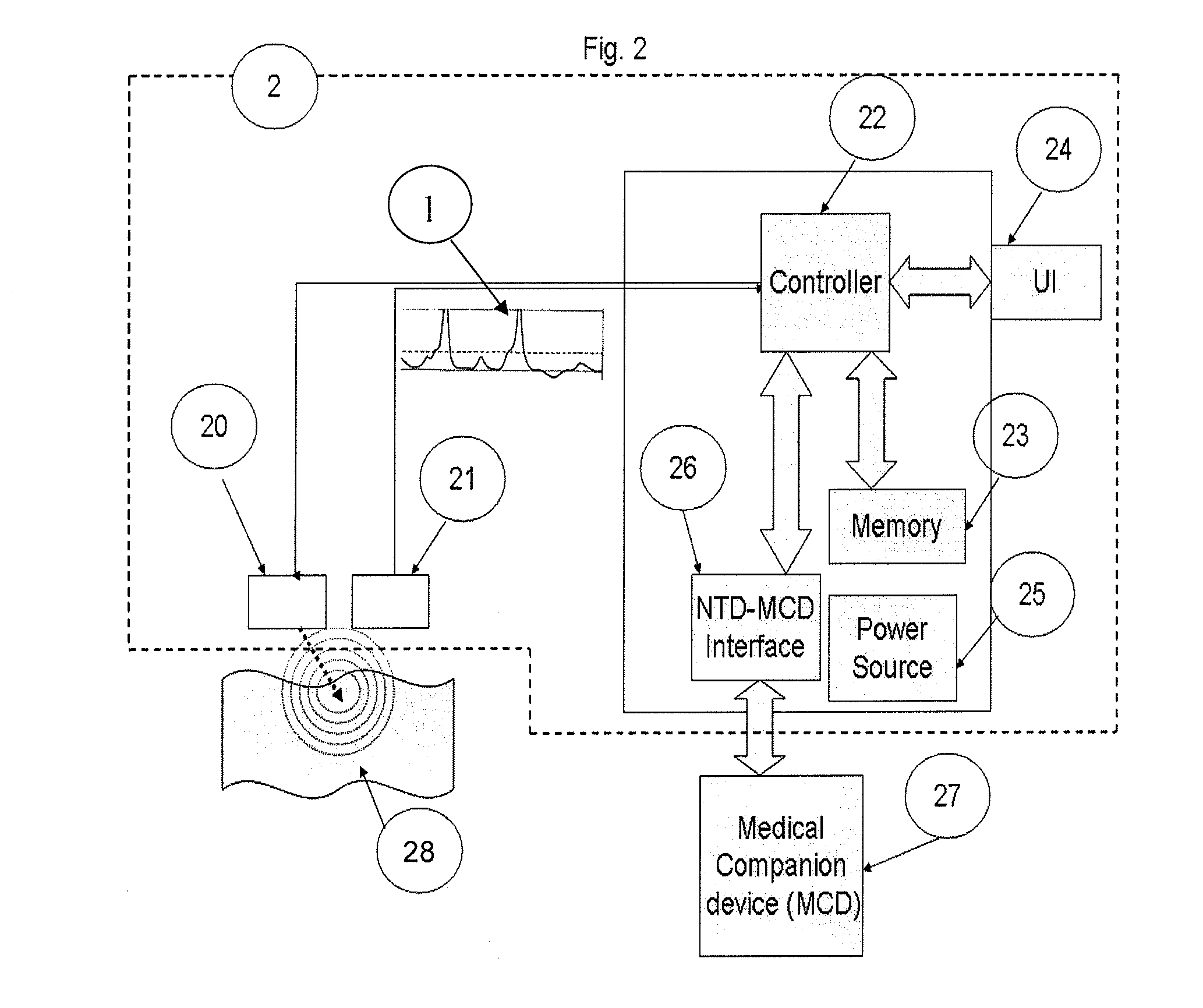
[0136]While in each of the previous embodiments the emitter 20 and the APD 21 require both physical contact with the targeted area 28 or lack thereof, in the third embodiment, the emitter 20 may require physical contact as in the second embodiment and the APD 21 may be such as not involving physical contact with the targeted area 28 or vice-versa. Apart from the mixed typology of the emitter 20 and the APD 21, this third preferred embodiment is similar in the other aspects to the first and / or the second embodiments.

fourth exemplary embodiment

[0137]In this fourth embodiment, the NTD 2 may be mounted on a surgical tool which is manually held and used by the surgeon without the mediation of a robot or any similar device. It should be noted that although the medical companion device (MCD) in previous embodiments was illustrated using a surgical robot, the MCD 27 may also be a non-robotic manually operated device or system and, more generally, any appropriate surgical tool. This consideration regarding the MCD 27 applies to all embodiments of the invention.

[0138]Typical surgical tools that fall in the category of MCD 27 in this embodiment are: scalpels, scissors, electrosurgical forcipes, ultrasonic surgical dissector and aspirator and syringes. Said tools may also optionally be used in a laparoscopic setting.

[0139]In this embodiment, as in the previous ones, the NTD 2 may be combined with a manually held surgical tool in any suitable manner which is desirable for a specific implementation of the invention. The NTD 2 may be ...

fifth exemplary embodiment

[0143]The fifth embodiment of this invention relates to a NTD 2 which is meant solely to detection of neural tissue and which constitutes a standalone device. In this case, the NTD 2 serves only the purpose of detecting the presence of neural tissue in the course of a surgery but it is not mounted or combined or otherwise used in conjunction, and does not communicate or affect in any manner whatsoever the functioning of any medical surgical tool—that is an MCD 27—that the surgeon uses in the course of the operation and does not interact in any way with the MCD 27.

[0144]Apart from the fact of being physically and functionally detached from the MCD 27, the NTD 2 in this embodiment may retain some or all of the NTD 2 functionalities described in the previous embodiments, as it may be advantageous for specific medical requirements.


PUM


Abstract

The invention relates to a method and apparatus for neural tissue detection carried out using the Neural Tissue Detector (NTD), which is the apparatus that embodies the hardware aspect of the invention. The NTD enables neural tissue detection by stimulating a small tissue area and by measuring possible occurrences of induced response from neural tissue, if neural tissue is present in said small stimulated tissue area. The information gathered by the measurement provides a real-time assessment of the nature of the tissue which is targeted by the NTD. The invention is applicable to all types of neural tissues, including motor, and / or sensory and / or other types of neural tissues. The invention offers particular advantages in robot based surgical procedures or intravascular catheter based procedures but can also be used for manual surgery, according to different specific embodiments.

Description

FIELD OF THE INVENTION[0001]The invention disclosed in this patent application relates to the field of electrophysiology and, more specifically, to the field of neural tissue detection. The invention relates to a variety of uses and applications for said neural tissue detection, including neural tissue sparing or neural tissue denervation.[0002]This invention relates to a novel method and apparatus for neural tissue detection carried out using the Neural Tissue Detector (NTD), which is the apparatus that embodies the hardware aspect of the invention, described hereafter in this patent application.[0003]The NTD enables neural tissue detection by stimulating a small tissue area and by measuring possible occurrences of induced response from neural tissue, if neural tissue is present in said small stimulated tissue area.[0004]The information gathered by said measurement provides a real-time assessment of the nature of the tissue which is targeted by the NTD.[0005]The invention is applic...

Claims


Application Information

Patent Timeline
IPC IPC(8): A61B5/04A61B5/00
CPCA61B5/0048A61B5/04001A61B5/4893A61B5/24A61B5/246
Inventor SZTRUBEL, GENEVIEVEMAYER, ARNALDO
Owner SZTRUBEL GENEVIEVE
Learn More