Determining a Tissue Parameter

a tissue parameter and parameter technology, applied in image data processing, diagnostics, applications, etc., can solve the problems of not always desirable three-dimensional perfusion recordings, inability to reliably display tissue parameters or for the entire x-ray image, and inability to achieve three-dimensional perfusion recordings, etc., to achieve the highest contrast agent concentration, the effect of high intensities and easy determination

Active Publication Date: 2015-07-30
SIEMENS HEATHCARE GMBH
View PDF12 Cites 7 Cited by
  • Summary
  • Abstract
  • Description
  • Claims
  • Application Information

AI Technical Summary

Benefits of technology

[0018]During segmentation of the vessels, at least one intensity threshold value may be used to differentiate tissue intensities and vessel intensities. This provides that intensity threshold values are determined, by way of which the vessel filling may be differentiated from the tissue filling. In one embodiment, the intensity threshold value may be determined at least partially (e.g., completely automatically and/or relative to a maximum intensity in a supplying artery). A supplying artery (e.g., in the case of recordings of the human brain, the carotid artery) may easily be located automatically and has the highest contrast agent concentration that will occur. The intensity threshold value may therefore be determined particularly ea

Problems solved by technology

However, the options known in the prior art do not allow tissue parameters to be displayed reliably or for the entire x-ray image, as the large vessels of the vessel tree overlay the parenchyma, and high cont

Method used


Image

  • Determining a Tissue Parameter
  • Determining a Tissue Parameter
  • Determining a Tissue Parameter

Examples


Embodiment Construction

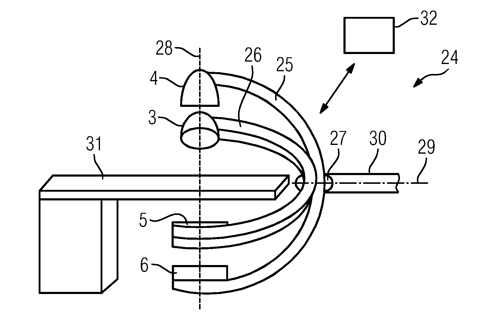
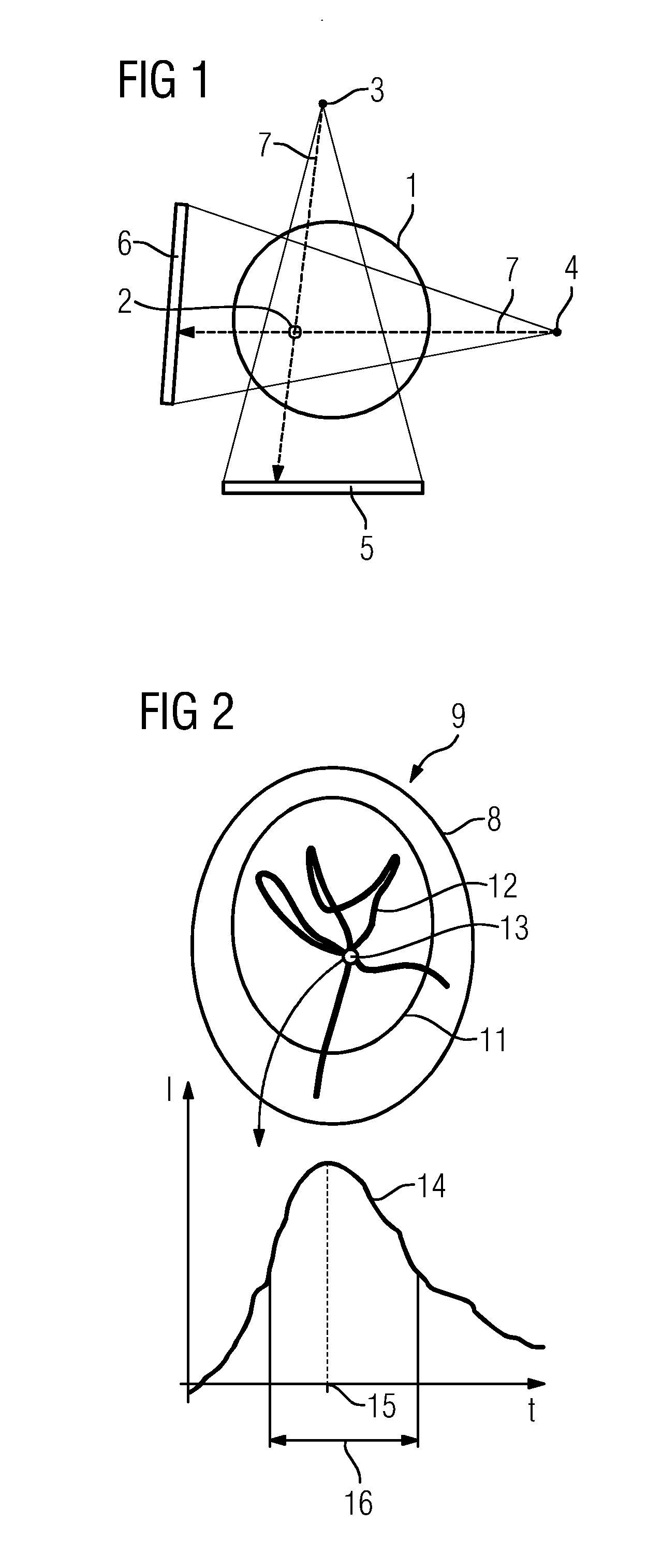
[0041]Exemplary embodiments of the method to be discussed in the following are for the determination of tissue parameters in the human brain (e.g., a relative cerebral blood volume and a relative cerebral blood flow (rCBV and rCVF)). An examination of the head of a patient is therefore considered here. The object of the examination is the parenchyma. As a basis, the method uses a series of digital subtraction angiography x-ray images, with a mask image first being recorded for each considered projection direction (e.g., two projection directions in the case of biplane examinations), for example, before the administration of the contrast agent after the patient has been positioned. After the administration of the contrast agent, a time series of raw images is recorded, showing the propagation of the contrast agent in the vascular system (e.g., the vascular system of the brain) and in the tissue. In digital subtraction angiography, to determine the x-ray images, the mask image recorde...


PUM


Abstract

A method for determining a tissue parameter of tissue that may be determined from passage of a contrast agent through the tissue based on a series of temporally consecutive two-dimensional digital subtraction angiography x-ray images showing propagation of the contrast agent in the tissue over time and a vascular system present in a region of the tissue includes locating at least some of the vessels of the vascular system by segmentation in the x-ray images. The method also includes assigning pixels showing segmented vessels an interpolation intensity determined by interpolation from intensities of at least some of the pixels bordering the segmented vessel, so that x-ray images from which vessels have been eliminated result. The method includes determining tissue parameters for at least some of the pixels of the series of x-ray images from which the vessels have been eliminated.

Description

[0001]This application claims the benefit of DE 10 2014 201 556.1, filed on Jan. 29, 2014, which is hereby incorporated by reference in its entirety.BACKGROUND[0002]The present embodiments relate to determining a tissue parameter.[0003]In angiography, it is known to administer contrast agent that may be clearly identified in images (e.g., x-ray images) and therefore allows an assessment of the blood flow and the passage of blood through the vascular system of a patient and also through the tissue. One conventional method for tracking the propagation of the contrast agent is digital subtraction angiography. With this, a mask image is recorded without contrast agent. After this, raw images are recorded, frequently as a temporal series in a time range during which the contrast agent passes through the target region or vascular system of interest. The contrast agent may also be seen in these raw images. In order to remove the anatomy causing interference during the analysis, digital sub...

Claims


Application Information

Patent Timeline
IPC IPC(8): A61B6/00G06T7/40G06T7/00
CPCA61B6/504A61B6/481A61B6/5217G06T2207/30104G06T7/0081G06T7/408G06T2207/10116G06T7/0012A61B6/032A61B6/4441A61B6/486G06T7/0016G06T7/11G06T2207/10016
Inventor KYRIAKOU, YIANNIS
Owner SIEMENS HEATHCARE GMBH
Why Eureka
Try Eureka