Compositions and methods for treating and preventing tissue injury and disease

a technology of compositions and methods, applied in the direction of drug compositions, skeletal/connective tissue cells, prostheses, etc., can solve the problems of tissue dysfunction, achilles tendon tear, and about 15% of achilles tendon and 40% of two tendon rotator cuff repairs subsequently fail, so as to improve tissue growth and migration, promote angiogenesis, and improve the effect of tendon healing

Active Publication Date: 2015-08-20
MICROVASCULAR TISSUES
View PDF2 Cites 20 Cited by
  • Summary
  • Abstract
  • Description
  • Claims
  • Application Information

AI Technical Summary

Benefits of technology

[0007]A variety of different therapeutic methods to aid in tissue repair have been developed. These include physical structures, such as better sutures, bone anchors, and patches or implants to provide scaffolding for tissue ingrowth. In addition, a variety of growth factors have been used to improve tissue growth and migration to the wound site, as well as to promote angiogenesis. For example, there are reports of improved tendon healing using growth factors such as BMP-2, BMP-12, PDGF-BB, and bFGF in preclinical models.

Problems solved by technology

Such injuries typically result in tissue dysfunction characterized by pain, inflammation and internal tissue stress, and can ultimately result in a functional disability.
For example, while sprains to tendons will heal spontaneously, complete tears of a tendon will often lead to disability if not surgically treated.
Even despite surgical repair, about 15% of Achilles tendon and 40% of two tendon rotator cuff repairs subsequently fail.
Furthermore, the repaired tendon seldom returns to pre-injury strength and function levels.
However, due to concerns that transplanted allogeneic cells or tissue may invoke an immune response and ultimately rejection, or transfer harmful viruses or other pathogens, this work has focused on the use of autologous cells.
Unfortunately, however, the use of autologous stem cells is inconvenient.
It requires two distinct surgical procedures with associated pain, cost and morbidity, and there are also risks associated with shipping the tissue to a laboratory for processing and delays in treatment of the injured patient.

Method used


Image

  • Compositions and methods for treating and preventing tissue injury and disease
  • Compositions and methods for treating and preventing tissue injury and disease
  • Compositions and methods for treating and preventing tissue injury and disease

Examples


example 1

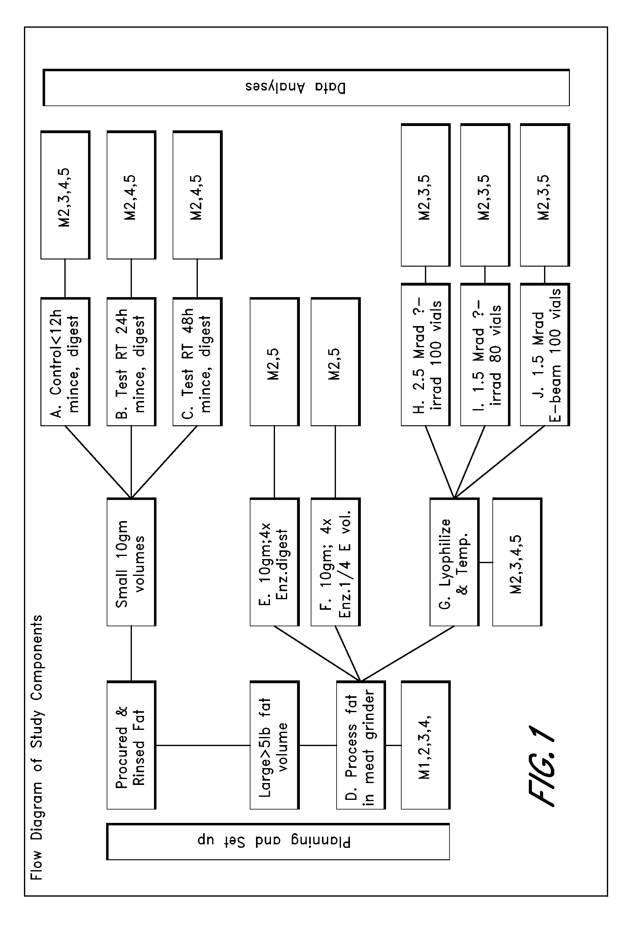
Microvascular Tissue Preparation and Characterization

[0140]In this study, microvascular tissue is prepared via different processes and then assayed. In brief, at least 5 to 10 lbs of subcutaneous fat is obtained from an organ donor and processed as follows: mince the tissue; enzymatically dissociate it; dilute (no quench), spin, decant and then wash the cell pellet; resuspend in lyophilization buffer; and lyophilization. The base conditions used for processing are as follows: adipose tissue is surgically recovered from a tissue donor. The tissue is minced with scissors, suspended in PBS with 0.2 U / ml CIzyme AS (Vitacyte, Indianapolis, Ind.) at 37° C. with gentle agitation for 60 min, then washed three times and resuspended at two million cells / ml in M3D. The cell suspension is held at room temperature until just prior to lyophilization, when the cells are diluted 50:50 with EZ-CPZ™ cryopreservation media (Incell Corp., San Antonio, Tex.), vialed, and loaded into the lyophilizer tray...

example 2

Treatment of Achilles Tendon Injury in Rats

[0159]This study demonstrates that microvascular tissue preparations of the present invention can be used to repair Achilles tendon injuries.

[0160]32 male Sprague Dawley rats (8 weeks old on DAY 1 and ˜250 g on DAY 1) are purchased from Harlan and acclimatized for at least 3 days. The rats are treated as summarized in the study design of Table 3.

TABLE 3Study Design#Animal / GroupgroupTreatmentRouteEndpoints18Achilles tendon is slightlyRightSurgery- implantabraded with mouse-toothTibiascaffold betweenforcepstibia and28Achilles tendon is slightlyAchilles tendonabraded with mouse-toothTerminationforceps + Tornier's GraftDay 7Material coated withCollagen38Achilles tendon is slightlyabraded with mouse-toothforceps + Tornier's GraftMaterial coated withCollagen + Processedmicrovasculartissue composition A48Achilles tendon is slightlyabraded with mouse-toothforceps + Tornier's GraftMaterial coated withCollagen + Processedmicrovascular tissuecompositi...

example 3

Treatment of Ischemia in Mice

[0187]This study demonstrates the effects of processed microvascular tissue compositions of the present invention in a murine model of limb ischemia. The murine model of limb ischemia is created as previously described (Jang J et al, Circulation 1999; Huang N et al, JOVE 2009), and is used to assess the effects of cell preparations in promoting angiogenesis after induction of hindlimb ischemia.

[0188]SCID mice 14-16 weeks old undergo surgically induced hindlimb ischemia. Immediately after surgery, processed microvascular tissue composition or vehicle control will be administered to the animals as detailed in Table 4 below. Briefly, three test cell articles (each at 0.5×10⁶ cells) or vehicle control are injected intramuscularly on day 0 after induction of hindlimb ischemia into the gastrocnemius. The three test articles include: processed microvascular tissue composition (Test Cells-I), processed microvascular tissue composition sterilized by E-beam (Test ...


PUM

PropertyMeasurementUnit
time pointsaaaaaaaaaa
pore sizeaaaaaaaaaa

Abstract

The present invention provides novel compositions comprising multipotent cells or microvascular tissue, wherein the cells or tissue has been sterilized and/or treated to inactivated virases, and related methods of using these compositions to treat or prevent tissue injury or disease in an allogeneic subject.

Description

RELATED APPLICATIONS[0001]This application claims the benefit of U.S. Provisional Application No. 61 / 703,203, filed on Sep. 19, 2012, the disclosure of which is expressly incorporated by reference herein.BACKGROUND[0002]1. Field[0003]Several embodiments of the present invention are directed to novel compositions comprising multipotent cells and / or microvascular tissue, which has been sterilized and / or treated to inactivate any viruses, and methods for their preparation and allogeneic or xenogeneic use in treating or preventing tissue injury and diseases, such as, e.g., arthritis.[0004]2. Description of the Related Art[0005]Injuries to soft tissues, such as muscles, tendons, ligaments, and joint capsules, occur quite frequently. Such injuries typically result in tissue dysfunction characterized by pain, inflammation and internal tissue stress, and can ultimately result in a functional disability. For example, while sprains to tendons will heal spontaneously, complete tears of a tendo...

Claims


Application Information

Patent Timeline
Patent Type & Authority Applications(United States)
IPC IPC(8): A61K35/44A61L27/54A61L27/38A61L27/36
CPCA61K35/44A61L27/3625A61L27/3691A61L27/54A61L2300/41A61L27/3834A61L2430/10A61L2430/34A61L2300/412A61L27/3687A61L2430/02A61L2430/06A61L2430/38C12N5/0653A61P17/00A61P17/02A61P19/00A61P19/02A61P19/08A61P29/00A61P43/00A61P9/00A61P9/10A61P9/14A61K35/12
Inventor PETERSON, DALE R.MATTERN, RALPH-HEIKOWILSON-WIRTH, COREYOHASHI, KEVIN L.
Owner MICROVASCULAR TISSUES
Why Eureka
Try Eureka