Porous solid phase for rapidly isolating biological molecules for nucleic acid amplification reaction from biological sample, and use thereof

Inactive Publication Date: 2015-09-10
BIO CUBE SYST
2 Cites 0 Cited by

AI-Extracted Technical Summary

Problems solved by technology

In the case when PCR is used for the diagnosis of pathogens, it may have a difficulty in examining a large amount of a sample at one trial although PCR has high specificity and detection sensitivity.
This is because the rapid separation of ...

Method used

[0032]In the method of the present invention, when the isolated biological molecule is gDNA it may be sucked by allowing a front end of a ceramic rod to contact with the biological sample. When the starting material is mRNA a front end of a ceramic rod is allowed to contact with the biological sample, and cDNA is synthesized using the total RNA adsorbed onto the front end as a template with reverse transcriptase. Since the total RNA is the one isolated from a plant or animal cell, there is a poly-A tail at the end of the mRNA, and cDNA can be easily synthesized using an oligo dT primer and a reverse transcriptase based on the characteristics of the sequence. Additionally, in the case of a virus, when there is a poly-A tail, cDNA can be synthesized in the same manner as described above, however, when the poly-A is absent (for example, tobacco mosaic virus), cDNA can be synthesized using a target RNA-specific antisense primer according to a method known in the art.
[0034]When a nucleic acid amplification technology is applied, it is important to design an appropriate primer for virus detection of the present invention. However, the amount of the template amplification can be increased when RT and PCR are performed separately in two different tubes rather than when they are performed in the same tube, thereby improving the reliability of virus verification results. According to a preferred embodiment of the present invention, there is provided a method for analyzing the presence/absence of virus within tissues using a genotyping primer designed to be matched with nucleotides sucked into the ceramic block.
[0040]When the ceramic of oxide material is used as a porous solid phase of the present invention its pore size can be adjusted. When a porous ceramic is manufactured using the same oxide material the size and number of pore can be adjusted according to the manufacturing temperature, and thus PCR or RT-PCR can be effectively performed using the optimized porous ceramic according to the type of biological molecules to be targeted. Regarding the pore size of such porous ceramic, the size of the porous solid phase may be appropriately adjusted so that it can selectively suck the targeted biological molecules. Additionally, although the...

Benefits of technology

[0011]The present invention enables a rapid diagnosis of the presence of a target sequence via rapid isolation of biological molecules present in a biological sample...

Abstract

The present invention provides a method for rapidly ascertaining whether or not a target sequence exists in a biological sample by: rapidly isolating biological samples for a nucleic acid amplification reaction from the biological sample by using a porous solid phase capable of rapidly sucking biological samples present in a biological sample into pores; and amplifying the same.

Application Domain

Nucleotide librariesLead monoxide +8

Technology Topic

Porous solidsSolid phases +3

Image

  • Porous solid phase for rapidly isolating biological molecules for nucleic acid amplification reaction from biological sample, and use thereof
  • Porous solid phase for rapidly isolating biological molecules for nucleic acid amplification reaction from biological sample, and use thereof
  • Porous solid phase for rapidly isolating biological molecules for nucleic acid amplification reaction from biological sample, and use thereof

Examples

  • Experimental program(7)

Example

Example 1
Adsorption of a Genetic Material Using a Porous Ceramic Cube and its Use as a Template for RT-PCR/PCR
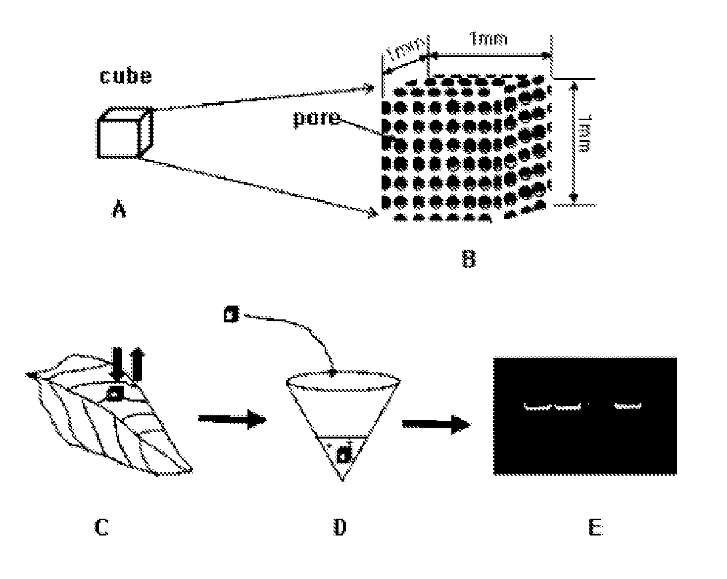
[0068]The adsorption of a genetic material using a porous ceramic cube of the present invention and a diagram illustrating its use as a template for RT-PCR/PCR is shown in FIG. 1. In the present invention, 14 kinds of cubes were manufactured by varying the manufacturing temperature using the main components described in Table 1 so that the size of pores on the surface of the 1 mm3 cube can vary, and the enlarged view of the porous ceramic cube is shown in FIG. 1B. Since cubes are porous smaller substances than the pores can be sucked. The present invention aims at selectively absorbing the desirable ones among various substances generated during rupture of tissues by varying the size of pores (In general, when ceramics are manufactured at high temperature the pore size becomes smaller), that is, providing a cube with an ultrafiltration function to be capable of maximally excluding PCR inhibiting substances depending on the pore size.
[0069]The present invention, a single porous ceramic cube manufactured above was placed on top of a plant leaf, and pressed with a flat portion on the rear side of metal tweezers so that the genetic substance can be simultaneously sucked into the pores of the cube while the pressed tissues are burst, and the result of using the single cube into which the genetic substance was sucked as a template for RT-PCR or PCR was confirmed in an agarose gel (FIG. 1E).

Example

Example 2
Isolation of a DNA/RNA Template for Nucleic Acid Amplification from a Biological Sample Using a Cut-Out Ceramic Fragment
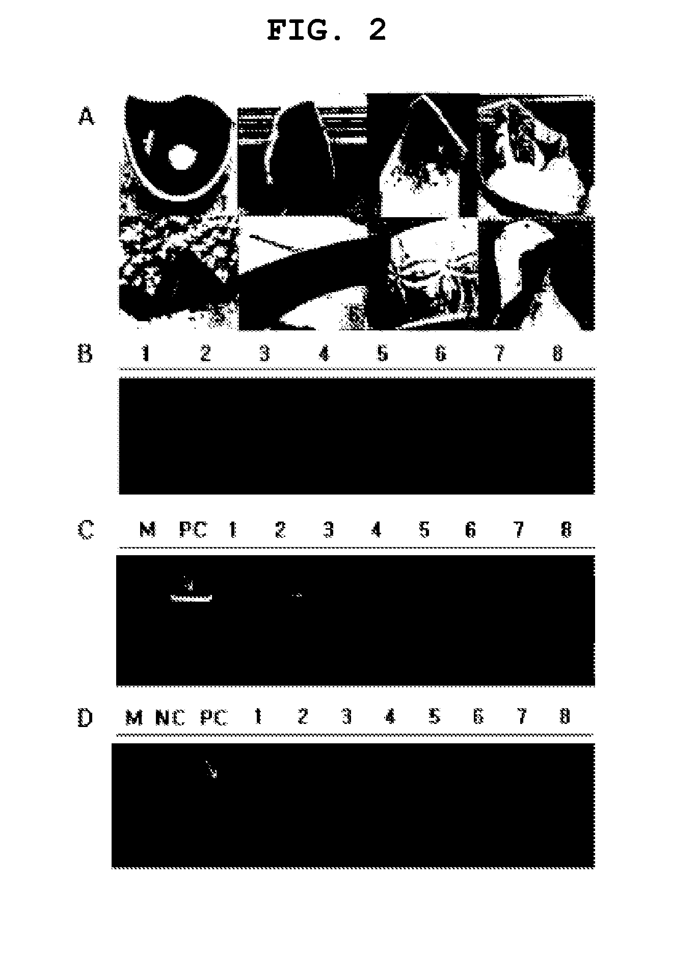
[0070]Seven kinds of cut-out ceramic fragments including blue and white porcelain was broken into small pieces using a nipper, and the fragments with a volume of about 1 mm3 selected from the unglazed area were used as an absorptive material for a genetic material. For the 5th ceramic fragment in FIG. 2A, it was divided into yellow and grey according to the area coated with glaze, and the yellow fragments and the grey fragments obtained respectively therefrom were indicated as 5 and 6, respectively. Each ceramic fragment was placed on top of Capsicum annuum CM334 pepper leaves, lightly pressed with a flat portion on the rear side of metal tweezers to obtain gDNA. One ceramic fragment keeping gDNA was added into each PCR tube, which was already added with a primer premix capable of verifying molecular markers related to the trichomes of pepper. The PCR premix consisted of 0.5 μL of a 10 pmol sense primer (Tsca-F: AAACGCCATCATTCGTTTTC: SEQ ID NO: 1), 0.5 μL of a 10 pmol antisense primer (Tsca-R: CATGAAAGTTGACCCGAACA: SEQ ID NO: 2), 4 μL of rTaq-Mix, and 15 μL of DW. The PCR product was denatured at 94° C. for 3 minutes, amplified under the set conditions (94° C./30 sec, 59° C./30 sec, 72° C./60 sec) via 35 amplification cycles, and reacted at 72° C./5 min, and the resultant was electrophoresed on a 1% agarose gel containing EtBr, and confirmed whether the target PCR product was amplified.
[0071]As a result, as shown in FIG. 2B, the amplification of PCR products in ceramic fragments 4, 5 and 6 were confirmed. Upon examination of the surface of the ceramic fragment 4, which showed the best genomic DNA amplification, under electronic microscope, it was confirmed that the pores in the ceramics were present in various sizes (FIG. 3A). However, there was a bit of difficulty in clearly distinguishing the kinds of ceramic fragments and amplification efficiency of PCR products when the surfaces or areas of ceramic fragments were not identically controllable.
[0072]Accordingly, in order to confirm to what extent each ceramic fragment can get the gDNA of pepper sucked and whether the sucked gDNA can be applied to PCR, the purified CM334 gDNA (1 μg/μL) was used as a material instead of leaves. The purified gDNA in an amount of 1 μL was dropped onto the surface of a plastic petri dish, and the gDNA was sucked into each ceramic fragment, which was used as a template for PCR. As shown in FIG. 2C, as is the case with a positive control group (PC) where 1 μL of CM334 gDNA was used as a template, it was confirmed that PCR products with the same size were amplified in all treated groups. Although there was a little difference in their concentrations the difference was speculated to be due to the difference in size or surface area of the ceramic fragments used therein. Meanwhile, it was confirmed that the ceramic fragments where the gDNA was sucked into can be used as a template for PCR amplification, and thus the possibility whether they can be used for the diagnosis of plant viruses was examined. In order to purify RNAs for negative and positive control using QIAGEN RNeasy Mini Kit, the total RNAs were isolated from sound Nicotiana rustica tobacco leaves (for NC) not infected with viruses and Nicotiana rustica tobacco leaves (for PC) artificially infected with Tomato spotted wilt virus (TSWV), and used them as templates for reverse transcription. Each of the ceramic fragments was placed on top of TSWV-infected tobacco leaves, pressed with a flat portion on the rear side of metal tweezers to get RNA or virus particles sucked thereinto, and each of the fragments was respectively added into a reverse transcription master premix (ELPIS-Biotech, Korea) already added with 0.5 μL of 10 pmol antisense primer TSNCPR (5′-TCAAGCAAGTTCTGCGAGTT-3′: SEQ ID NO: 3), one per each tube. After performing a reverse transcription at 42° C. for 1 hour, 1 μL of the resulting liquid was used as a template for PCR. The PCR premix consisted of 0.5 μL of 10 pmol sense primer (Tsca-F AAACGCCATCATTCGTTTTC: SEQ ID NO: 4), 0.5 μL of 10 pmol antisense primer (Tsca-R CATGAAAGTTGACCCGAACA: SEQ ID NO: 5), 4 μL of rTaq-Mix, and 15 μL of DW. The PCR product was denatured at 94° C. for 3 minutes, amplified under the set conditions (94° C./30 sec, 59° C./30 sec, 72° C./60 sec) via 35 amplification cycles, and reacted at 72° C./5 min, and the resultant was electrophoresed on a 1% agarose gel containing EtBr, and the presence/absence of the target PCR product (777 bp) was confirmed. As shown in FIG. 2D, it was confirmed that the expected RT-PCR products were amplified in the ceramic fragments-treated groups except the ceramic fragments 4 and 6. The results suggest that TSWV verification is possible with TSWV RNA or TSWV particles sucked into the ceramic fragments alone, and the higher amplification of PCR product in RT-PCR than gDNA shown in FIG. 2B appears to be due to the higher amount of PCR templates produced by the reverse transcription. From the foregoing, it was suggested that the manufacture of the ceramic fragments into a uniform size and its subsequent use for absorption of biological molecules will enable to obtain a better result.

Example

Example 3
Examination of an Absorption Rate of Biological Molecules According to the Types of Porous Ceramic Cubes of Oxide Material
[0073]The picture of the surfaces of the respective porous ceramic cube manufactured using the oxide described in Table as a main component enlarged under scanning electron microscope (SEM) is shown in FIG. 3. Since the surface and pore size vary according to the main components of ceramics and their manufacturing temperature, they should be manufactured to obtain the optimum pore size and number of the ceramic cubes in order to improve the absorption rate of the target biological molecules.
TABLE 1 Sign Main Component Manufacturing Temperature (° C.) 1 Al2O3 1450 2 Al2O3 1550 3 Fe2O3 800 4 Fe2O3 850 5 Fe2O3 900 6 LTCC 650 7 LTCC 750 8 LTCC 850 9 PbO 1000 10 PbO 1150 11 PbO 1250 12 ZnO 800 13 ZnO 900 14 ZnO 1000
[0074]① Regarding Total RNA Isolated from CMV-Infected Capsicum annuum CM334 Pepper Leaves and Genomic DNA Isolated from CM334 Pepper
[0075]In order to examine the absorption efficiency of biological molecules according to the type of porous ceramic cubes comprised of oxide material, the total RNA isolated from CMV-infected pepper leaves and genomic DNA isolated from CM334 pepper were sucked into each ceramic cube, and used as a template for RT-PCR and PCR, respectively. For CMV, a sense primer (5′-TACATTGAGTCGAGTCATG-3′: SEQ ID NO: 6) and an antisense primer (5′-TGGAATCAGACTGGGACA-3′: SEQ ID NO: 7) were respectively added at a concentration of 25 pmol to an RT-PCR premix, and amplified under the set conditions 50° C./20 min, 94° C./10 min, (94° C./30 sec, 55° C./30 sec, 72° C./60 sec) for 35 amplification cycles, 72° C./5 min, and electrophoresed on a 1% agarose gel containing EtBr, and the presence/absence of the target PCR product (670 bp) was confirmed (FIG. 4A). For gDNA, high concentration of gDNA (100 μg) isolated from Capsicum annuum CM334 pepper leaves was respectively sucked into each porous ceramic cube, and added one per each PCR tube. The PCR premix consisted of 0.5 μL of 10 pmol sense primer (Tsca-F: AAACGCCATCATTCGTTTTC: SEQ ID NO: 8), 0.5 μL of 10 pmol antisense primer (Tsca-R: CATGAAAGTTGACCCGAACA: SEQ ID NO: 9), 4 μL of rTaq-Mix, and 15 μL of DW. The PCR product was denatured at 94° C. for 3 minutes, amplified under the set conditions (94° C./30 sec, 59° C./30 sec, 72° C./60 sec) via 35 amplification cycles, and reacted at 72° C./5 min, and the resultant was electrophoresed on a 1% agarose gel containing EtBr, and the presence/absence of the target PCR product was confirmed. In the case of viral RNA, as shown in 4A, the 14 kinds of ceramic cubes showed similar results (FIG. 4A). In contrast, the efficiency in PCR reaction using the purified gDNA varied according to the cube material and cube manufacturing temperature (FIG. 4B). Lane 1 (Al2O3, 1450° C.) and Lane 2 (Al2O3, 1550° C.) showed no significant difference in an amount of amplification, and Lane 3 (Fe2O3, 800° C.) and Lane 4 (Fe2O3, 850° C.), which were manufactured at low temperature, showed almost no amplification, however, Lane 5 (Fe2O3, 900° C.), which was manufactured at high temperature, showed a slight amplification. Lane 6 (LTCC, 650° C.) showed no amplification but Lane 7 (LTCC, 750° C.) and Lane 8 (LTCC, 850° C.) showed a significant level of amplification. Lane 9 (PbO, 1000° C.) and Lane 11 (PbO, 1250° C.) showed a low level of amplification but Lane 10 (PbO, 1150° C.) showed a significant level of amplification. Lane 12 (ZnO, 800° C.) and Lane 14 (ZnO, 1000° C.) showed almost no amplification, but Lane 13 (ZnO, 900° C.) showed a low level of amplification. Conclusively from the above results, genomic DNA absorption rate varied according to cube material and cube manufacturing temperature, and in structures with almost no pores as in Lane 8 (LTCC, 850° C.), Lane 11 (PbO, 1250° C.), and Lane 14 (ZnO, 1000° C.), only a small amount of genomic DNA, which is not detectable under the PCR conditions used in the present invention, was supposed to be sucked.
[0076]② Regarding CMV-Infected Pepper Leaves and Purified CMV Particles
[0077]In order to examine the absorption efficiency of biological molecules according to the type of the porous ceramic cubes, the CMV-infected pepper leaves and CMV particles were sucked into ceramic cubes and used as templates for RT-PCR and PCR. For CMV, a sense primer (5′-TACATTGAGTCGAGTCATG-3′: SEQ ID NO: 10) and an antisense primer (5′-TGGAATCAGACTGGGACA-3′: SEQ ID NO: 11), as a CMV particles-specific primer set, were added respectively at a concentration of 25 pmol to RT-PCR premix (RPampl, Biocubesystem, Korea), and reacted under the conditions of 50° C./20 min, 94° C./10 min, (94° C./30 sec, 55° C./30 sec, 72° C./60 sec) 35 amplification cycles, and 72° C./5 min, and the resultant was electrophoresed on a 1% agarose gel containing EtBr, and the presence/absence of the target PCR product (670 bp) was confirmed. When the purified virus was sucked into a porous ceramic cube there was a higher amplification in the PCR product but the PCR product amplification feature between the two treated sections were similar (FIG. 5).
[0078]③ Sucking Rate of Biological Molecules According to Manufacturing Temperature and Material of the Porous Ceramic Cube
[0079]In FIG. 5A, Lanes 1 and 2 have the same main component of Al2O3, but their manufacturing temperatures are 1450° C. and 1550° C., respectively, and Lanes 3, 4 and 5 have the same main component of Fe2O3, their manufacturing temperatures are 800° C., 850° C., and 900° C., respectively, and the PCR efficiency was high when DNA was isolated using porous ceramic cubes composed of Al2O3 and Fe2O3 manufactured at 1550° C. and 900° C., respectively. Lanes 6, 7, and 8 have the same main component of Low temperature co-fired ceramic (LTCC), and their manufacturing temperatures were 650° C., 750° C., and 850° C., respectively, wherein Lane 6 showed no amplification at all, and Lane 8 showed a higher amplification than Lane 7.
[0080]④ Analysis of Pepper DNA Amplification Efficiency Using Porous Ceramic Cubes
[0081]A single porous ceramic cube was placed on top of a Capsicum annuum sr10 pepper leaf, pressed with a flat portion on the rear side of metal tweezers to get gDNAsucked thereinto, and the resultant was added into a PCR tube, one per each tube. The solution for PCR reaction was prepared by adding 0.5 μL of 10 pmol sense primer (Primer 10-F: 5′-TGGCTTATCGAAGGAGCCAT-3′: SEQ ID NO: 12), 0.5 μL of 10 pmol antisense primer (Primer 10-R: 5′-AGATGAAACCAAAGCCTCCA-3′: SEQ ID NO: 13), a cube with gDNA, and 9 μL of DW to 10 μL to a 2×PCR premix (gDamp1, Biocubesystem, Korea), and that for a positive control group was prepared by adding 2 μL of purified gDNA (20 ng/μL) and 7 μL of DW instead of a cube. The PCR product was denatured at 94° C. for 3 minutes, amplified under the set conditions (94° C./30 sec, 58° C./30 sec, 72° C./60 sec) via 35 amplification cycles, and reacted at 72° C./5 min, and the resultant was electrophoresed on a 1% agarose gel containing EtBr, and confirmed whether the PCR product was amplified. As a result, Lane 5 (Fe2O3, 900° C.), Lane 8 (LTCC, 850° C.), Lane 9 (PbO, 1000° C.), Lane 10 (PbO, 1150° C.) and Lane 14 (ZnO, 1000° C.) showed good amplifications, whereas Lane 6 (LTCC, 650° C.), Lane 7 (LTCC, 750° C.) and Lane 8 (LTCC, 850) and Lane 12 (ZnO, 800° C.), Lane 13 (ZnO, 900° C.) and Lane 14 (ZnO, 1000° C.) were apparently distinguished according to the manufacturing temperature (FIG. 6)

PUM

PropertyMeasurementUnit
Shape

Description & Claims & Application Information

We can also present the details of the Description, Claims and Application information to help users get a comprehensive understanding of the technical details of the patent, such as background art, summary of invention, brief description of drawings, description of embodiments, and other original content. On the other hand, users can also determine the specific scope of protection of the technology through the list of claims; as well as understand the changes in the life cycle of the technology with the presentation of the patent timeline. Login to view more.

Similar technology patents

Macrocyclic picolinamides as fungicides

ActiveUS20140187588A1good performanceeasy to use
Owner:CORTEVA AGRISCIENCE LLC

System and method of monitoring temperature

InactiveUS7140768B2less susceptible to tampereasy to use
Owner:COLD CHAIN TECH

Wireless charging transmitter, receiver, charging device and wireless charging method

ActiveCN104158269ASimple codingeasy to use
Owner:湖南长城科技信息有限公司 +1

Classification and recommendation of technical efficacy words

  • easy to use

Laser assisted total joint arthroplasty

InactiveUS20090234360A1easy to use
Owner:ALEXANDER VLADIMIR

Enclosure for a portable hemodialysis system

ActiveUS20090114582A1easy to use
Owner:DEKA PROD LLP
Why Eureka
Try Eureka