Detection of carious dentin tissue and removal thereof by means of a dental instrument

a technology of dentin tissue and dental instrument, which is applied in the field of dentin caries to achieve the effect of reducing the aggressiveness of active components and high viscosity

Inactive Publication Date: 2015-11-05
RLS GLOBAL AB
View PDF4 Cites 2 Cited by
  • Summary
  • Abstract
  • Description
  • Claims
  • Application Information

AI Technical Summary

Benefits of technology

[0060]In a further aspect, there is provided a preparation containing an active, caries-dissolving two-component liquid in the form of a first active, caries-dissolving component and the a second component which reduces the aggressiveness of the active component to mucous membranes, and a gel substance for use in removal of smear layer(s) from healthy or carious dentin tissue. The preparation may an aqueous composition compr

Problems solved by technology

Surprisingly, the inventors have found that some dental instruments such as some dental drills do not yield dentin tissue comprising smear layer(s) upon treatment of the

Method used


Image

  • Detection of carious dentin tissue and removal thereof by means of a dental instrument
  • Detection of carious dentin tissue and removal thereof by means of a dental instrument
  • Detection of carious dentin tissue and removal thereof by means of a dental instrument

Examples


example 1

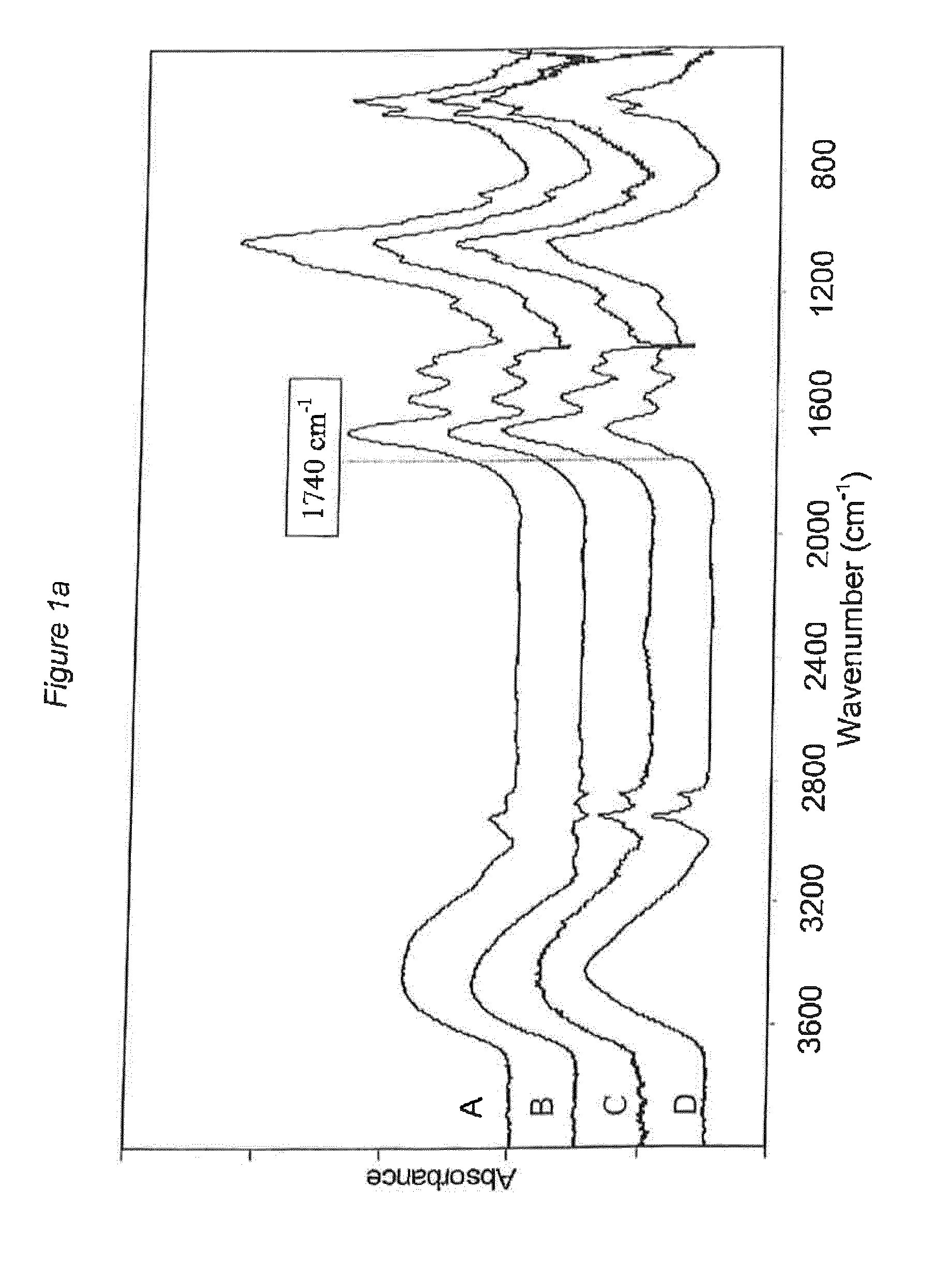
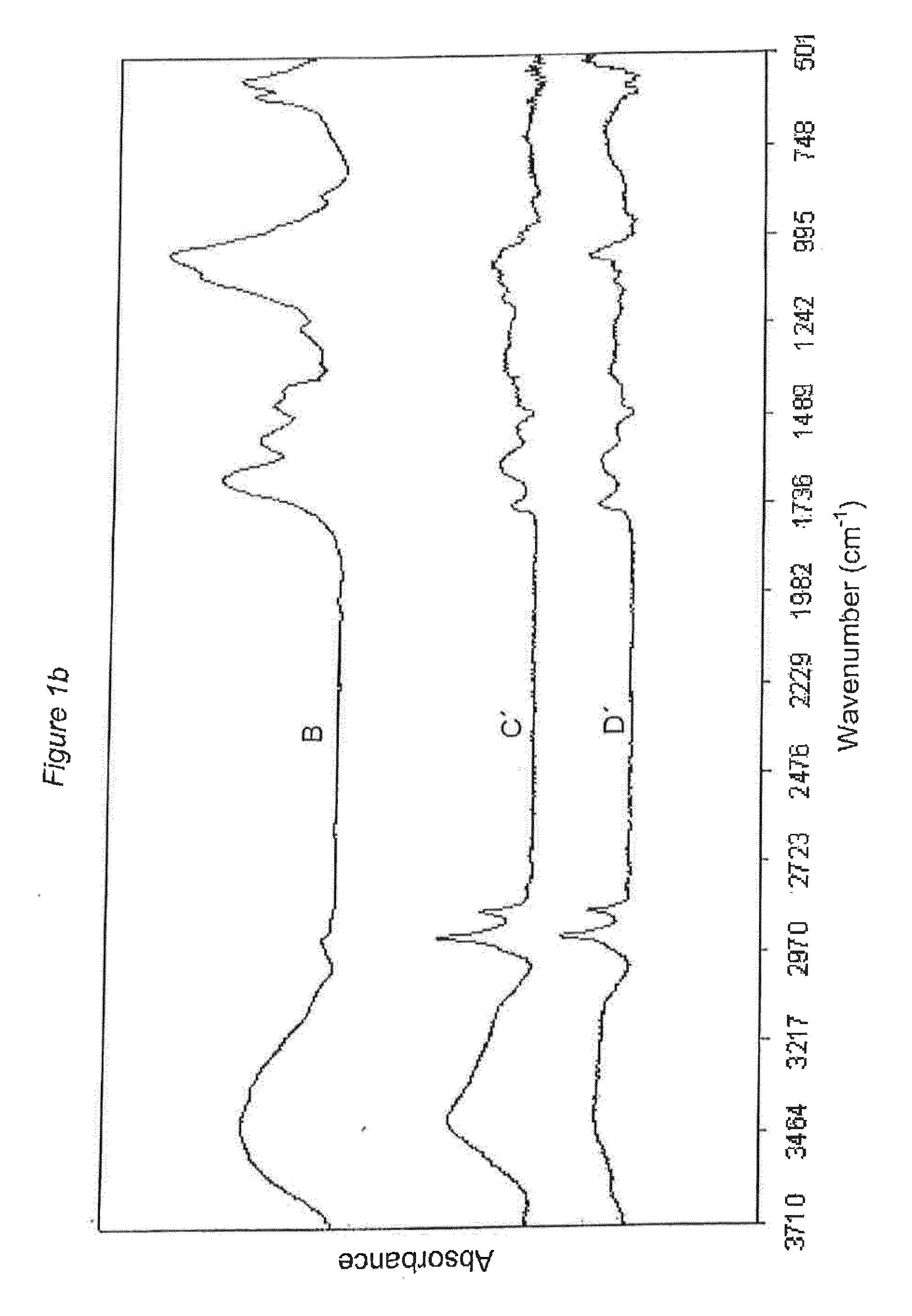
[0217]Two extracted permanent human teeth with no previous dental restorations were selected due to their severe carious tissue status and analysed with FTIR within one week after extraction. The outermost part of the carious dental lesions was removed. The remaining dental carious tissue was divided into two layers; one outer layer with discoloured, soft and infected dental carious tissue and one inner layer that was seen as uncoloured and excavated down to the estimated hardness of the remaining healthy dentin by tactile procedures. For each tooth, one sample was taken from healthy dentin and another sample was taken from the inner layer of carious tissue. After incubation in purified water they were left to dry at ambient temperature. The dry weight of each tooth sample was approximately 1 mg. Each sample was then mixed with potassium bromide (KBr) before subsequent FTIR examination with a total pellet weight of 100 mg. The IR analyses were performed using a Mattson Cygnus 100 FT...

example 2

[0220]This experiment aimed to see if the added colours SEEK, Acid red 1, Lucifer Yellow, a combination of Lucifer Yellow and Acid red 1, or 5-(2-2-hydrazinyl-2-oxoethylthio)acetamido)-2-(3-hydroxy-6-oxo-6H-xanthen-9-yl)benzoic acid, Alexa Fluor® 594 and Alexa 350 bind to the surface of caries or not, after excavation down to a clinically caries free denoted surface. Experiments were performed during 4 days.

[0221]Whole caries infected tooth was added to an ortho acryl gel, which after hardening was used as a holder prior to sawing (saw of brand Zaw Micro Tone, German). Slices, having a thickness of 150 μm, were left over night in each colour to be tested (Day one). On the second day, the colours were washed out by rinsing the slices with MQ water and the teeth sections were photographed under either a microscope (6.7×; visible light), in UV (no microscope) or under a fluorescence microscope (100×). Thereafter, an aqueous salt solution (NaCl, 1 M) was added to the slices and incubate...

example 3

[0227]Extracted permanent human teeth with no previous dental restorations were selected due to their severe carious tissue status and analysed with FTIR within one week after extraction. The outermost part of the carious dental lesions was removed. The remaining dental carious tissue was divided into two layers; one outer layer with discoloured, soft and infected dental carious tissue and one inner layer that was excavated down to the observed uncoloured hard surface of the remaining healthy dentin controlled by tactile procedures.

[0228]Healthy dentin tissue and carious dentin tissue from the innermost layer were collected. This healthy dentin tissue sample and carious dentin tissue sample were then each divided into three samples.

[0229]The healthy dentin tissue sample (26 mg) was divided and treated as follows. Sample one, hereinafter denominated S1, (11 mg) was repeatedly washed in purified water and put under vacuum and defined as untreated sample, i.e. the reference sample. The...


PUM


Abstract

The application relates to a method for testing a dental instrument comprising the steps of: a) subjecting healthy dentin tissue free from smear layer(s) to treatment with a dental instrument, thereby yielding treated dentin tissue, b) addition of a composition comprising one or more hydrazine derivative(s) to the treated dentin tissue obtained in step a), c) examining if the treated dentin tissue in step b) is stained by the one or more hydrazine derivative(s), and d) determining whether or not the treated dentin tissue contains a smear layer, wherein said one or more hydrazine derivatives is a compound of formula (I) RNHNH2 wherein R is a chemical group containing a chromophore or forming a chromophore with NHNH2. Further, the application relates to a kit of parts for detection and removal of carious dentin tissue and to a method for detection and removal of carious dentin tissue.

Description

TECHNICAL FIELD[0001]The present invention concerns dental caries. In particular, the present invention concerns detection of carious dentin tissue using hydrazine derivative(s), and selective removal of detected carious tissue.BACKGROUND[0002]In the treatment of carious lesions the major part of the carious tissue can be examined by judging the hardness of the affected surface using tactile instruments (so-called probing) and / or by visually observing the well-known discoloration known as Maillard products. However, the caries affected surface near the healthy dentin, which is called the transparent zone, is not discoloured.[0003]For early stage caries where the caries only affects the enamel of the tooth different therapies are used. In one therapy one removes the caries affected tissue using mechanical means such as dental drill tools. Another therapy uses a preventive methodology such as fluoridation.[0004]Initial caries, i.e. caries affecting only the enamel of the tooth, is nor...

Claims


Application Information

Patent Timeline
IPC IPC(8): A61K6/00A61C5/02G01N23/225A61B5/00G01N33/483G01N21/35A61C3/02A61C19/06A61C5/42
CPCA61K6/0061A61C3/02A61C5/023A61C19/063G01N2021/3595G01N33/4833G01N21/35G01N23/2258A61K6/002A61B5/4547A61B5/0088A61C5/42A61K6/66A61K6/25
Inventor ALMHOJD, ULRICANILSSON, KEFLORBERGER, FREDRIKHANSSEN, KENTH
Owner RLS GLOBAL AB
Why Eureka
Try Eureka