System and method for three dimensional printed implantation guides

a three-dimensional printing and implantation guide technology, applied in the field of three-dimensional printing implantation guides, can solve the problems of lack of a reproducible process that may be used to fabricate size-specific customized bio-printed musculoskeletal tissue for use in various orthopedic applications, inability to precisely size, shape and contour the osteochondral and chondral graft material to be placed, and inability to develop the articular surface of joints

Inactive Publication Date: 2015-12-10
KOSAREK FR J +1
View PDF2 Cites 4 Cited by
  • Summary
  • Abstract
  • Description
  • Claims
  • Application Information

AI Technical Summary

Benefits of technology

[0009]The problems associated with conventional means of creating osteochondral and chondral grafts is addressed by the present invention by providing a reproducible process that may be used to fabricate size-specific, customized bio-printed musculoskeletal tissue in particular for use in various orthopedic applications.
[0010]The present invention also includes a custom, patient specific, 3-D printed guide for precise placement of osteochondral graft material that utilizes data obtained from radiologic imaging of the subject joint.
[0011]In one aspect of the invention, a method of repairing a defect on an articular surface of a bone within a joint is provided. The method includes acquiring a data set of a defect on an articular surface of a bone within a joint to be repaired by radiological imaging; evaluating the data set for the location of the defect; marking said defect with computer software; transferring said data with said marked defect to a 3-D printer and printing out a guide that demarcates said defect as a cut-out portion in said guide, said guide including a first guide reference element thereon; performing an osteochondral biopsy on an area of the damaged joint away from the defective area to obtain osteochondral cells; culturing the osteochondral biopsy cells to create a biogel; loading the biogel into a 3-D bioprinter to create an osteochondral tissue plug and hardening sai

Problems solved by technology

While great strides have been made in this area, a reproducible process that may be used to fabricate size-specific, customized bio-printed musculoskeletal tissue for use in various orthopedic applications is lacking.
Specifically, a method to create precisely sized, shaped and contoured osteochondral and chondral graft material to be placed in defects on the articular surface of joints has not been developed.
In addition, no precise method or guide to aid in the exact placement of these osteochondral or chondral grafts on joint surfaces exists.
Known technologies do not address this need.
Both of these osteochondral ALLOgraft materials are problematic for several reasons.
Fresh cadaveric ALLOgraft material is difficult to obtain from a donor patient as it has to be acquired and implanted in a short period of time from the deceased donor to the recipient.
This leads to logistic problems related to speedy harvest and delivery of the fresh cadaveric ALLOgraft material.
To address this problem, fresh frozen ALLOgraft cadaveric material has been used, however, it is problematic because it has been frozen, the cartilage and osseous cell viability within the tissue is decreased.
Further, both frozen and fresh osteochondral ALLOgraf

Method used


Image

  • System and method for three dimensional printed implantation guides
  • System and method for three dimensional printed implantation guides
  • System and method for three dimensional printed implantation guides

Examples


Embodiment Construction

[0028]Unless defined otherwise, all technical and scientific terms used herein have the same meaning as is commonly understood by one of skill in the art to which this invention belongs.

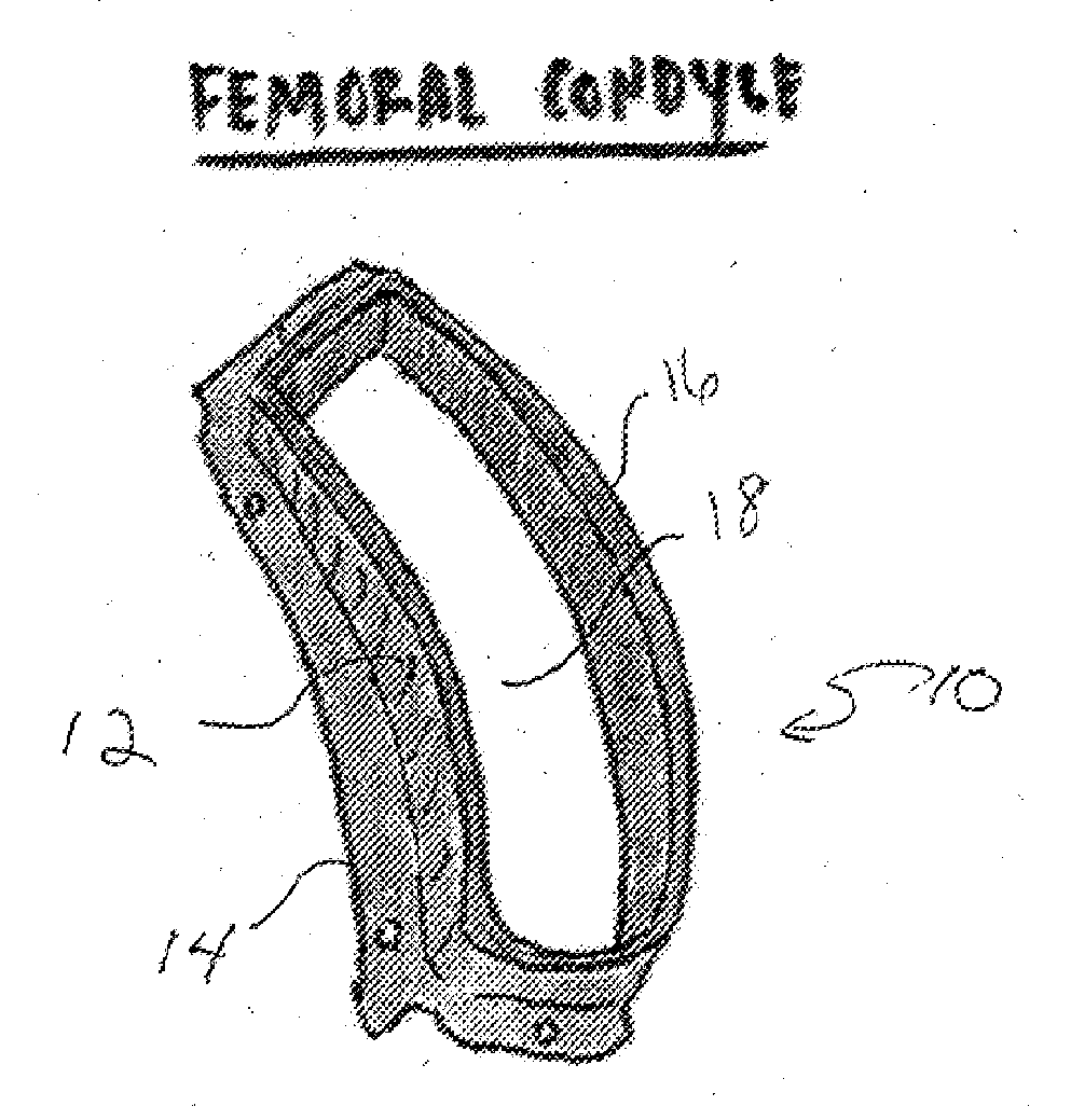
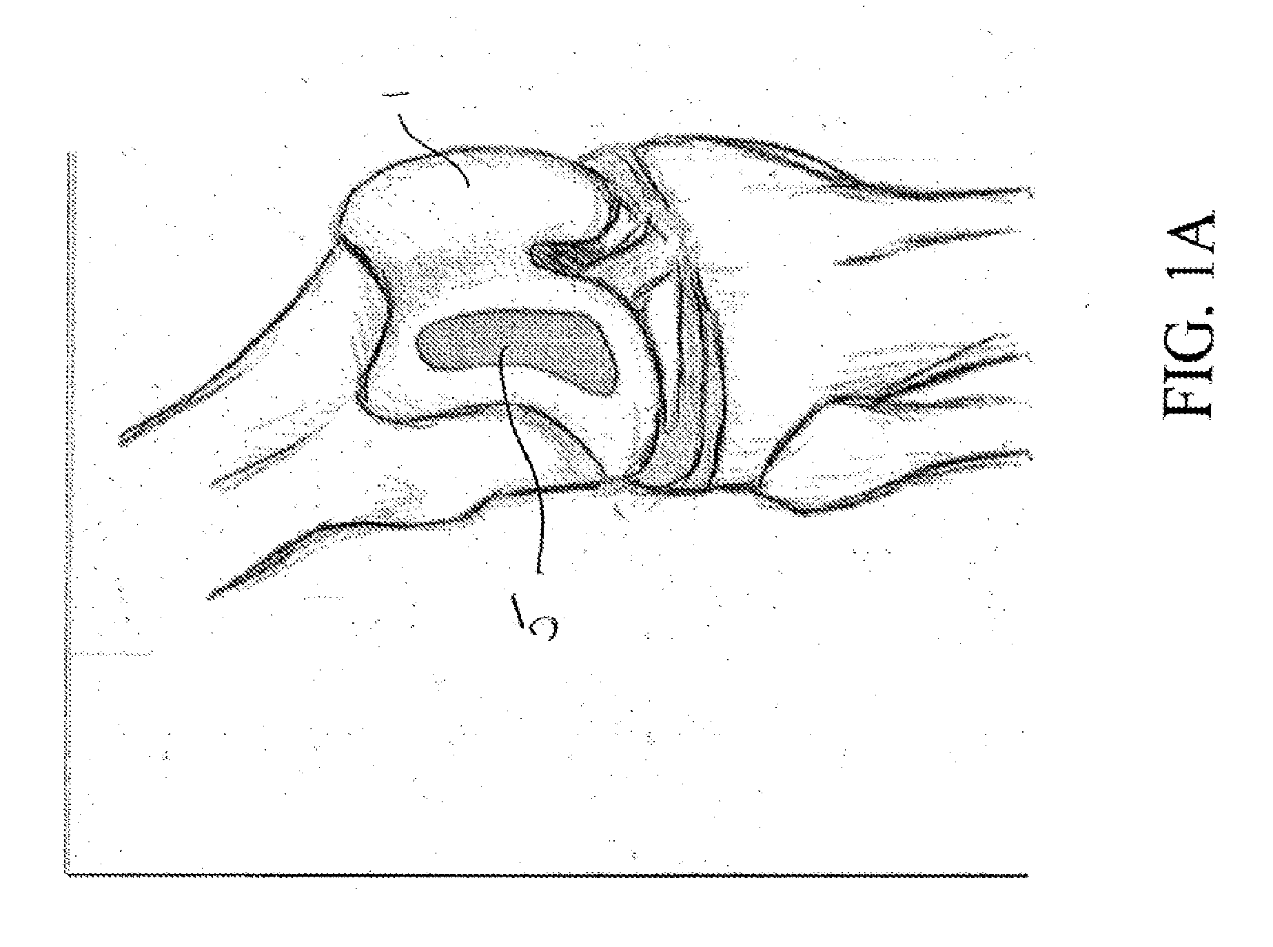
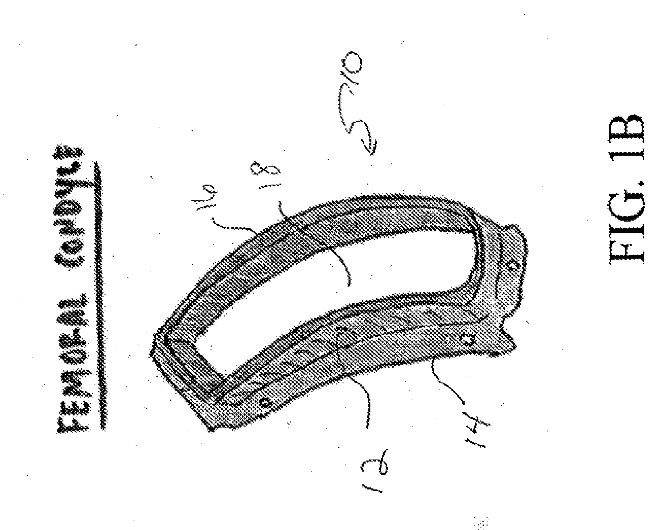
[0029]In one aspect the present invention relates to a reproducible process that may be used to fabricate size-specific, customized bio-printed musculoskeletal tissue for use in various human and veterinary orthopedic applications. Articular cartilage grafts and osteochondral grafts for implantation into any joint are described herein as exemplary. However, the present invention may also be utilized with bone grafts, labral grafts, meniscal grafts, spine disc grafts and ligament grafts among other tissues. The process of the present invention will now be described.

[0030]Acquisition of Data:

[0031]Precise data is needed for creation of orthopedic tissue. We propose the use of high resolution CT arthrography of the body part in question. Conventional CT, electron beam CT or CT arthrography, Ultrasound, ...


PUM

PropertyMeasurementUnit
Widthaaaaaaaaaa
Areaaaaaaaaaaa
Depthaaaaaaaaaa

Abstract

A process for fabricating size-specific, customized bio-printed musculoskeletal tissue using three dimensional data collected from radiologic imaging is provided. Also, provided is a guide that is created from radiological imaging that demarcates the area of surgical interest. The guide is 3D printed according to guide dimensions collected from radiological imaging, including, but not limited to, CT imaging scans, CT arthrography, ultrasound, MRI, MR arthrography, or any other imaging modality used to image the musculoskeletal system.

Description

FIELD OF THE INVENTION[0001]The present invention relates generally to a process for fabricating size-specific, customized bio-printed musculoskeletal tissue using three dimensional data collected from radiologic imaging. The present invention also relates to a guide that is created from radiological imaging that demarcates the area of surgical interest. The guide is 3D printed according to guide dimensions collected from radiological imaging, including, but not limited to, CT imaging scans, CT arthrography, ultrasound, MRI, MR arthrography, or any other imaging modality used to image the musculoskeletal system.BACKGROUND OF THE INVENTION[0002]Bioprinting is a novel science which produces the automated fabrication of human tissue and organs using a three-dimensional (“3D”) bioprinter. In this field, tissues are created by using living cells as tiny building blocks and printing these blocks along with matrix on to sheets of biopaper. Rather than the more antiquated system of building...

Claims


Application Information

Patent Timeline
IPC IPC(8): A61F2/30A61B17/17A61B8/08A61B10/02A61B5/055A61B6/03A61B17/88A61F2/46
CPCA61F2/30942A61B17/8808A61B17/17A61F2/4603A61B10/025A61B5/055A61F2002/30948A61B8/0875A61F2/4657A61B2017/00526A61F2002/4662A61F2002/30985A61B6/032A61F2/30756A61F2/4618A61F2002/30762A61F2002/30764A61B17/1764A61B17/1767A61B17/1771
Inventor KOSAREK, FRANK J.RIVERA, MICHELLE J.
Owner KOSAREK FR J
Why Eureka
Try Eureka