Culture medium and method for inducing differentiation of pluripotent stem cells into neuroepithelial cells
a technology of neuroepithelial cells and culture medium, which is applied in the field of stem cell biology, can solve the problems of low neural production efficiency, contamination of non-neural cells within the cultured population, and low purity of esc-derived neural cells, and achieve the effect of high differentiation
- Summary
- Abstract
- Description
- Claims
- Application Information
AI Technical Summary
Benefits of technology
Problems solved by technology
Method used
Image
Examples
example 1
Generation of Embryoid Bodies from ESCs
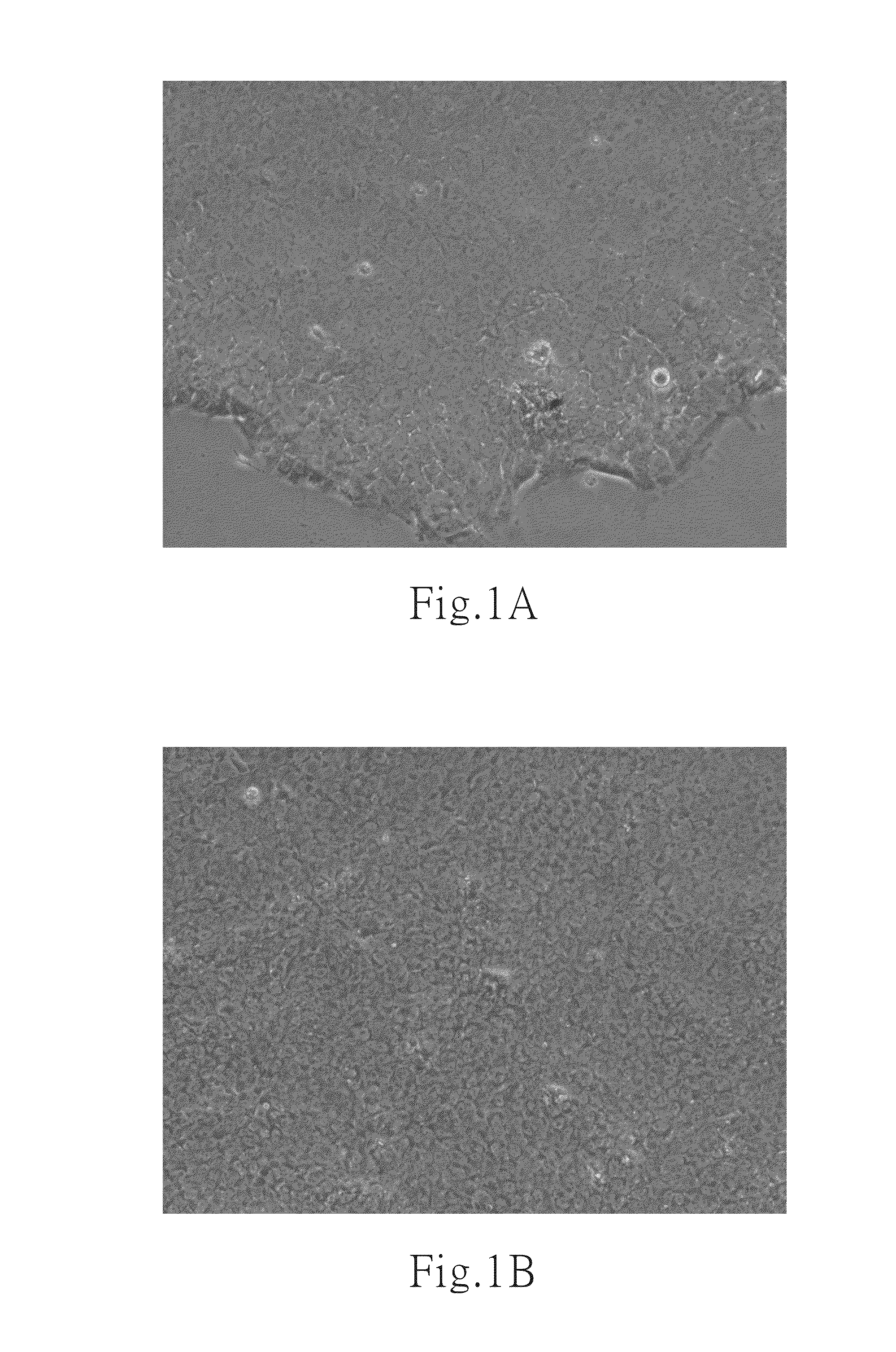
[0074]The TW1 cells, showing the classic morphology of hESCs (FIG. 1), were cultured and maintained in mTESR1 medium at 37° C. and 5% CO2. After the stage of cell expansion, the ESCs were dissociated with collagenase I and dispase for 5 mins at 37° C. The suspended cells were cultured in DMEM-F12 medium containing 20% knock-out serum replacement (KSR, Invitrogen, USA) at 37 and 5% CO2. These suspended ESCs gradually aggregated together and formed cell clusters, named as embryoid bodies, within 2 days culture.
example 2
Induction of the Neural Differentiation of Pluripotent Stem Cells into Neuroepithelial Cells
[0075]The embryoid bodies in example 1 were collected into a 15 ml centrifuge tube and placed for 10 mins until the embryoid bodies decended to the tube bottom. The supernatant was removed and replaced with neural induction medium, containing 0.5 μM BIO, 10 μM SB431542 and 10 ng / ml FGF2. The constitutions of the neural induction medium are listed in Table 1.
[0076]Notably, an announcement has to be emphasized here is that the working concentration of these additive drugs in the culture medium are not restricted on what we indicated. The working concentration of BIO is between 0.05 μM to 50 μM; the working concentration of SB431542 is between 1 μM to 100 μM; and the working concentration of FGF2 is between 1 ng / ml to 100 ng / ml.
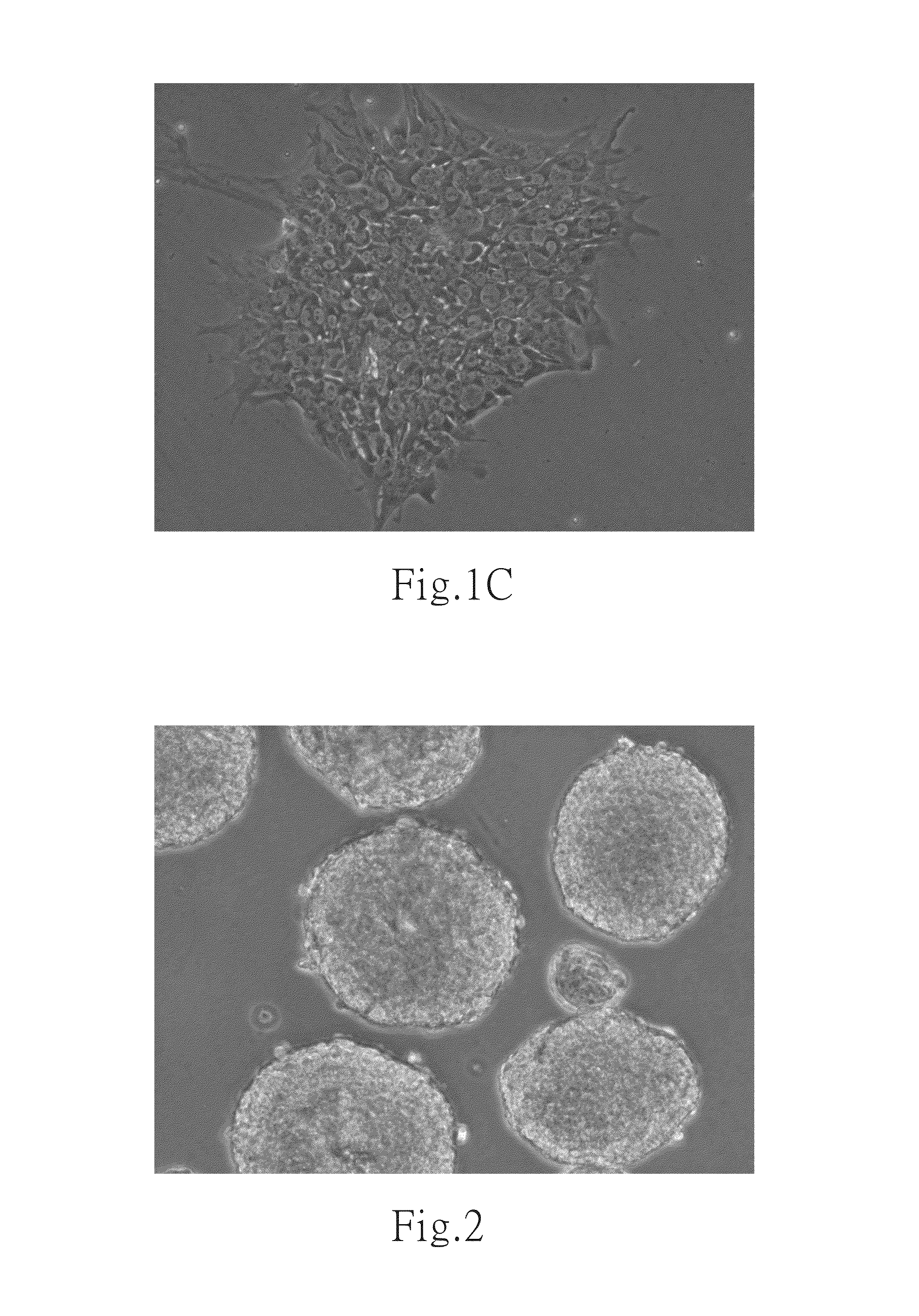
[0077]The collected embryoid bodies were further cultured with the neural induction medium for 2 days to become neuroepithelial cells as shown in FIG. 2. In FIG. 2, the c...
example 3
Further Culturing the Neuroepithelial Cells
[0078]The culture medium of the neuroepithelial cells in example 2 was switched from the neural induction medium to next medium which comprise 10 ng / ml FGF2. The constitutions of the new medium are listed in Table 2 as below. The culture medium were renewed every two days to maintain the stemness of the neuroepithelial cells.
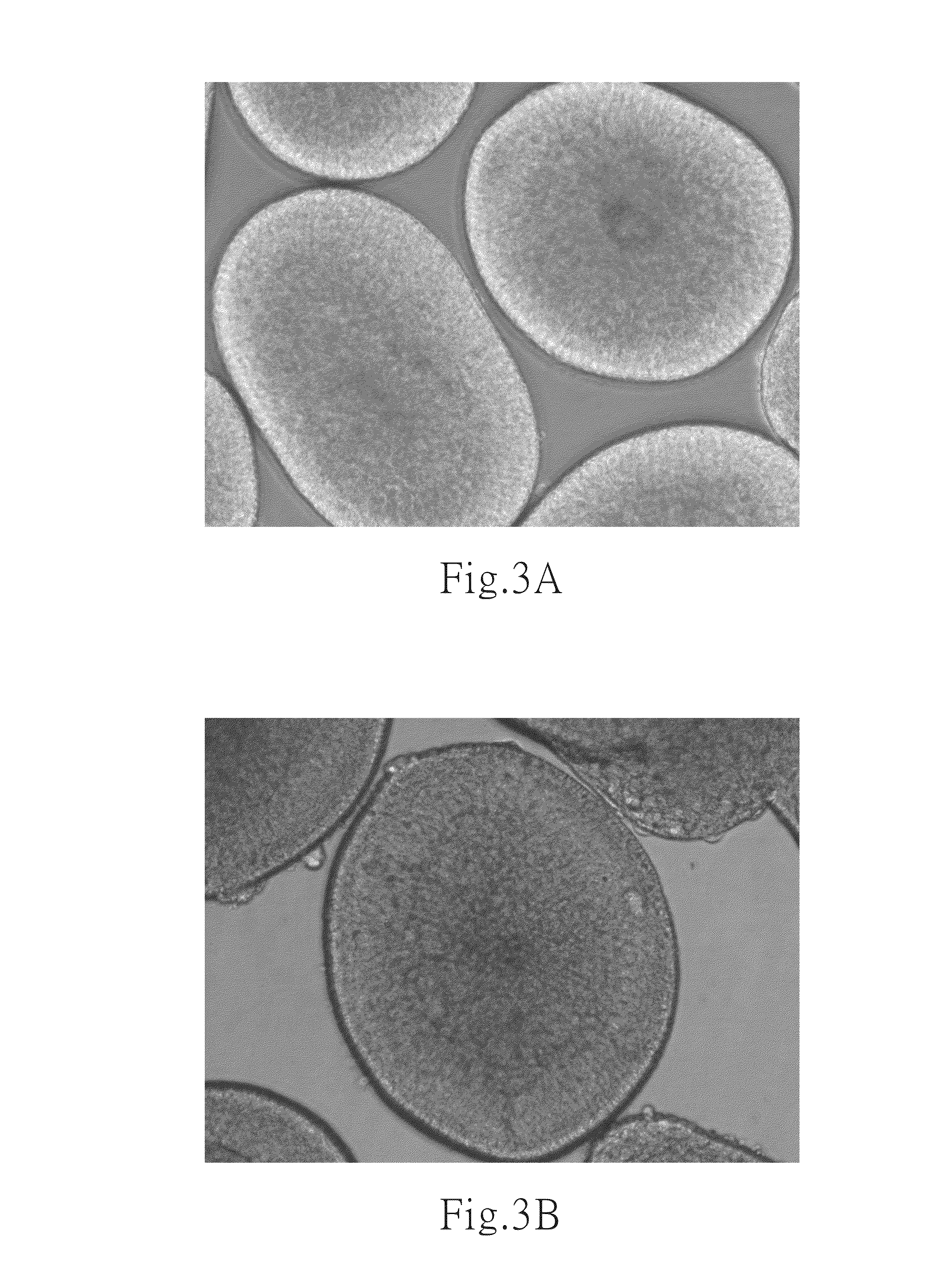
[0079]The resulting cells were shown in FIG. 3 and FIG. 4 with 100×, 200×, and 400× magnifications under a microscope. In FIG. 3A, the resulting cells formed numerous columnar structure units among the embryoid bodies and shared with homogenous morphology. The FIGS. 3B and 3C revealed the columnar structure unit which contained the tightly tubular aggregation at the edge and rosette formations, resembling the early neural tube of embryo. The FIGS. 4A and 4B showed the classical morphology of neural rosettes within attached embryoid bodies on a culture dish at culture day 6 and day 8. Therefore, according to the examinat...
PUM
 Abstract
Description
Claims
Application Information
 - R&D
- Intellectual Property
- Life Sciences
- Materials
- Tech Scout
- Unparalleled Data Quality
- Higher Quality Content
- 60% Fewer Hallucinations
Browse by: Latest US Patents, China's latest patents, Technical Efficacy Thesaurus, Application Domain, Technology Topic, Popular Technical Reports.
© 2025 PatSnap. All rights reserved.Legal|Privacy policy|Modern Slavery Act Transparency Statement|Sitemap|About US| Contact US: help@patsnap.com


