

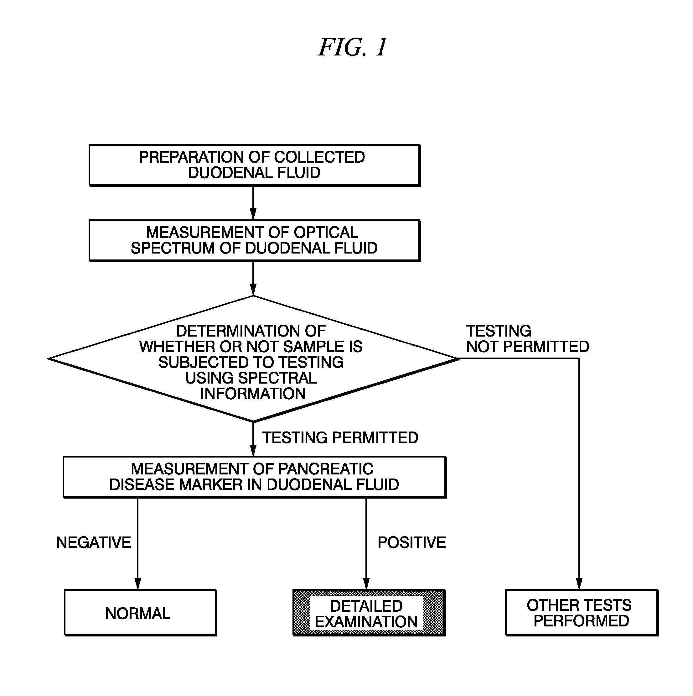
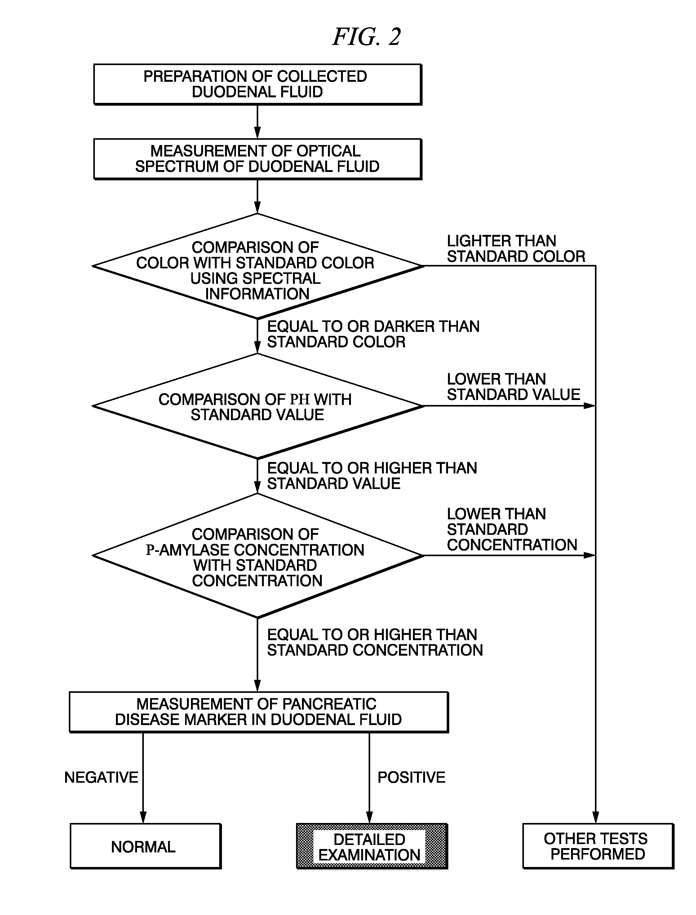
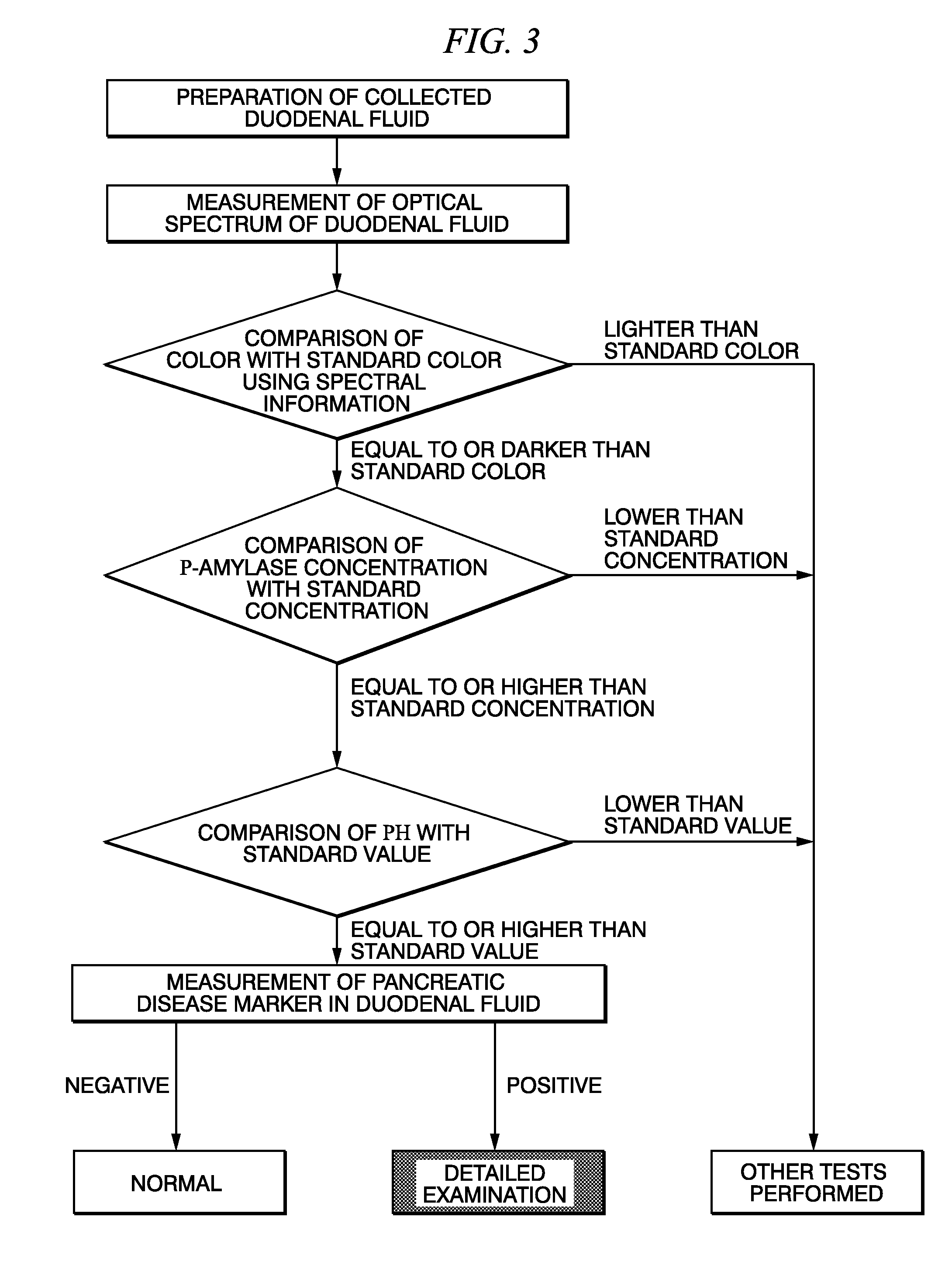
Method for selecting duodenal fluid sample for detecting pancreatic disease marker and method for detecting pancreatic disease marker
a duodenal fluid and pancreatic cancer technology, applied in the field of pancreatic cancer marker detection methods, can solve the problems of poor prognosis, high skill requirements, and pancreatic cancer
- Summary
- Abstract
- Description
- Claims
- Application Information
AI Technical Summary
Benefits of technology
Problems solved by technology
Method used
Image

Examples
reference example 1
[0096]Duodenal fluid samples were collected transendoscopically from 49 pancreatic cancer cases followed by measuring the CEA concentrations in the samples. CEA concentrations were measured by ELISA using a commercially available kit (IBL International Corp.). The samples were judged to be negative or positive based on the measured CEA concentrations using a cutoff value of 126 ng / mL.
[0097]Sensitivity and the ratios of the number of positive specimens to the total number of specimens as determined from the test results are shown in Table 1 according to T classification representing the stage of localization of the primary lesion in the pancreas. Tis represents noninvasive cancer, T1 represents that having a tumor diameter of 2 cm or less that is localized in the pancreas, T2 represents that having a tumor diameter of greater than 2 cm that is localized in the pancreas, T3 represents cancer that has invaded any of the pancreatic bile duct, duodenum or peripancreatic tissue, and T4 re...
example 1
[0099]The absorption spectra were measured for duodenal fluid samples collected transendoscopically from 49 cases of pancreatic cancer and 7 cases of benign pancreatic diseases. The absorption spectra of all specimens are shown in FIG. 4. An enlarged view of the vicinity of the absorption peak of FIG. 4 (300 nm to 560 nm) is shown in FIG. 5. The duodenal fluid samples clearly demonstrated an absorption peak wavelength range at 350 nm to 540 nm, and particularly 400 nm to 460 nm.
[0100]Moreover, the color depth of each duodenal fluid sample (specimen) was evaluated. More specifically, the absorption spectrum of each specimen at 380 nm to 780 nm was measured using a spectrophotometer at an optical path length of 10 mm (A10). Next, the resulting absorption spectra were converted to transmission spectra (T10) based on the following equation followed by further converting to an optical path length of 5 mm (T5).
T10=10−A10
T5=(−Log(T10, 10) / 10)*5 [Equation 2]
[0101]Chromaticity composed of...
reference example 2
[0111]Duodenal fluid samples were collected transendoscopically from 11 pancreatic cancer cases followed by measuring the CEA concentrations in the samples. CEA concentrations were measured by ELISA using a commercially available kit (IBL International Corp.). The samples were judged to be negative or positive based on the measured CEA concentrations using a cutoff value of 126 ng / mL. Furthermore, specimens M7 and M11 among these 11 cases were the same as specimens M7 and M11 in Reference Example 1 and Example 1.
[0112]The results of measuring CEA concentration in each sample are shown in Table 6. As a result, 5 of 11 cases (specimens F67, S136, F9, M7 and M11) were false negatives (cases of pancreatic cancer having CEA concentrations below the threshold value), and sensitivity was 54.5%.
PUM
| Property | Measurement | Unit |
|---|---|---|
| Density | aaaaa | aaaaa |
| Density | aaaaa | aaaaa |
| Nanoscale particle size | aaaaa | aaaaa |
Abstract
Description
Claims
Application Information