Diagnostic histopathology using multiplex gene expression FISH

a histopathology and gene expression technology, applied in the field of diagnostic histopathology using multiplex gene expression fish, can solve the problem that the method was formally not compatible with fixed treated cell lines or tissues, and achieve the effect of high probability of turning cancerous

Inactive Publication Date: 2006-09-07
AUREON LAB INC +2
View PDF5 Cites 23 Cited by
  • Summary
  • Abstract
  • Description
  • Claims
  • Application Information

AI Technical Summary

Benefits of technology

[0027] Another advantage of the invention's method is that it can be used on a wide variety of samples including cultured cells, fixed treated tissues of any age (from freshly made fixed tissue to tissues fixed over 10, over 20, over 30, over 40 or over 60 years old. The tissue sample may be stained, such as hematoxylin and eosin (H&E) stained or the tissue may be previously stained but destained. The tissue sample may also be a paraffin embedded tissue sample, a frozen sample, cells which have been placed on a slide by centrifugation (i.e., cytospin (Thermo Electron Corporation, Waltham, Mass. USA), or a sample on a slide. Some sample may have a combination of the above listed characteristics. For example, a fixed tissue may be paraffin embedded and H&E stained and destained. Such samples are also suitable as a starting material for the methods of the invention.
[0028] The methods of the invention are particularly suitable for the detection of nascent RNA (also called nascent transcripts) for the diagnosis of neoplastic or preneoplastic tissues. Preneoplastic tissues would include tissues which has a high probability to turn cancerous. These tissues include, for example, tissue with hyperplasia and hypertrophy.
[0029] Sample preparation (autofluorescence reduction) may be performed by pressure cooking the tissue sample to produce a pressure cooked sample and then treating the sample with ammonia ethanol and sodium borohydride. For example, a tissue may be pressured cooked, then contacted with ammonia ethanol, and then contacted with sodium borohydride. Contacting may be performed, by dipping a slide containing the tissue sample into a container containing ammonia ethanol and then dipping the slide into a container containing sodium borohydride.
[0030] Pressure cooking may be performed, for example, by incubating a glass slide with sections of the sample tissue in a chamber at about 125° C., at a pressure of between about 20 to about 24 PSI. This can be performed in a commercially available decloaking chamber or other chamber that can maintain these conditions. During pressure cooking, the sample tissue may be contacted with a solution that has deparaffinization and / or antigen retrieval activities. Examples of such solutions include, for example, “Declere™” solution from Cell Marque (Hot Springs, Ariz., USA) or “Reveal™” solution from BioCare Medical (Concord, Calif., USA). Under these conditions, pressure cooking may be performed for between 10 minutes and 1 hour such as, for example, for 30 minutes.
[0031] Following pressure cooking, the sample may be contacted to ammonia-ethanol. For example, the sample may be dipped in a tank of ammonia-ethanol. The ammonia-ethanol may be at a concentration of about 0.1% to 0.5%. In a preferred embodiment, the ammonia-ethanol may be about 0.25%. The length of contact may be between about 10 to 50 minutes, between 10 to 30 minutes or about 40 minutes.
[0032] Following ammonia-ethanol treatment, the sample may be treated by contact with sodium borohydride at a concentration of about 1% to about 5%. This contact may last between 10 and 50 minutes such as, for example, for about 20 minutes.

Problems solved by technology

Such methods were formally not compatible with fixed treated cell lines or tissue.

Method used


Image

  • Diagnostic histopathology using multiplex gene expression FISH
  • Diagnostic histopathology using multiplex gene expression FISH
  • Diagnostic histopathology using multiplex gene expression FISH

Examples


example 1

Material and Methods

Samples

[0140] Microarrays (TMA) consisting of 0.6 mm core biopsies of formalin-fixed, paraffin-embedded tissues were created using a tissue arrayer (Beecher Instrument). Cohorts of 59 anonymized prostate samples present in the TMA in triplicate were analyzed. Of the 59 patients, 20 were prostate adenocarcinoma (PCa), 12 were benign prostatic hyperplasia (BPH), 13 were prostatic intraepithelial neoplasia (PIN), 14 were prostate metastasis (PCaMet).

Sample Preparation—Pretreatment

[0141] 5 μm paraffin-embedded sections were dried at 37° C. for about 1 hour and then transferred to a decloaker chamber for 30 minutes where they were deparaffinized and antigen-retrieved using a solution that allows both steps to occur at the same time. The slides were then washed in PBS for 10 minutes, incubated for 20 minutes in 0.25% ammonia-ethanol at room temperature (RT), and then incubated for 50 minutes in 5% sodium borohydride in PBS at RT. The slides were then washed twice...

example 2

Patient Analysis by Microarray

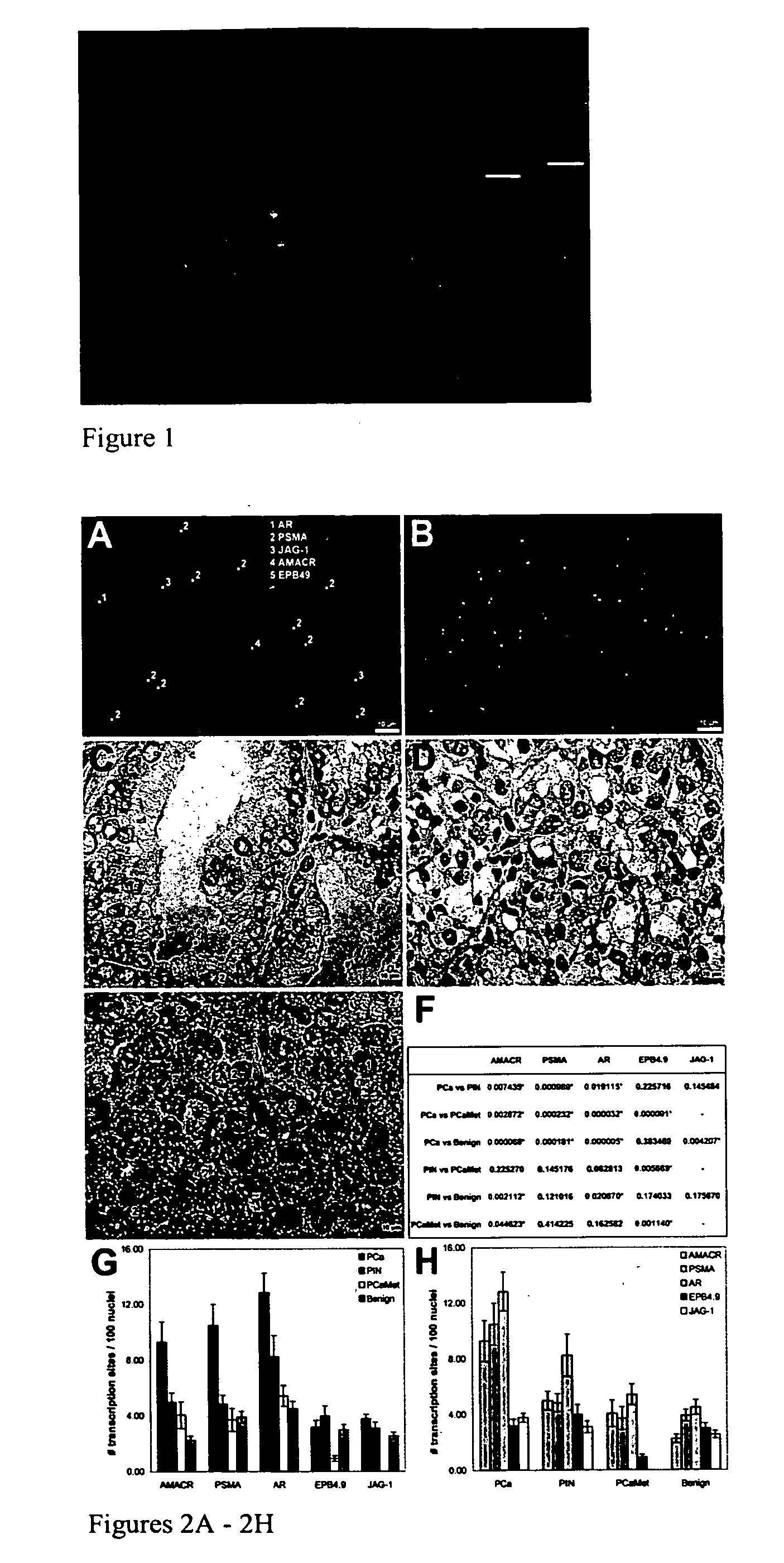
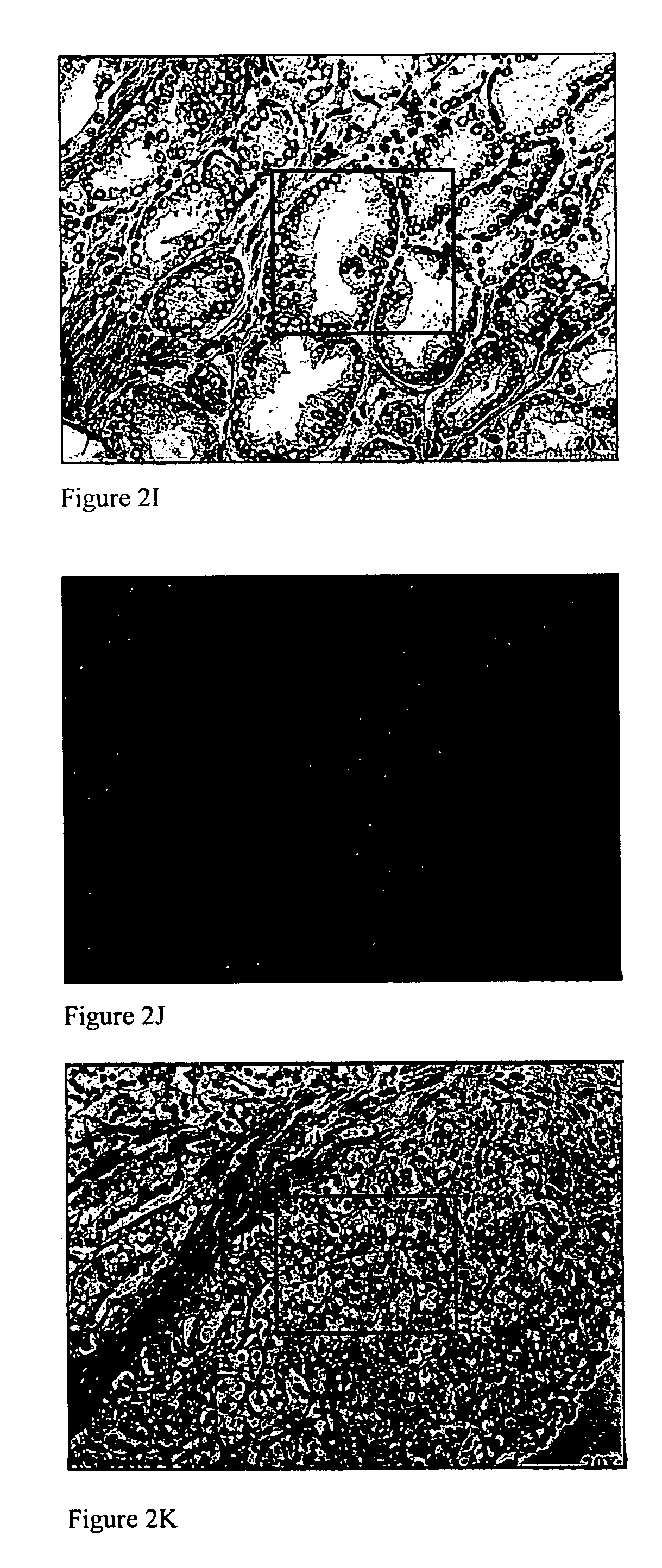
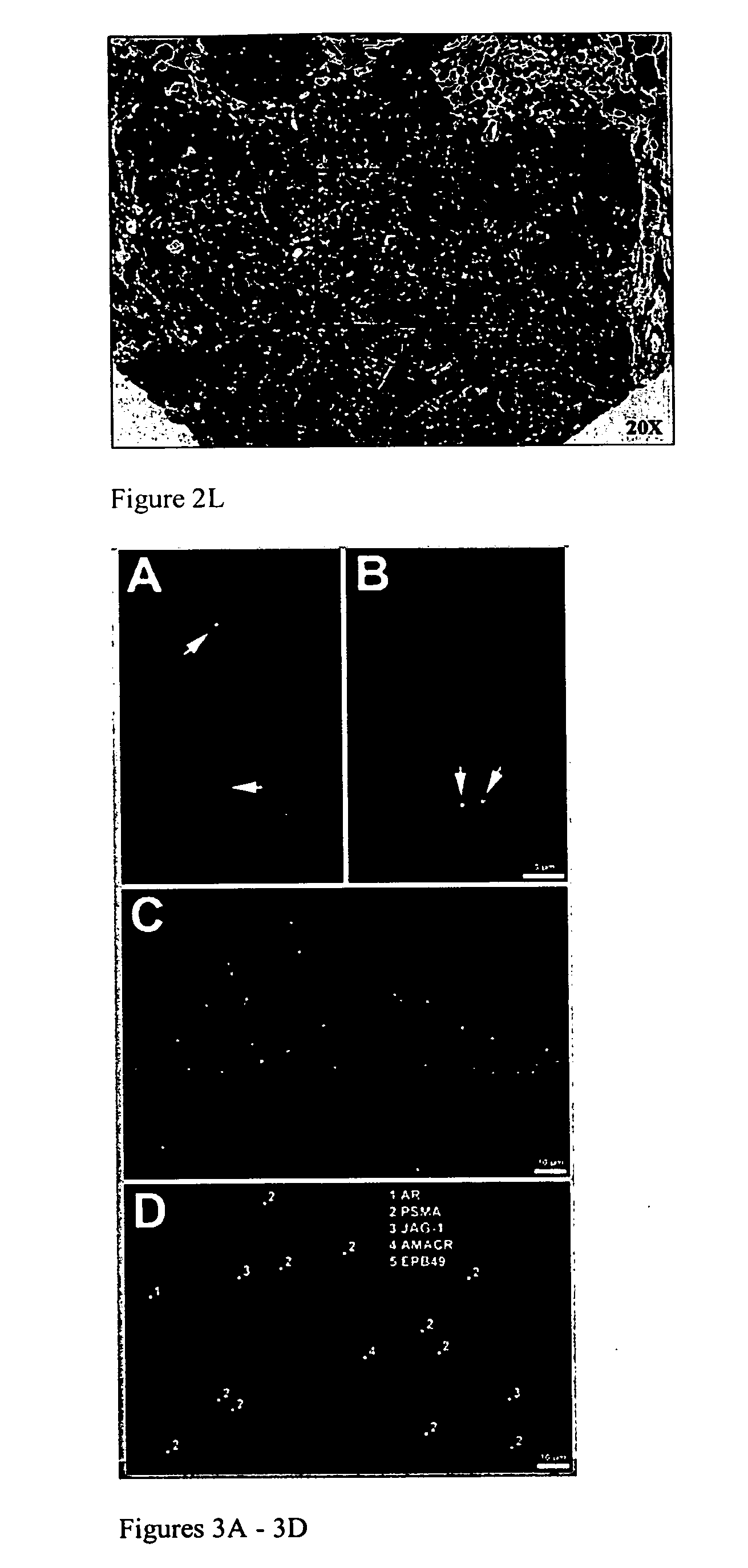
[0157] To investigate how peT-FISH data correlate with histopathologic features, epithelial cells with established pathologic morphologies: benign prostate tissue, prostatic intraepithelial neoplasia (PIN), prostate cancer (PCa) and prostate cancer metastasis (PCaMet, Glinsky G., et al. J Clin. Invest. 113, 913-923 (2004)) was examined. peT-FISH was performed on ‘de-stained’ hematoxylin and eosin (H&E) tissue sections (FIG. 3). The ability to use destained section is an important capability because in many cases, patient tissue blocks are no longer available for analysis. The panels show overlays of peT-FISH results on morphology image data. The procedure was modified for high-throughput sampling of patient material in TMA format (Capodieci P., et al. J Natl Cancer Inst. 96, (2004)).

[0158] Random fields were chosen from each individual from tissue microarray (TMA) blocks representing tissue samples derived from patients that had undergone therapeutic ...

example 3

Analysis of Tissue Sections

[0160] We have determined that the resolution provided by current microscopes is sufficient for detecting the expression of up to 40,000 transcription sites in a cell simultaneously. Our reasoning is as follows. The nucleus is approximately 10 μm in diameter which corresponds to a volume of approximately 500 μm3 (V= 4 / 3(πr2)). A voxel is approximate 0.1 μm by 0.1 μm by 0.25 μm or 2.5×10⁻³ μm3. Thus, there are 200,000 voxels per nucleus which corresponds to a detection ability of about 40,000 transcription sites. Approximately 10,000 genes are expected to be transcribed in a nucleus simultaneous. Thus, the microscopic technique of the invention is capable of detecting all the expressed genes in a cell at the same time.

[0161] Our experimental strategy is based on detection of nascent RNA. Nascent RNAs are RNAs that are being transcribed from genomic DNA (i.e., chromosome). Since the RNAs are still being transcribed, they are associated with their correspon...


PUM

PropertyMeasurementUnit
temperatureaaaaaaaaaa
diameteraaaaaaaaaa

Abstract

The present invention relates to a method for detecting one or more nascent RNAs in a tissue sample using FISH. In the method, a plurality of probes (8 to 82) may be used to detect a single species of nascent RNA. Further, a plurality of nascent RNA species may be detected simultaneously using between 8 and 82 probes for each nascent RNA. The invention comprises, in addition, methods of preparing a sample for nascent RNA detection by reducing autofluorescence of the tissue sample. These techniques may be synergistically combined to achieve significantly improved results.

Description

RELATED APPLICATIONS [0001] This application is a continuation-in-part of U.S. Ser. No. 11 / 283,062 filed Nov. 17, 2005, which is a continuation of U.S. Pat. No. 6,995,020, U.S. Ser. No. 10 / 624,233, filed Jul. 21, 2003 and issued Feb. 7, 2006. This application also claims the benefit of priority from U.S. 60 / 671,549, filed Apr. 15, 2005, and U.S. 60 / 707,612, filed Aug. 12, 2005. All patents, patent applications and references cited in this specification are hereby incorporated by reference.BACKGROUND OF THE INVENTION [0002] Advancements in the understanding of gene expression and epidemiology combined with developments in technology have allowed for the correlation of genetic expression with, for example, disease states. An accurate correlation may enable risk assessment for an individual based on the expression profile of their individual cells. Further, drug screening and other research based protocols may quickly generate data in cell lines or tissue samples that can be extended t...

Claims


Application Information

Patent Timeline
Patent Type & Authority Applications(United States)
IPC IPC(8): C12Q1/68C12N1/08
CPCC12Q1/6841C12Q1/6876Y10S436/80Y10T436/143333C12Q2527/125C12Q2537/143C12Q2527/109C12Q2527/101C12Q2600/158
Inventor CAPODIECI, PAOLADONOVAN, MICHAEL
Owner AUREON LAB INC
Why Eureka
Try Eureka