Diagnosing and treating movement disorders

a movement disorder and diagnosis technology, applied in the field of medicine, can solve the problems of not being validated, emotionally distress, substantially disabled, etc., and achieve the effects of reducing the severity of abnormal movements, increasing the amount of time required, and reducing the magnitude of motions

Active Publication Date: 2016-07-14
MDDT
View PDF2 Cites 22 Cited by
  • Summary
  • Abstract
  • Description
  • Claims
  • Application Information

AI Technical Summary

Benefits of technology

[0019]It is possible to compile data representing the motion along a sufficient number of degrees of freedom for each joint by placing sensors at a plurality of joints and measuring the motion at each joint independently along a desired number of degrees of freedom (e.g. three degrees of freedom) around that joint. Because any given abnormal movement may comprise a contribution from any number of muscles, such measurement of motion around each of the joints that may be implicated in the abnormal movement permits decomposition of the sensor data into the particular motions, and therefore the particular joints, muscle group composition and directional bias, involved in a single abnormal movement. The distribution of composition among joints (e.g. fingers, wrist, elbow, shoulder neck, etc.), the muscle group composition (e.g. F / E, U / R, P / S, A / A) and the directional bias within each joint muscle group provides information to determine the individual muscles involved in and their respective contributions to the abnormal movement. This permits accurate targeting of the muscles for therapy for the abnormal movement in question. Thus, therapy may be based on relative contribution of each muscle to the abnormal movement, the contribution of each muscle being determined from the analyzed sensor data showing the muscle group composition for the movement and the directional bias within each of the muscle groups that are part of the composition.
[0023]The dosage of drug injected into each muscle may be determined by following a procedure whereby amplitude of an abnormal movement at each joint guides the maximum dose of the drug to be injected into the muscles involved with joint, the muscle composition of the abnormal movement guides how the maximum dosage is divided between muscle groups, and the directional bias within each muscle group guides how the dosage given to a muscle group is divided between individual muscles. The procedure may be followed manually by a clinician, or the procedure may be embodied in a computer program or software and the dosages for each individual muscle determined from calculations based on sensor data and pre-set or inputted parameters. For example, total dosage for a joint may be correlated to amplitude data for the abnormal movement at the joint, whereby the amplitude may be compared to a standard curve of amplitude vs. total dosage or to a standard dosage for a range of amplitudes. More severe abnormal movements would warrant a greater total dosage. Once the total dosage for a joint is determined, composition data showing the relative contribution of each muscle group (e.g. abductors / adductors, flexors / extensors, etc.) may permit dividing the total dosage for that joint between the various muscle groups on a pro-rated basis, for example if abductor muscles were found to contribute 40% to the abnormal movement at the joint then abductor muscles would receive 40% of the total dosage of drug for that joint. Once the division of total dose between muscle groups has been determined, the dosage for each individual muscle in the muscle group can be determined from the directional bias on a pro-rated basis, for example within the abductor / adductor muscle group if abductors are responsible for 80% of the abnormal movement caused by the abductor / adductor muscle group then the abductor muscles would receive 80% of the dosage and the adductor muscles would receive 20%. Where one muscle falls within two or more muscle groups, that muscle would receive an amount of drug related to the sum of the dosages calculated for the muscle's contribution to the abnormal movement for each muscle group. In some cases, one muscle may be a contributor to abnormal movement at different joints, in which case the dosage received by that muscle would also be related to the sum of the dosages calculated for the muscle's contribution to the abnormal movement for each joint. Automation of the calculations in a computer program or software based on sensor data for amplitude, composition and directional bias and on standard total dosage correlations to the amplitude could standardize treatment decisions, improve the accuracy of treatment decisions and permit non-expert clinicians to make treatment recommendations.
[0024]Dosages of drugs will depend to a certain extent on the particular drug being used. For example, a dose range of: 10U-60U, especially 20U-40U or 10U-30U, of BoNT A can be used for each shoulder tremor contributing muscle; 10U-50U, especially 20U-40U or 10U-30U, of BoNT A for every elbow tremor contributing muscle; and 5U-25U, especially 5U-15U, of BoNT A for every wrist tremor contributing muscle (e.g. forearm and wrist muscles). All dosages are adjusted based on amplitude of the tremor. Likewise, if the composition and directional bias in one patient shows that the tremor is predominantly flexor at the wrist, a physician can optimize dosage based on medical experience to inject a higher dose in the flexor carpi radialis and flexor carpi ulnaris, and give lower doses to the extensor carpi radialis and extensor carpi ulnaris muscles. By Further, the present invention makes it possible to avoid injecting drugs into muscles that have no contribution to the abnormal movement. This reduces side effects and the amount of drug that is needed to be effective at treating the movement disorder.
[0025]In addition, and very importantly, the concentration of the drug may be increased, for example doubled, to reduce the occurrence of volume-dependent weakness in the muscles injected and also in the adjacent muscles due to the spread of the toxin. Thus, the same dose may be delivered in a smaller volume, for example half the volume.
[0030]In a rehabilitation therapy, a time course of treatment may be prescribed based on kinematic assessment and recurrence of the abnormal movements after initial treatment. Initial treatments may result in a decrement in the magnitude and / or frequency of the abnormal motions observed, and repeated application of the therapy guided by kinematic assessment may reduce the magnitude of the motions and / or increase the amount of time required between therapy events. This leads to long term suppression of the disorder. Further, movement disorders may also comprise a neurological component in which the brain has been conditioned over time to trigger the abnormal movements associated with the movement disorder. Treatment of the muscle involved to reduce the severity of the abnormal movements may help re-condition the brain providing a feedback loop to reduce abnormal movement without the aid of therapy. Therefore, accurate muscle selection and treatment parameters (e.g. dosing) would help re-condition the brain providing a long-term solution to the movement disorder. Only optimal treatment regimens would be useful in this regard, so developing treatment regimens based on kinematic assessment as disclosed herein would be greatly preferred over the currently employed visual assessment strategies. In some treatment regimes, treating only one side of a subject's body may lead to benefits for the other side of the body.
[0031]Subtle motions and the muscles involved in movement disorders are generally too difficult for clinicians to visually assess. Visual assessment is therefore a subjective and clinician-experience dependent assessment tool. Although all movement disorder may be visually diagnosed, current visual assessment is generally limited because: human eyes are not at a level needed to properly pin-point the location of symptoms; movement disorders change from task to task, which his not traceable by human eyes; and, movement disorders change over time, which is not traceable and comparable through notes and visual assessment. The present system and methods are able to provide a breakdown of motions that is accurate and objective. Such accuracy leads to more appropriate treatment at the correct muscles involved in the movement disorder, which leads to greater efficacy of treatment, reduction in the amount of therapy required and fewer side effects. For example, use of the present system and methods in conjunction with BoNT A injection therapy increases the effectiveness of BoNT A, reduces the dose required, increases functionality of treated limbs and reduces muscle weakness, muscle weakness having been a side effect of BoNT A therapy. Further, objective sensor data analyzed in a consistent way permits accurate tracking of the progression of the movement disorder following medical intervention.

Problems solved by technology

However, detailed kinematic assessment of both tremor types to deconstruct tremor dynamics, including both muscle composition and directional bias have not been done to validate these assumptions.
Subsequent functional impairment is a result of tremor, and this can be substantially disabling if the dominant arm is affected.
In addition, the presence of tremor is an obvious visible symptom, which can be cosmetically disabling, making patients feel as though they “stand out” causing emotionally distress.
While options exist for management of ET and PD tremor, the therapeutic efficacy can still be quite poor with the side effects of medication and danger in brain surgery pose considerable risk, especially in the older age group.
Although tremor has been treated with BoNT A, the studies have been open-label, or small and the results of BoNT A have not generally been particularly favorable.
However, all patients had some degree of weakness as a side effect, and functional disability and action tremor did not improve significantly.
It is possible that despite the reduction in tremor, weakness overshadowed the improvement and resulted in a lack of significant functional improvement seen.
However, this has not been largely accepted as a primary treatment option by clinicians, approved by health regulatory bodies, nor is it reimbursed by insurance companies for off-label use.
The lack of functional improvement using BoNT A is a side effect profile produced by the injection.
Intramuscular injections can produce substantial weakness in the muscles due to the toxin's well-known action.
This weakness is in the muscles injected and also in the adjacent muscles due to the spread of the toxin.
It is known that this weakness and spread is dose and volume dependent.
However, when tremor is superimposed on say cervical dystonia, the assessment of the movement and the subsequent injection pattern determination becomes that much more difficult.
Tremor in the upper limb can be complex to assess visually simply because of the number of body parts involved.
In most cases this is a very difficult task and may over- or under-estimate the movement subcomponents.
If this happens, the injections of BoNT A may be given in incorrect muscle groups resulting in suboptimal benefit and increased side effects.

Method used


Image

  • Diagnosing and treating movement disorders
  • Diagnosing and treating movement disorders
  • Diagnosing and treating movement disorders

Examples


example 1

Kinematic Assessment of Tremor Composition and Directional Bias in a Wrist

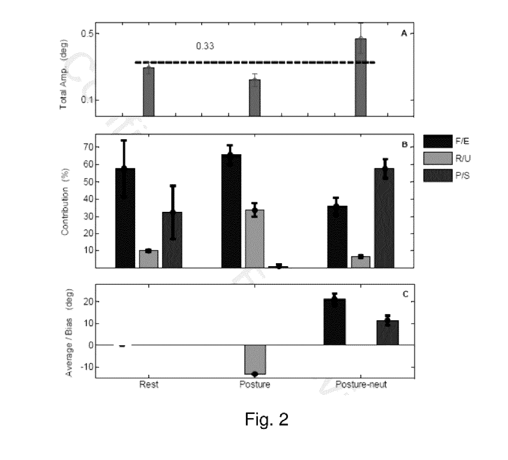
[0049]Kinematic methodology is well established for studying the dynamics of movement in the upper limb. Technological advances have made this a reliable and viable option in the characterization of complex movements such as tremor. Wrist tremor, for example, is variable and has three directions of movement: flexion / extension (F / E), radial / ulnar (R / U), pronation / supination (P / S). Hence, visually-guided judgment of the complexity of movement over time may be difficult and inaccurate. Further, kinematic studies to date have not deconstructed the complex movements into their muscle compositions and directional biases within muscle groups. As described herein, kinematic methodology can accurately allow for assessment of all these variables, leading to improved characterization of tremor dynamics. In order to understand the biomechanics of tremor in both ET and PD, the composition of these tremor types in a wrist w...

example 2

Treating Arm Limb Tremor and Deviation Using Kinematic Analysis and Botulinum Neurotoxin Type A (BoNT A) Injection for Wrist, Elbow and Shoulder

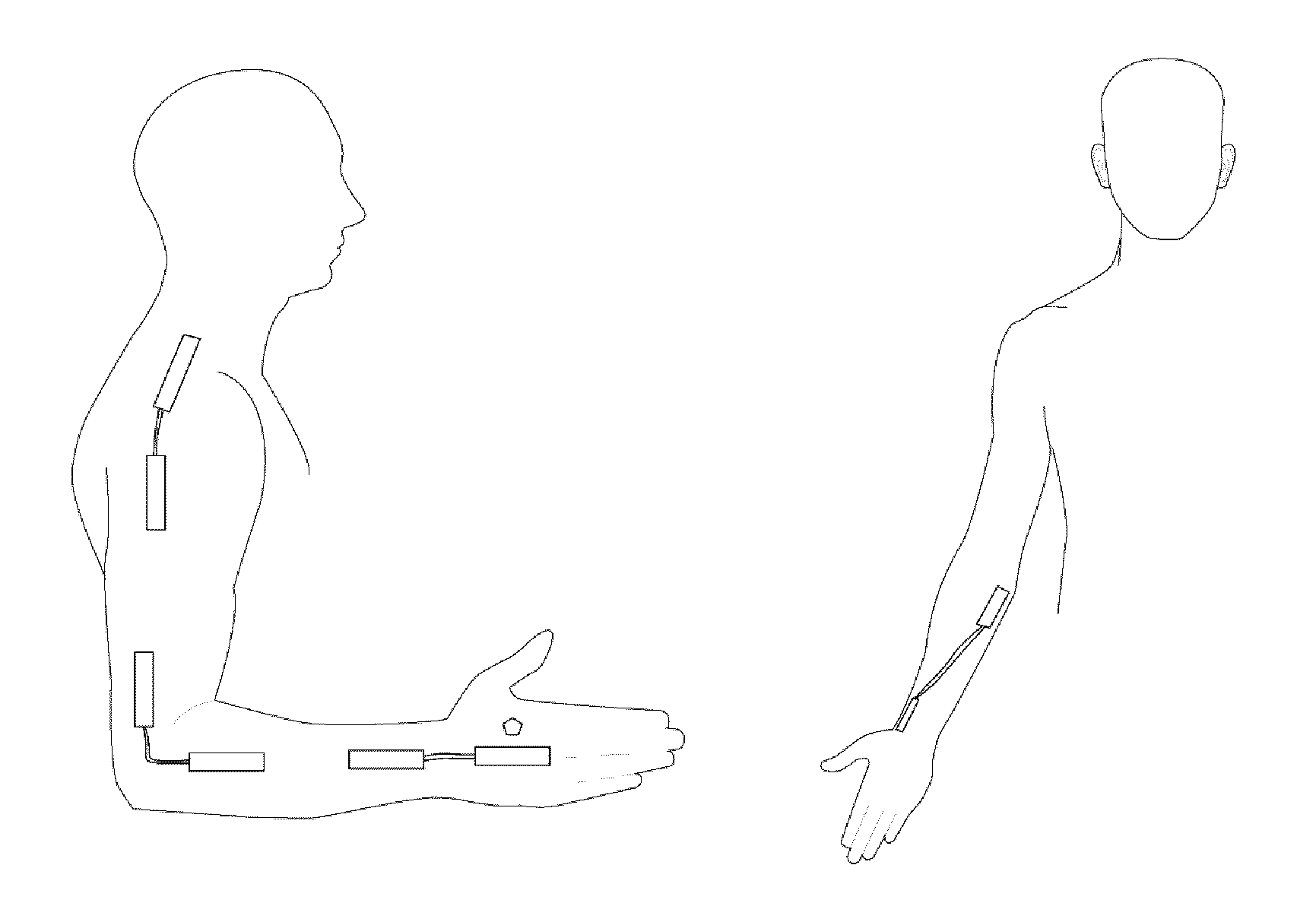
[0076]In order to capture the accurate representation of tremor in the upper limb, measurements were done on the entire arm on all major joints; wrist, elbow and shoulder. Wrist tremor is highly variable and has three directions of movement: flexion / extension (F / E), radial / ulnar (R / U), and pronation / supination (P / S), as mentioned in Example 1. Elbow tremor has one direction of movement done by flexion / extension (F / E), while shoulder tremor has three directions of movement: flexion / extension (F / E), abduction / adduction (A / A), and internal / external rotation. With same criteria as Example 1, 18 ET and 23 PD patients, different recruits from Example 1, were enrolled into a 8 month long arm tremor study with baseline data collected (Table 4).

TABLE 4Subject Demography for ET and PDETPDIDAgeGenSideIDAgeGenSideMT-0776MRMT-0171FLMT-0874FRMT-0235MRMT-0...

example 3

Treating Head and Neck Tremor with Torticollis Using Kinematic Analysis and Botulinum neurotoxin type A (BoNT A) Injection

[0086]A subject's head and neck tremor was measured and analyzed generally in accordance with the kinematic method described in Examples 1 and 2. To accomplish this, sensors were placed on the subject's body as depicted in FIG. 8A, with sensors on the head and neck of the subject. FIG. 8A depicts sensors on the head, shoulder and neck. The torsiometer was placed at the back of the neck while two inclinometers are placed on both shoulders and one inclinometer is placed on the side of the patient's head. The torsiometer was able to detect rotational tremor and dystonic movements along with the rotational range of movement. The two inclinometers on the shoulders are able to capture shoulder elevation. The inclinometer located on the side of the patient's head was used to measure tremor and dystonic movements for lateral left-right tilts and forward-backward head sag...


PUM

PropertyMeasurementUnit
degrees of freedomaaaaaaaaaa
movementsaaaaaaaaaa
movement disorderaaaaaaaaaa

Abstract

A system for obtaining and analyzing data for overall joint motion from a plurality of joints of a subject experiencing a movement disorder involves a plurality of kinematic sensors configured to be placed on a body of a subject experiencing a movement disorder proximal a plurality of joints of the subject. The kinematic sensors are selected to measure overall joint motion with sufficient degrees of freedom for individual joints so that data collected by the sensors can be deconstructed into multiple degrees of freedom for individual joints and analyzed to provide amplitude of the movements caused by the movement disorder and / or relative contributions from and / or directional bias for each muscle group that may be implicated in the movement of each joint. Such a system permits methods for determining a treatment regimen for treating the movement disorder, whereby the treatment regimen is based on the amplitude of the movements and / or the relative contribution and / or directional bias of each muscle group to the movements caused by the movement disorder.

Description

CROSS-REFERENCE TO RELATED APPLICATIONS[0001]This application is a continuation-in-part of International Patent Application PCT / CA2013 / 000804 filed Sep. 20, 2013, and further claims the benefit of United States Provisional Patent application U.S. Ser. No. 61 / 993,489 filed May 15, 2014.FIELD OF THE INVENTION[0002]The present invention relates medicine, particularly to methods and systems for diagnosing and treating movement disorders.BACKGROUND OF THE INVENTION[0003]Tremor is a relatively treatment-resistant symptom of various movement disorders, for example Parkinson's disease (PD) and Essential Tremor (ET), and Essential Tremor is one of the most common movement disorders. Tremor is assumed to be visually easy to assess and therefore should be relatively easy to treat ET action tremors (postural or kinetic) and PD rest tremors. However, detailed kinematic assessment of both tremor types to deconstruct tremor dynamics, including both muscle composition and directional bias have not ...

Claims


Application Information

Patent Timeline
Patent Type & Authority Applications(United States)
IPC IPC(8): A61B5/00A61M5/172A61N7/00A61F5/01A61B5/11A61N2/00
CPCA61B5/4839A61B5/11A61B5/4082A61N2/006A61N7/00A61B2562/0223A61M5/1723A61B5/4848A61B5/4519A61B2562/0219A61F5/01A61N1/36067A61N2/002A61B5/1101A61B5/1127A61K38/4893A61N5/0613A61H23/0245A61H39/08A61H2201/10A61H2201/5058A61H2201/5061A61H2201/5069A61H2201/5084A61H2201/5092A61H2230/60A61H2230/62A61P21/00A61P25/00A61P25/08A61P25/14A61P25/16
Inventor RAHIMI, FARIBORZJOG, MANDAR
Owner MDDT
Why Eureka
Try Eureka