Measurement of tissue structures

a tissue structure and tissue technology, applied in the field of tissue structure measurement and classification, can solve the problems of significant tissue destruction and dysfunction, inequitable health service provision, additional morbidity for bcc sufferers, and deferred cost to the nhs

Active Publication Date: 2016-10-06
ROYAL HOLLOWAY UNIV OF LONDON +1
View PDF4 Cites 25 Cited by
  • Summary
  • Abstract
  • Description
  • Claims
  • Application Information

AI Technical Summary

Benefits of technology

[0023]The locations identified within each region are optionally at least a predetermined distance away from an outer edge of each region. This reduces t

Problems solved by technology

However, identifying the tumour margins intra-operatively remains one of the key challenges in tissue-conserving surgery.
BCC is a major health problem as it accounts for ˜75% of skin cancer cases worldwide with more than 60,000 cases being diagnosed each year in the UK.
Although BCC rarely spreads to distant sites in the body, these cancers can lead to significant tissue destruction and dysfunction.
However, the high costs have limited the availability of MMS throughout the UK, leading to an inequitable health service provision.
This is not the case at present due to limited availability of histopathology technicians skilled in frozen section preparation and skilled surgeons.
Failure to provide MMS for difficult cases of BCC results in additional surgery for removal of recurrent tumours, causing additional morbidity for BCC sufferers and deferred costs to the NHS.
In many cases, the traditional methods of histopathological examination of resection specimens require tissue preparation procedures which, for practical reasons, cannot be performed intra-operatively.
For example, breast conserving surgery (BCS) relies on visual or X-ray inspection of excised tissues, techniques which have significantly lower diagnosis accuracy compared to histopatholo

Method used


Image

  • Measurement of tissue structures
  • Measurement of tissue structures
  • Measurement of tissue structures

Examples


Embodiment Construction

[0035]Aspects and embodiments of the invention are described in further detail below by way of example and with reference to the enclosed drawings in which:

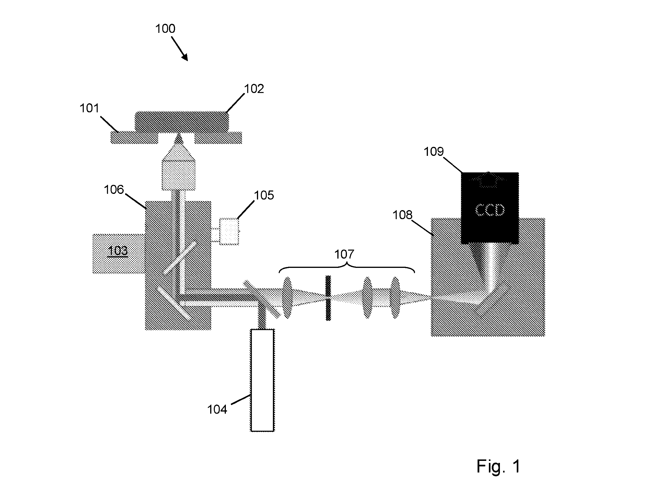
[0036]FIG. 1 is a schematic diagram of an apparatus according to an embodiment of the invention;

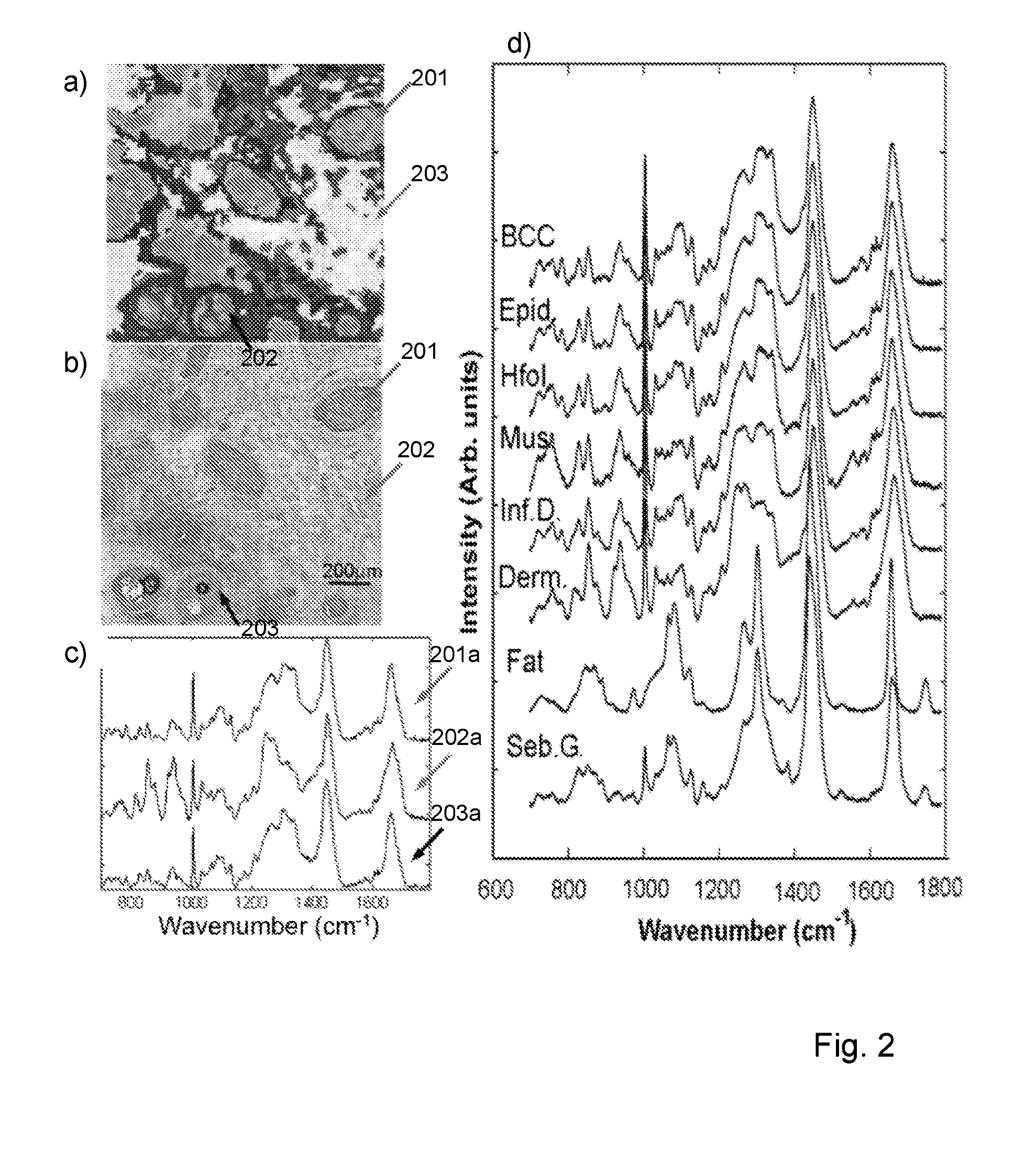
[0037]FIGS. 2a and 2b are images of a tissue sample derived from Raman spectra assignment (FIG. 2a) and conventional HE staining (FIG. 2b);

[0038]FIG. 2c is a plot of mean spectra for three different tissue structures in the sample of FIGS. 2a and 2b;

[0039]FIG. 2d is a series of plots for mean Raman spectra for various tissue structures;

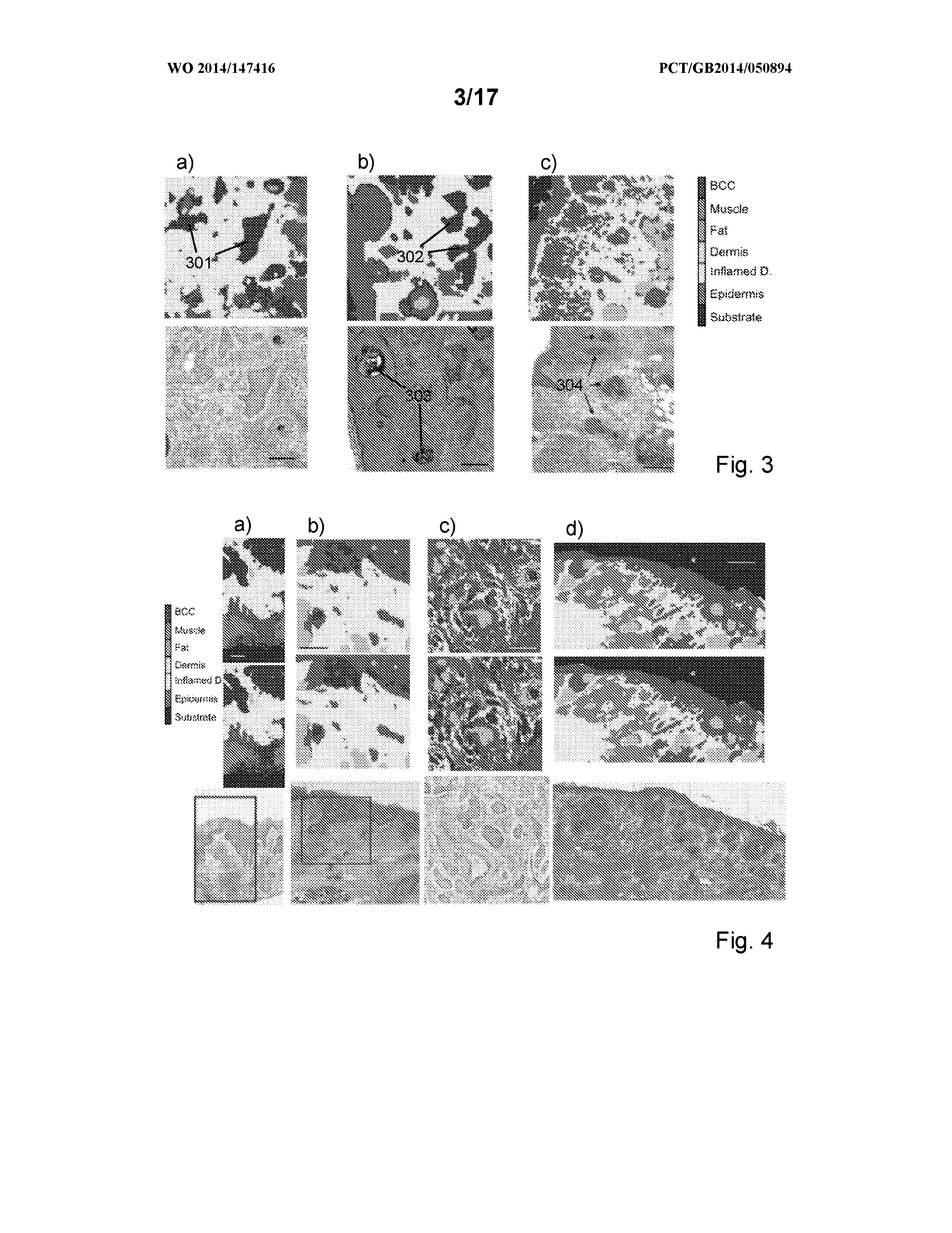
[0040]FIGS. 3a to 3c are raster scanned Raman spectrometry images and corresponding HE stained histopathology images of tissue sections (scale bar=200 μm);

[0041]FIGS. 4a to 4d are raster scanned Raman spectrometry images and corresponding HE stained histopathology images of unsectioned tissue blocks (scale bar=200 μm);

[0042]FIG. 5a is an autofluorescence intensity image of a sample taken with 377 nm inc...


PUM


Abstract

The disclosure relates to measurement and classification of tissue structures in samples using a combination of light imaging and spectroscopy, in particular although not necessarily exclusively for detection of tumours such as basal cell carcinoma or breast tumours in tissue samples. Embodiments disclosed include a method of automatically identifying tissue structures in a sample, the method comprising the steps of: measuring (1702, 1703) a response of an area of the sample to illumination with light; identifying (1704) regions within the area having a measured response within a predetermined range; determining (1705) locations within the identified regions; performing (1706) spectroscopic analysis of the sample at the determined locations; and identifying (1707) a tissue structure for each region from the spectroscopic analysis performed on one or more locations therein.

Description

FIELD OF THE INVENTION[0001]The invention relates to measurement and classification of tissue structures in samples using a combination of light imaging and spectroscopy, in particular although not necessarily exclusively for detection of tumours such as basal cell carcinoma or breast tumours in tissue samples.BACKGROUND[0002]In tissue-conserving surgery sequential layers of tissues are excised to ensure the removal of all tumour cells while maintaining as much healthy tissue as possible. Negative margins (in which all cancerous cells are removed) have been directly associated with lower local recurrence rates in many cancer types. Conservation of healthy tissue is important for tissue function preservation, wound healing and cosmesis. However, identifying the tumour margins intra-operatively remains one of the key challenges in tissue-conserving surgery.[0003]Histopathology is generally considered to be the “gold standard” method for evaluation of tissues for many diseases, includi...

Claims


Application Information

Patent Timeline
IPC IPC(8): G01N21/64G01N35/00G01N21/65G01N33/483
CPCG01N21/6486G01N33/4833G01N35/00G01N21/65G01N2021/6417G01N21/6458G01N2800/7028G01N2201/06113G01N21/6408G01J3/027G01J3/28
Inventor NOTINGHER, IOANKONG, KENNYROWLANDS, CHRISTOPHERWILLIAMS, HYWELLEACH, IAINVARMA, SANDEEPPERKINS, WILLIAMKOLOYDENKO, ALEXEY
Owner ROYAL HOLLOWAY UNIV OF LONDON
Why Eureka
Try Eureka