
System and method for providing assessment of tumor and other biological components contained in tissue biopsy samples

a tissue biopsy and tumor technology, applied in the field of biopsies, can solve the problems of less progress in optimizing the tissue acquisition process, insufficient conventional methods to ensure the success of intended molecular diagnostic tests, and insufficient standard approaches to on-site biopsy screening

Active Publication Date: 2016-12-22
MEMORIAL SLOAN KETTERING CANCER CENT
View PDF3 Cites 5 Cited by
  • Summary
  • Abstract
  • Description
  • Claims
  • Application Information

AI Technical Summary

Benefits of technology

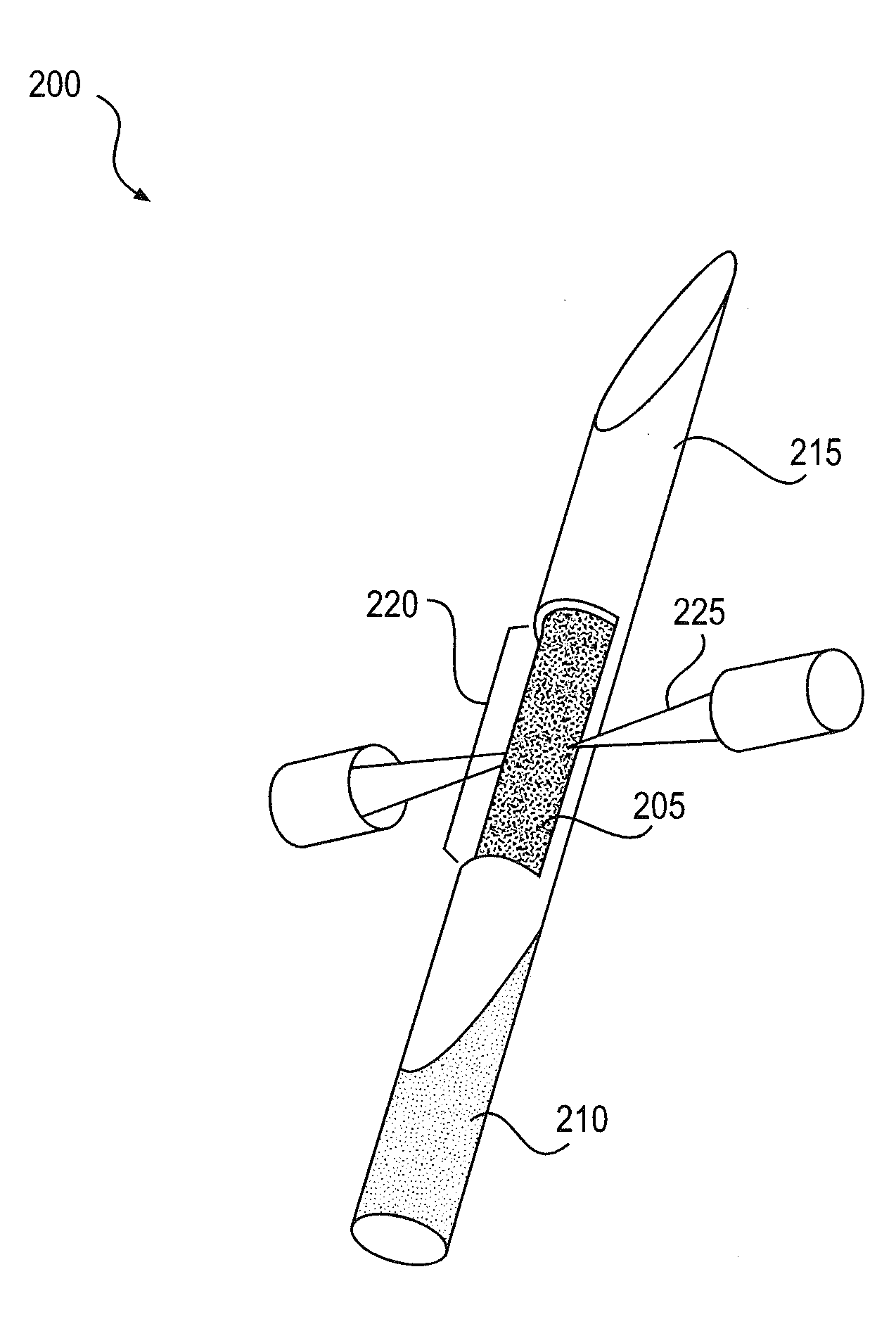
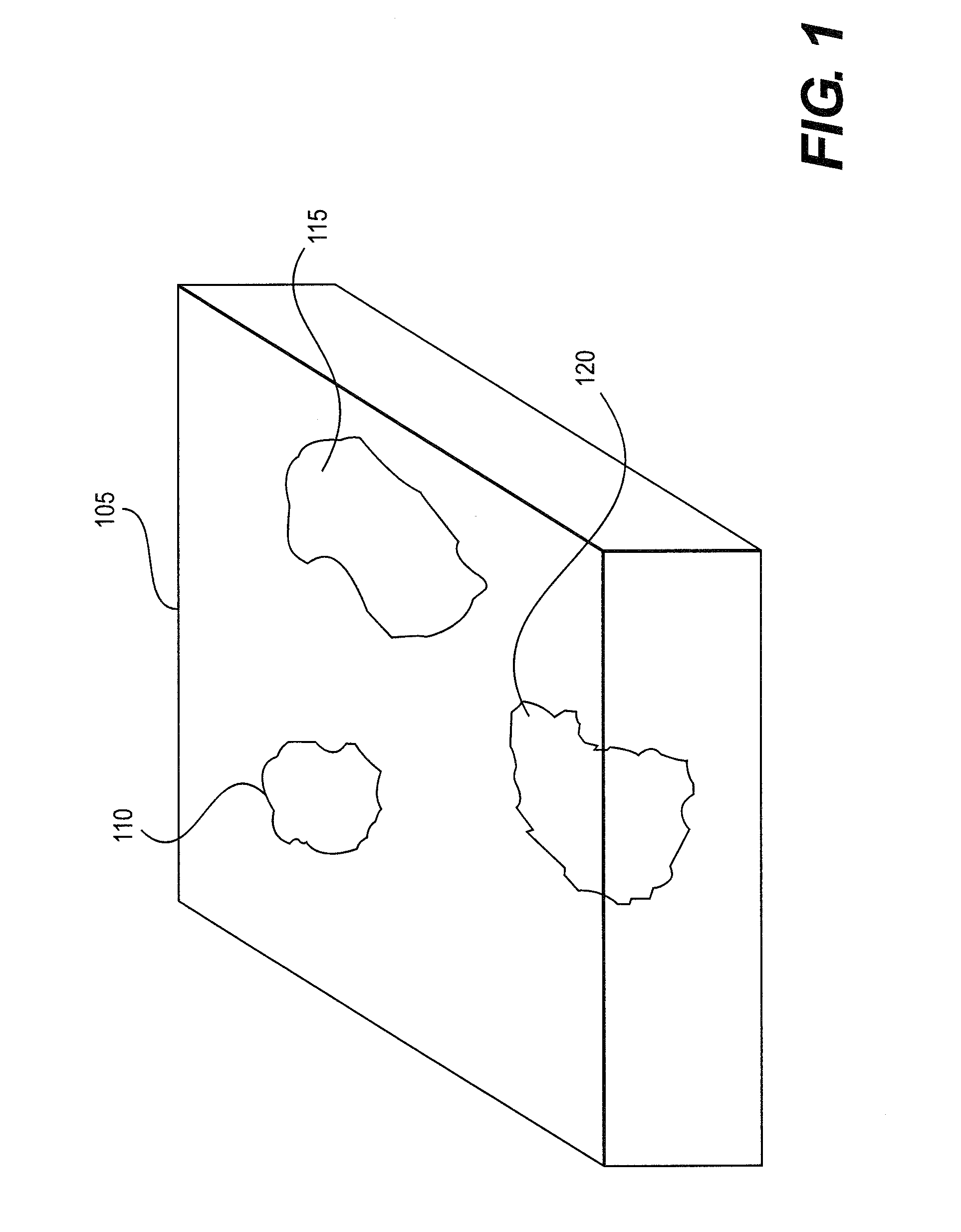
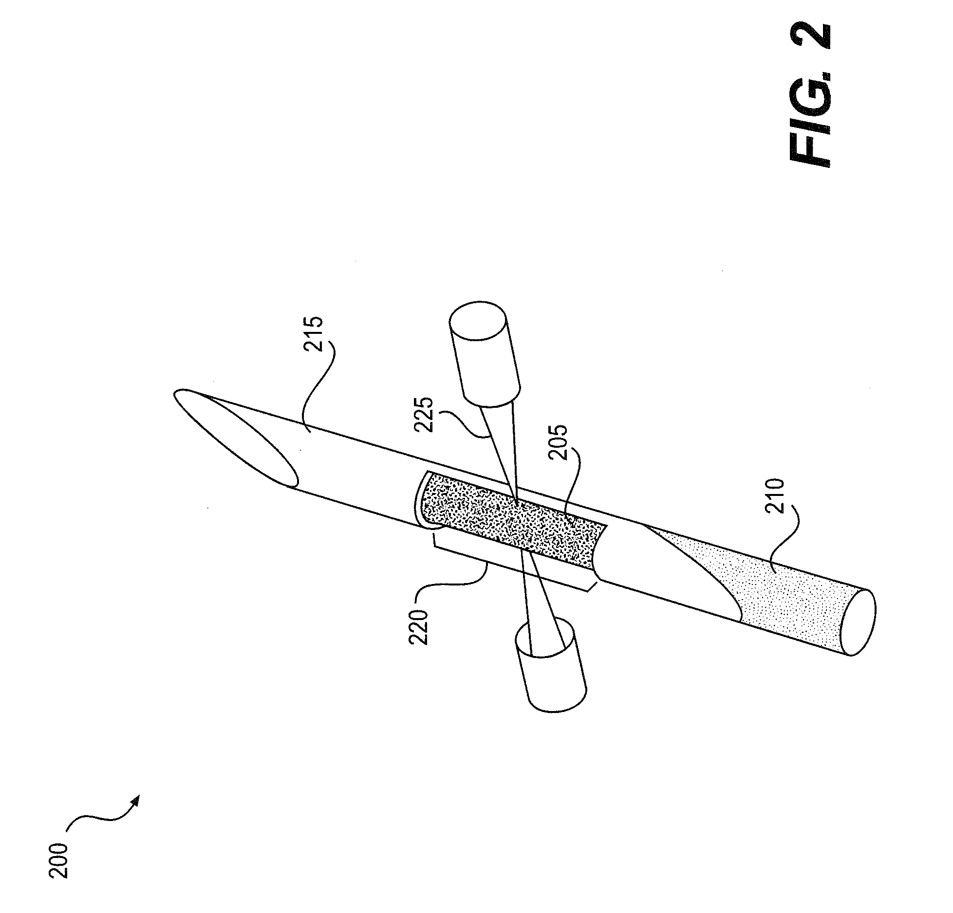
The patent describes a system, method, and computer-accessible medium for identifying and excising cancerous tissue from a patient. The system receives information related to a scan of the patient's tissue, which can include a three-dimensional scan using computed tomography. The scan can include information about the tissue types of cancerous and non-cancerous tissue. The system identifies the tissue types and sends the information to a system for quantifying the amount of biological components, such as DNA, RNA, proteins, or metabolites, in the tissue. The system can also receive further information about the biological components and excise the cancerous tissue based on the quantification. The method can involve inserting a biopsy device into the patient to remove a tissue sample, labeling the sample with a dye, stain, or other marker, and scanning the labeled sample for information about the biological components. The system can also use a paraffin block or other tissue fixation block to identify the tissue types. Overall, the system and method can improve the accuracy and effectiveness of cancer diagnosis and treatment.

Problems solved by technology

Unfortunately, the standard approach to on-site biopsy screening can fail to identify sample deficiencies in a significant number of patients, particularly when genetic testing can be essential.
While targeted therapies have improved, less progress has been made to optimize the process of the tissue acquisition (e.g., the tissue biopsy).
Preliminary studies examining results after image-guided percutaneous needle biopsies revealed that conventional methods can be insufficient to ensure the success of the intended molecular diagnostic tests.
However, this practice may not be ubiquitously utilized due to resource burden and cost, and additionally, can negatively impact the biopsy sample prior to histopathologic analysis.
It is known that touch-preparations of core needle biopsy samples on glass slides can (i) misrepresent the quantity of neoplastic cells in the specimen by depleting the core sample of cancer cells, and (ii) disrupt tissue architecture and fragment core samples destined for diagnostic and molecular analysis.
However, cancer patients themselves suffer the most critical costs of needle biopsy failures.
As biopsy results frequently determine cancer treatment alternatives, a biopsy failure can result in delay of appropriate care.
In addition to care delays, patients can be subject to increased risks from repeated biopsy attempts related to multiple needle insertions (e.g., bleeding, infection, or seeding of cancer cells along the needle path), as well as from sedation or anesthesia.
Unfortunately, deficiencies in the current standard practice for on-site assessment of biopsy adequacy can often lead to failure of subsequent specimen analysis.
These deficiencies can be particularly apparent during attempts to acquire the tissue from metastatic disease in bone, as existing techniques and assays may only be optimized for soft tissues.

Method used


Image

  • System and method for providing assessment of tumor and other biological components contained in tissue biopsy samples
  • System and method for providing assessment of tumor and other biological components contained in tissue biopsy samples
  • System and method for providing assessment of tumor and other biological components contained in tissue biopsy samples

Examples


Embodiment Construction

[0007]An exemplary system, method and computer-accessible medium can include, for example, receiving information related to a scan(s) of a three-dimensional structure(s) containing a first tissue(s) and a second tissue(s), and providing the information to a system for identifying respective tissue types of the first tissue(s) and the second tissue(s). The structure(s) can be scanned to generate the information. The structure(s) can include a paraffin block(s). The first tissue(s) or the second tissue(s) can be excised. The tissue types can include a cancerous tissue and a non-cancerous tissue, and the first tissue(s) can be identified as the cancerous tissue and the second tissue(s) can be identified as the non-cancerous tissue. Further information from the system can be received which can include an identification of the tissue type of the first tissue(s) and the second tissue(s). The scan(s) can be a three-dimensional scan of the three-dimensional structure(s), which can be perfor...


PUM

PropertyMeasurementUnit
wavelengthsaaaaaaaaaa
oblique angleaaaaaaaaaa
structureaaaaaaaaaa

Abstract

An exemplary system, method and computer-accessible medium can include, for example, receiving information related to a scan(s) of a three-dimensional structure(s) containing a first tissue(s) and a second tissue(s), and providing the information to a system for identifying respective tissue types of the first tissue(s) and the second tissue(s). The structure(s) can be scanned to generate the information. The structure(s) can include a paraffin block(s). The first tissue(s) or the second tissue(s) can be excised. The tissue types can include a cancerous tissue and a non-cancerous tissue, and the first tissue(s) can be identified as the cancerous tissue and the second tissue(s) can be identified as the non-cancerous tissue.

Description

CROSS-REFERENCE TO RELATED APPLICATION(S)[0001]This application relates to and claims priority from U.S. Patent Application No. 61 / 940,330, filed on Feb. 14, 2014, the entire disclosure of which is incorporated herein by reference.FIELD OF THE DISCLOSURE[0002]The present disclosure relates generally to biopsies, and more specifically, to exemplary embodiments of instrument, system and method for a rapid assessment of the presence of cancer tissue and / or Deoxyribonucleic acid (“DNA”) presence and quantity of a tumor, as well as the presence and quantity of biological components relevant to medical diagnostics or biomedical research (e.g., genetic material, proteins, and metabolites) in tissue biopsy samples.BACKGROUND INFORMATION[0003]For cancer patients undergoing a needle biopsy, a primary concern can be biopsy safety, as well as obtaining a diagnosis that can enable a rapid and effective treatment, if treatment can be necessary. Unfortunately, the standard approach to on-site biop...

Claims


Application Information

Patent Timeline
Patent Type & Authority Applications(United States)
IPC IPC(8): A61B10/02A61B5/00G01N33/483G01N21/27G01N24/08A61B6/00G01N21/64
CPCA61B10/0275A61B6/00A61B5/0075A61B5/0066G01N33/4833G01N21/6428G01N21/27G01N24/08A61B5/0071G01N21/17
Inventor SOLOMON, STEPHEN B.DURACK, JEREMY C.
Owner MEMORIAL SLOAN KETTERING CANCER CENT
Learn More