Method for generating synthetic electron density information for dose calculations based on MRI

a synthetic electron density and dose calculation technology, applied in the field of generating synthetic electron density information for dose calculations based on mri, can solve the problems of low contrast, high image contrast, and limit the accuracy by which target and risk organs can be identified, and achieve the effect of reducing one, alleviating one, and eliminating on

Active Publication Date: 2017-03-16
SPECTRONIC
View PDF0 Cites 13 Cited by
  • Summary
  • Abstract
  • Description
  • Claims
  • Application Information

AI Technical Summary

Benefits of technology

The present invention is about a method for generating synthetic CT image stacks for use in radiation treatment planning. This method aims to address the drawbacks of current techniques, which result in low accuracy and difficulty in handling information. The method involves segmenting the MR image stack and aligning it with a template electron density information map, which can be acquired from a different person with different geometries. The transformation of the template map allows for precise spatial correspondence with the acquired MR image stack. The method also includes using a set of templates for forming synthetic electron density information, which can be compared to an acquired MR image stack of a patient to determine the synthetic information. This approach allows for accurate and efficient treatment planning even when multiple templates are used.

Problems solved by technology

Both between and within different types of soft-tissues (such as organs, muscles and fat), the image contrast is highly limited.
During radiation treatment planning, this low contrast is a disadvantage, since it limits the accuracy by which target and risk organs can be identified.
However, the disadvantage is that otherwise healthy tissue is thereby also unnecessarily irradiated.
However, even such combined CT / MRI workflow has several disadvantages.
First, acquiring both an MRI and a CT is both expensive and time consuming, and it also adds additional discomfort to the patient.
Secondly, the registration between MRI and CT is rarely perfect, which adds an uncertainty to the identification of both the target and the risk organs.
In addition, the CT itself adds a significant radiation dose to the patient, which limits its repeated use in following tumor changes between fractions.
However, this method requires extensive user interaction in order to generate desired electron density information.

Method used


Image

  • Method for generating synthetic electron density information for dose calculations based on MRI
  • Method for generating synthetic electron density information for dose calculations based on MRI
  • Method for generating synthetic electron density information for dose calculations based on MRI

Examples


Embodiment Construction

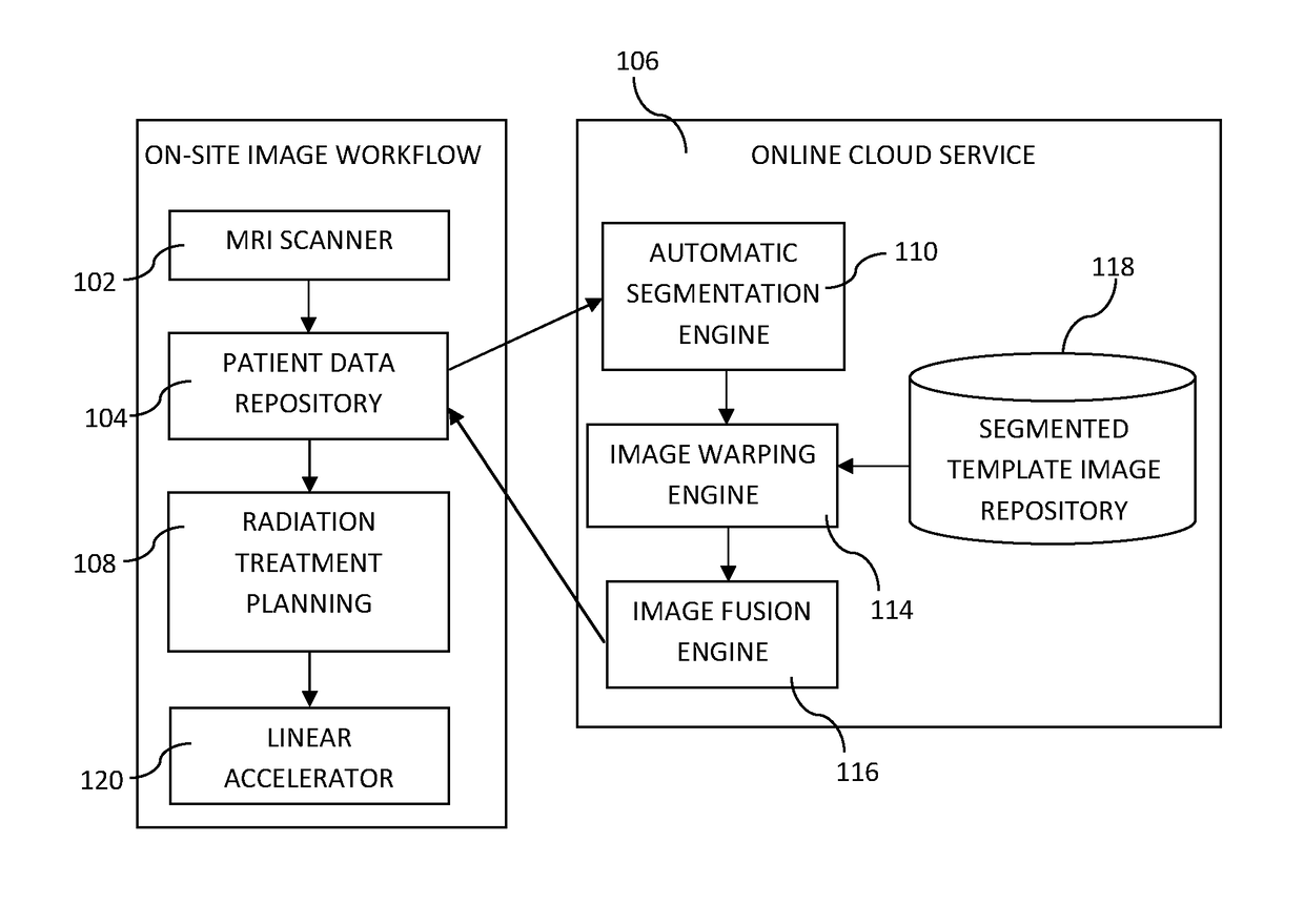
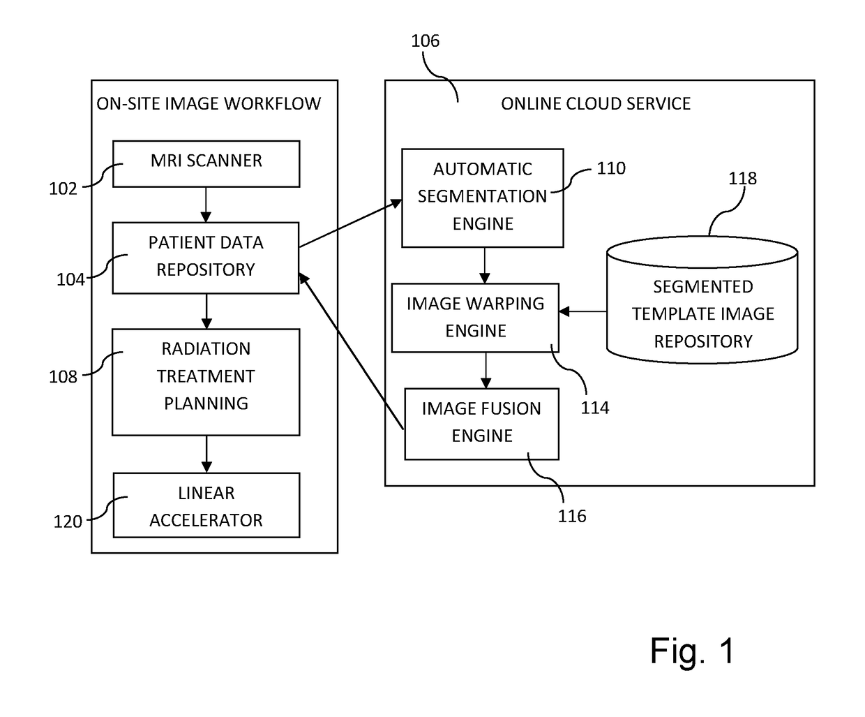
[0067]Embodiments of the present invention relate, in general, to the field of generating synthetic electron density information. The forming of synthetic electron density information may be particularly useful for MRI based radiation treatment planning. However, it should be realized that it may be used in other applications as well. For instance, the synthetic electron density information may be used as attenuation information for a positron emission tomography (PET) camera or a single-photon emission computed tomography (SPECT) camera.

[0068]A preferred embodiment relates to generating synthetic CT image stacks for radiation dose calculation in cancer radiotherapy, such as prostate treatments, based on one or more MRI image stacks. However, it should be appreciated that the invention is as such equally applicable to radiotherapy in any other anatomical area, such as brain, head / neck, lungs, uterus, abdomen, or any other region in which radiation treatment planning is required. Lik...


PUM


Abstract

A method for generating synthetic electron density information based on an acquired magnetic resonance, MR, image stack is disclosed. The method comprises: analyzing the acquired MR image stack to automatically segment the acquired MR image stack; generating warp information for a template dataset comprising at least one template electron density information map and at least one template structure information, said generating comprising utilizing structure information of the acquired MR image stack and said template structure information in order to determine transformations bringing the template structures in spatial correspondence with the structures in the acquired MR image stack, wherein said generated warp information describes at least one transformation bringing the at least one template electron density information map in spatial correspondence with the acquired MR image stack; and forming synthetic electron density information corresponding to the acquired MR image stack based on at least part of a warped template electron density information map.

Description

TECHNICAL HELD[0001]The following disclosure relates generally to the fields of medical imaging and radiation therapy and, more specifically, to the application of image processing techniques in a computer software tool for creating a synthetic electron density information from a magnetic resonance image (MRI), e.g. for use in radiation treatment planning for cancer.BACKGROUND[0002]Tomography refers to a technique for capturing a two-dimensional (2D) or three-dimensional (3D) cross-sectional image of an object, through the use of radiation or any kind of penetrating wave. Computed Tomography (CT) refers to a medical imaging technique that uses X-rays and computer processors to collect and display 2D or 3D images or tomograms of an object. Magnetic Resonance imaging (MRI) refers to a tomographic technique that uses a powerful magnetic field and a radio frequency transmitter to capture detailed images of organs and structures inside the body.[0003]Radiation therapy refers to a techniq...

Claims


Application Information

Patent Timeline
Patent Type & Authority Applications(United States)
IPC IPC(8): A61N5/10G06T7/00A61B6/00G01R33/48A61B5/055A61B6/03
CPCA61N5/1039A61B5/055A61B6/032A61B6/5247G06T2207/10081G01R33/4808G06T7/0012G06T7/0081G06T2207/10088A61B6/563G06T2200/04G06T2207/20128G06T2207/30004A61B6/037G06T7/337G06T7/11
Inventor SIVERSSON, CARL
Owner SPECTRONIC
Why Eureka
Try Eureka