Multi-Modality Image Fusion for 3D Printing of Organ Morphology and Physiology

a multi-modality, image fusion technology, applied in the field of 3d printing of organs, can solve problems such as limited printing capabilities

Active Publication Date: 2017-08-03
SIEMENS HEALTHCARE GMBH
View PDF5 Cites 33 Cited by
  • Summary
  • Abstract
  • Description
  • Claims
  • Application Information

AI Technical Summary

Benefits of technology

[0004]The present invention provides a method and system for multi-modality image fusion for 3D printing of organ morphology and physiology. Different image modalities provide different information on an organ. It is therefore desirable to go beyond the current mono-modality status quo and provide a tool that enables fusing of information from multiple medical imaging modalities into a single model and printing such a fused model using 3D printing. The present inventors have recognized that generating a 3D printed model that combines morphological, structural, dynamic, and functional information from various medical imaging modalities would be a great use in the clinical workflow, facilitating decision making, therapy planning, discussions during clinical board meetings, teaching to patients, and device development. Embodiments of the present invention utilize a framework that relies on image analytics, semantic image fusion, and advanced materials for 3D printing of organ models. Embodiments of the present invention combine the information from multiple imaging sources to create a holistic model of an organ, including morphology, substrate, and physiology, and prints the holistic model using 3D printing with advanced materials.

Problems solved by technology

However, the printed models come from a single medical imaging modality and only limited printing capabilities are available.

Method used


Image

  • Multi-Modality Image Fusion for 3D Printing of Organ Morphology and Physiology
  • Multi-Modality Image Fusion for 3D Printing of Organ Morphology and Physiology
  • Multi-Modality Image Fusion for 3D Printing of Organ Morphology and Physiology

Examples


Embodiment Construction

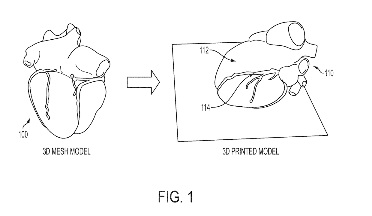
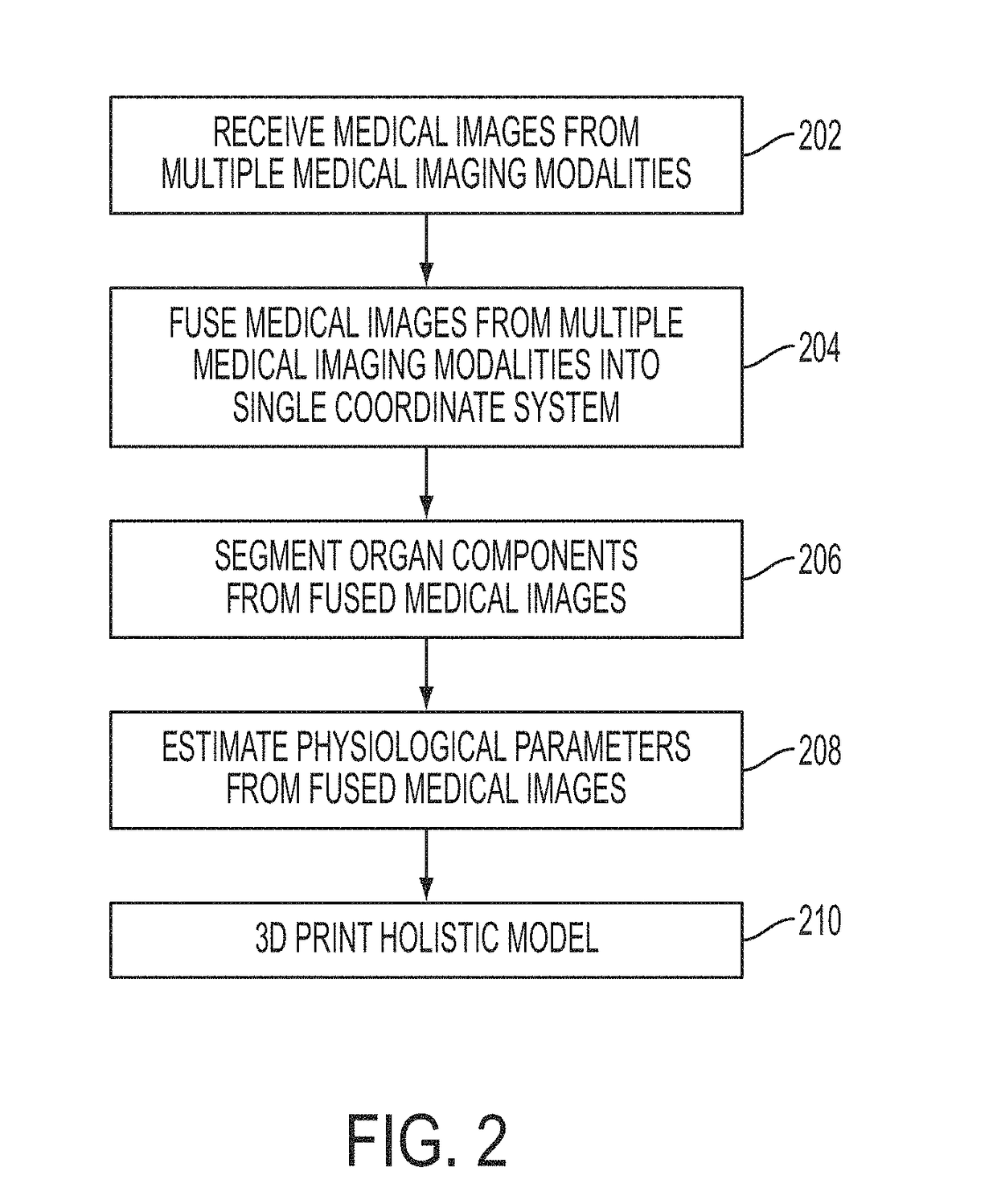
[0016]The present invention relates to multi-modality image fusion for 3D printing of organ morphology and physiology. A digital image is often composed of digital representations of one or more objects (or shapes). The digital representation of an object is often described herein in terms of identifying and manipulating the objects. Such manipulations are virtual manipulations accomplished in the memory or other circuitry / hardware of a computer system. Accordingly, is to be understood that embodiments of the present invention may be performed within a computer system using data stored within the computer system or available through a network system.

[0017]Different image modalities provide different information on an organ. In cardiology for instance, ultrasound is typically the modality of choice for cardiac function, computed tomography (CT) for cardiac morphology, and magnetic resonance (MR) for tissue substrate. Similarly for the liver, ultrasound can provide flow and elasticity...


PUM

PropertyMeasurementUnit
strengthaaaaaaaaaa
velocityaaaaaaaaaa
stiffnessaaaaaaaaaa

Abstract

A system and method for multi-modality fusion for 3D printing of a patient-specific organ model is disclosed. A plurality of medical images of a target organ of a patient from different medical imaging modalities are fused. A holistic mesh model of the target organ is generated by segmenting the target organ in the fused medical images from the different medical imaging modalities. One or more spatially varying physiological parameter is estimated from the fused medical images and the estimated one or more spatially varying physiological parameter is mapped to the holistic mesh model of the target organ. The holistic mesh model of the target organ is 3D printed including a representation of the estimated one or more spatially varying physiological parameter mapped to the holistic mesh model. The estimated one or more spatially varying physiological parameter can be represented in the 3D printed model using a spatially material property (e.g., stiffness), spatially varying material colors, and / or spatially varying material texture.

Description

[0001]This application claims the benefit of U.S. Provisional Application No. 62 / 288,481, filed Jan. 29, 2016, the disclosure of which is herein incorporated by reference in its entirety.BACKGROUND OF THE INVENTION[0002]The present invention relates to 3D printing of an organ based on medical image data, and more particularly to multi-modality image fusion for 3D printing of organ morphology and physiology from multiple imaging modalities.[0003]3D printing has seen tremendous progress in recent years. Recent advances in material design. Current medical applications rely on simple anatomical modeling based on a single medical imaging modality, most commonly computed tomography (CT). 3D printing can be performed locally, via 3D printers such as MakerBot or more advanced devices, or via services (e.g., Materialise or ShapeWays). However, the printed models come from a single medical imaging modality and only limited printing capabilities are available.BRIEF SUMMARY OF THE INVENTION[000...

Claims


Application Information

Patent Timeline
Patent Type & Authority Applications(United States)
IPC IPC(8): B29C67/00A61B8/08A61B6/03B33Y50/02G06F19/00G05B19/4099B33Y10/00B33Y30/00A61B5/055A61B34/10G16H30/20G16H30/40G16H50/50
CPCB29C67/0088B29L2031/7532A61B8/485A61B6/032A61B8/5261A61B34/10G06F19/3406G06F19/321G06F19/3437G05B19/4099B29C67/0059B29C67/0085B33Y10/00B33Y30/00B33Y50/02A61B2034/105G05B2219/49023A61B5/055B29L2031/7534A61B5/0044A61B2576/023G05B2219/45172A61B5/0035B33Y80/00B33Y50/00B29C64/386G16H40/63G16H50/50B29C64/112B29C64/20G16H30/20G16H30/40
Inventor MANSI, TOMMASOHOULE, HELENEGRBIC, SASAMILKOWSKI, ANDRZEJ
Owner SIEMENS HEALTHCARE GMBH
Why Eureka
Try Eureka