

Composition and method of using mir-302 precursors as drugs for treating alzheimer's diseases
- Summary
- Abstract
- Description
- Claims
- Application Information
AI Technical Summary
Benefits of technology
Problems solved by technology
Method used
Image

Examples
Embodiment Construction
[0112]Referring particularly to the Examples provided for the purpose of practical demonstration only and not limitation.
1. Bacterial Cell Culture and Chemical Treatments
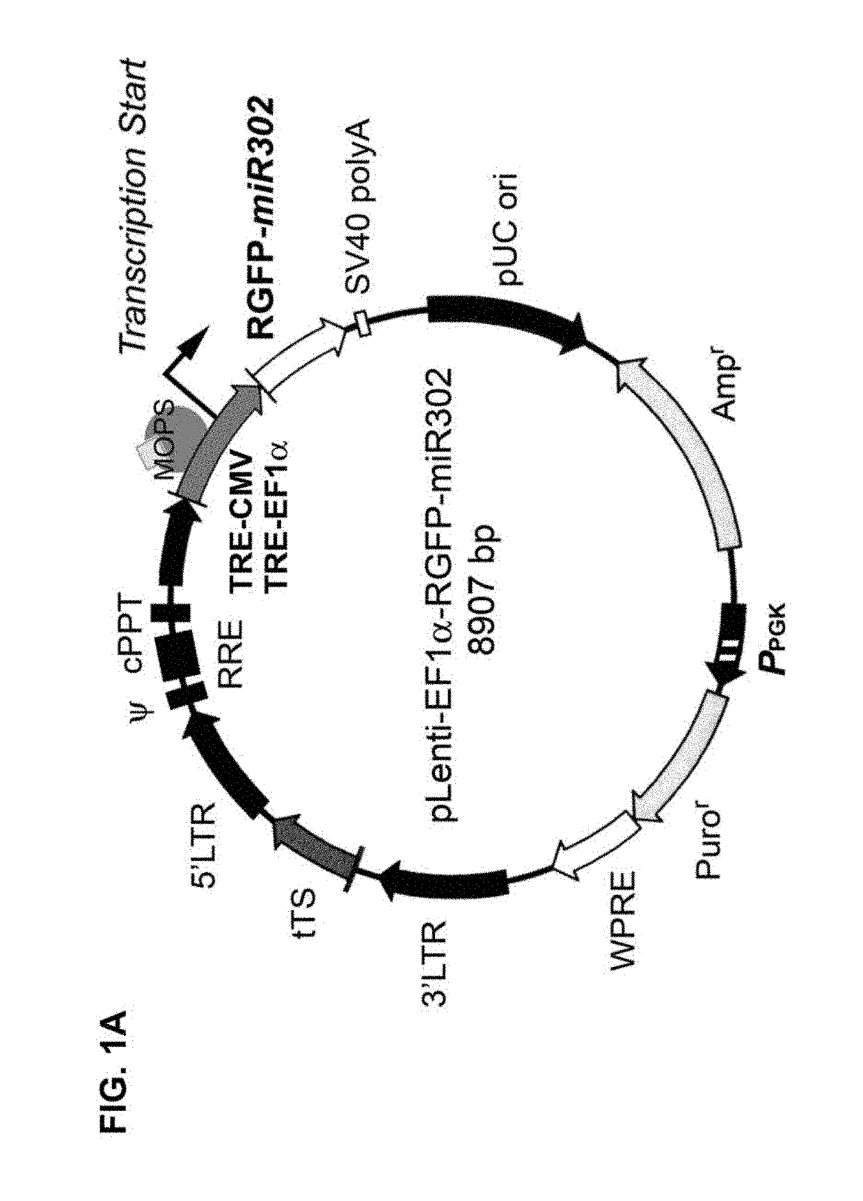
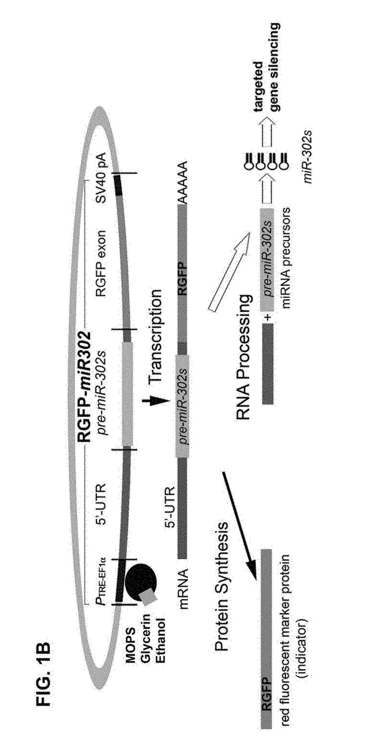
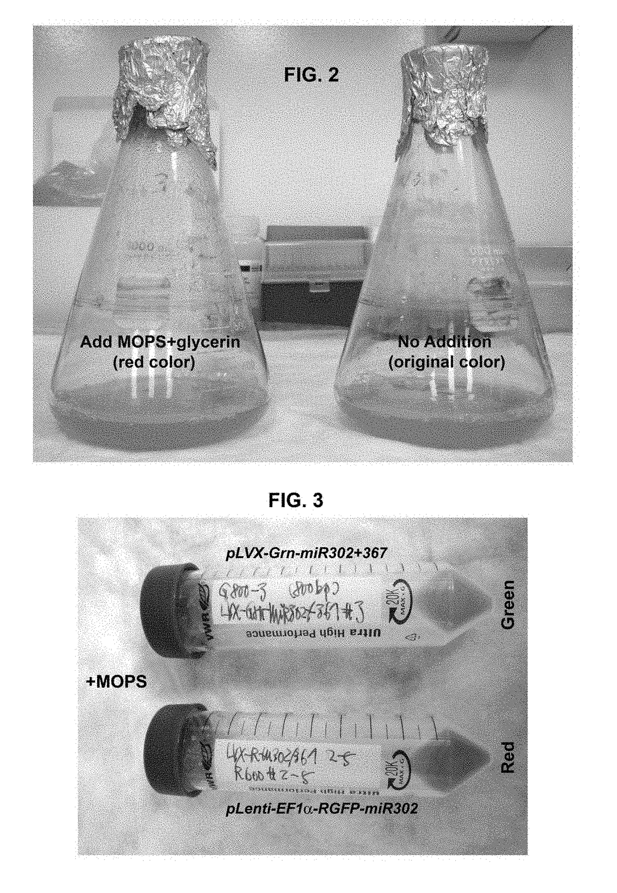
[0113]Competent cells of E. coli DH5alpha strain were acquired from the z-competent E. coli transformation kit (Zymo Research, Irvine, Calif.) and transformed by mixing with about 1˜10 μg of a desired plasmid vector such as pLVX-Grn-miR302+367 and / or pLenti-EF1alpha-RGFP-miR302 vectors. Non-transformed bacterial cells were normally grown in Luria-Bertani (LB) broth supplemented with 10 mM MgSO4 and 0.2 mM glucose at 37° C. with frequent agitation at 170 rpm, whereas the transformed bacterial cells were cultivated under the same condition with further addition of 100 μg / mL ampicillin. For chemical induction, about 0.1˜10 mL of MOPS, glycerin, and / or ethanol, respectively or in combination, was added into per litter of LB broth supplemented with 10 mM MgSO4 and 0.2 mM glucose in the presence of 100 μg / mL ampicillin. A...
PUM
| Property | Measurement | Unit |
|---|---|---|
| Fraction | aaaaa | aaaaa |
| Composition | aaaaa | aaaaa |
| Electrical resistance | aaaaa | aaaaa |
Abstract
Description
Claims
Application Information