Spherical nucleic acids (SNA) flare based fluorescence in situ hybridization

a technology fluorescence in situ, which is applied in the field of spherical nucleic acids (sna) flare based fluorescence in situ hybridization, can solve the problems of increasing cost and complexity

Inactive Publication Date: 2017-08-24
AURASENSE
View PDF4 Cites 7 Cited by
  • Summary
  • Abstract
  • Description
  • Claims
  • Application Information

AI Technical Summary

Benefits of technology

[0014]In other aspects the invention is compositions and kits for performing the methods described herein. A kit may include a lipid for producing a liposome, a fluorescent dye or plurality of fluorescent dyes, one or ...

Problems solved by technology

In order to increase the fluorescence intensity for each mRNA molecule, more unique...

Method used


Image

  • Spherical nucleic acids (SNA) flare based fluorescence in situ hybridization
  • Spherical nucleic acids (SNA) flare based fluorescence in situ hybridization

Examples


example 1

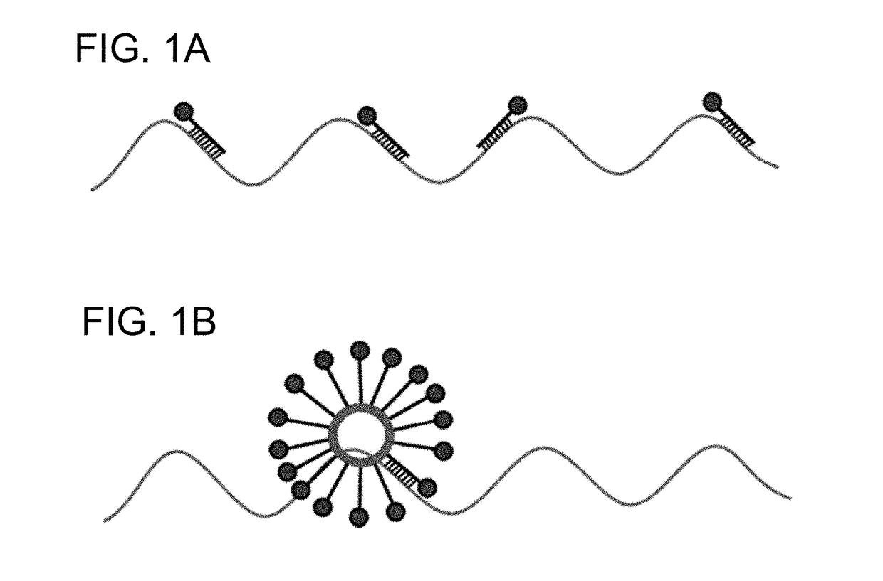
[0084]A panel of oligonucleotides were 5′-modified with Cy5 and 3′-modified with a di-stearyl group and incorporated into small unilamellar vesicles (55 nm in diameter) composed of 1,2-dioleoyl-sn-glycero-3-phosphatidylcholine (DOPC) (FIG. 1) at a density of 100 oligonucleotides per vesicle. These L-SNAs FISH probes were tested for their ability to bind (β-Actin mRNA transcripts in fixed human cell lines for fluorescence in situ hybridization (FISH) imaging. Commercially available FISH probes (BioSearch Technologies) were used as a control.

Methods

A. Adherent Cell Fixation

[0085]Adherent cells are first grown on 12 mm round #1 Poly-L-Lysine coated coverslips in a 12-well cell culture plate for 24 hours prior to fixation. Growth medium is then aspirated, cells washed with 1 mL of 1× Phosphate Buffered Saline (PBS) RNAse free, and fixed for 10 minutes at room temperature in 1 mL of fixation buffer composed of 1:1:8 ratio of 37% formaldehyde solution, 10× PBS RNAse free, and nuclease fre...


PUM

PropertyMeasurementUnit
Temperatureaaaaaaaaaa
Fractionaaaaaaaaaa

Abstract

The invention relates to a method of performing in situ hybridization such as fluorescence in situ hybridization (FISH) using liposomal spherical nucleic acids (L-SNAs) nanoparticles labeled with dye molecules. The nanoparticles contain one or more nucleic acids that recognize a target of interest in a sample.

Description

RELATED APPLICATIONS[0001]This application claims the benefit under 35 U.S.C. §119 of U.S. provisional application 62 / 299,454, filed Feb. 24, 2016 the entire contents of which is incorporated herein by reference.FIELD OF INVENTION[0002]The present invention generally relates to methods of using nanoparticles containing oligonucleotides labeled with dye molecules to detect a target nucleic acid, and kits and compositions thereof.BACKGROUND[0003]Fluorescence in situ hybridization (FISH) for the detection of mRNA is a widely used technique to image mRNA molecules in cells, or tissue sections, that are fixed, then permeabilized to allow probe access to the gene target. Current technology utilizes multiple fluorescently labeled oligonucleotide probes with unique sequences to recognize a single mRNA target. After the probes are hybridized to the target, the samples can be imaged using fluorescence microscopy. The maximum potential signal intensity using individually fluorescently labeled ...

Claims


Application Information

Patent Timeline
IPC IPC(8): C12Q1/68
CPCC12Q1/682C12Q1/6841C12Q2525/30C12Q2537/143C12Q2565/30
Inventor GILJOHANN, DAVID A.KANG, RICHARDPATEL, PINAL
Owner AURASENSE
Why Eureka
Try Eureka