
Cardiac phase-resolved non-breath-hold 3-dimensional magnetic resonance angiography

a phase-resolved, non-breath-hold technology, applied in the field of angiography, can solve the problems of limiting the useful time window of extracellular contrast agents, and achieve the effect of high quality delineation

Active Publication Date: 2017-09-28
RGT UNIV OF CALIFORNIA
View PDF2 Cites 6 Cited by
  • Summary
  • Abstract
  • Description
  • Claims
  • Application Information

AI Technical Summary

Benefits of technology

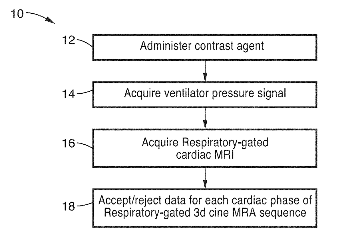
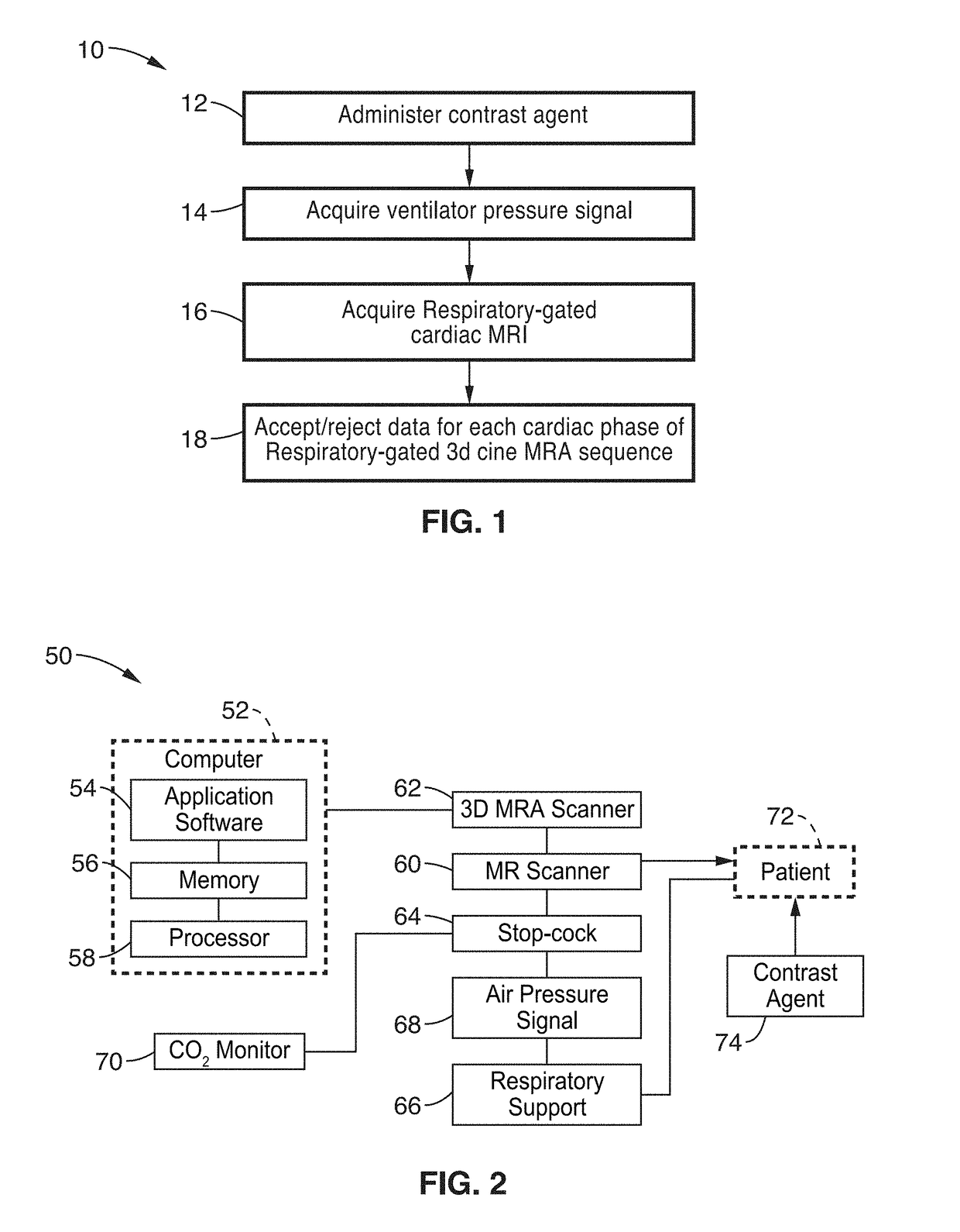
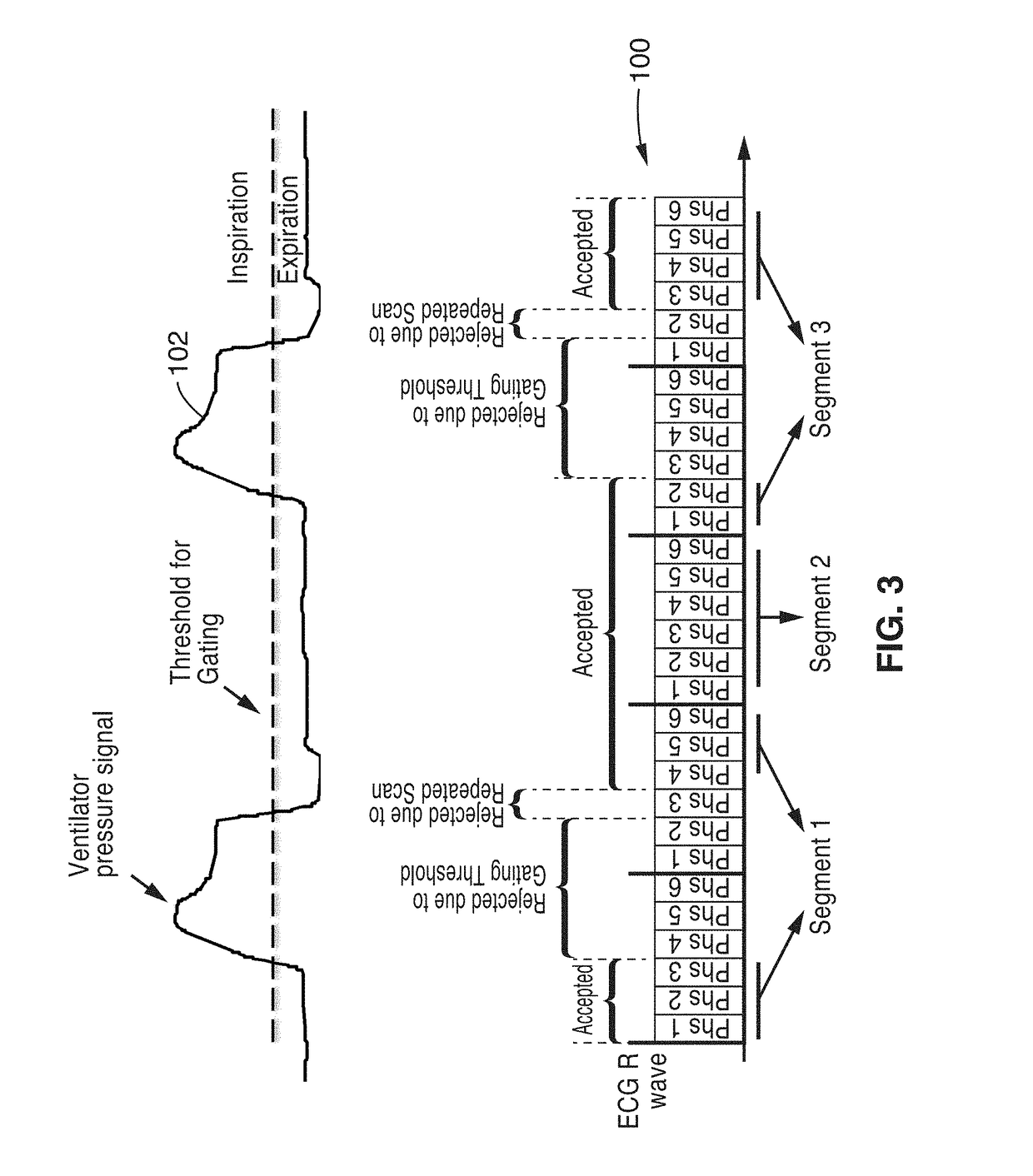
The patent describes using ferumoxytol as a contrast agent to improve the delineation of cardiovascular anatomy in children who are under general anesthesia and controlled mechanical ventilation. By combining this with a 3D cine MRA sequence and ventilator respiratory gating, high-quality imaging of the heart can be achieved without the need for breath-holding or the need to acquire data during the first-pass of the contrast agent. This allows for a segmented acquisition that can provide both anatomic and dynamic, functional information about the heart in three dimensions with unprecedented spatial resolution.

Problems solved by technology

These attributes of the extracellular contrast agents limit their useful angiographic time window to the first-pass and very early distribution phases.

Method used


Image

  • Cardiac phase-resolved non-breath-hold 3-dimensional magnetic resonance angiography
  • Cardiac phase-resolved non-breath-hold 3-dimensional magnetic resonance angiography
  • Cardiac phase-resolved non-breath-hold 3-dimensional magnetic resonance angiography

Examples


example 1

[0037]Six patients (mean age 2.4±2.1 y.o., range 3 days to 5 years, 4 male) underwent cardiovascular MRI with controlled mechanical ventilation. Conventional breath-held contrast-enhanced MRA (CE-MRA) was performed during the first-pass in comparison to delayed steady state distribution phases of ferumoxytol, which was used in place of gadolinium based contrast agents (GBCA).

[0038]Four patients underwent general anesthesia and 2 patients were transferred already intubated from the neonatal intensive care unit (NICU) and the patients were monitored by pediatric anesthesiologists or NICU staff respectively, who monitored the patients continuously throughout the imaging exam. As appropriate, anesthesia was maintained with inhalation of a mixture of oxygen and savoflurane and patients from the NICU were sedated with fentanyl. In all cases, 0.2 mg / kg of Rocuronium Bromide was administrated as a muscle relaxant. An MR compatible ventilator (Fabius MRI, Drager Medical, Telford, Pa.) was us...

example 2

[0059]Use of ferumoxytol as a contrast agent was also evaluated as a non-gadolinium alternative for high-resolution CEMRA in renal failure. 9 patients aged 6 days to 14 years were evaluated with first pass and steady state CEMRA following ferumoxytol (Feraheme, AMAG) infusion at a dose of 0.05 mmol / kg to 0.07 mmol / kg. All patients were studied on a Siemens Magnetom TIM Trio system. Coil configurations included combinations of head-neck, body array and spine array, depending on patient size. Two patients had complex congenital heart disease and 8 were being considered for organ transplantation. The patients with CHD had supplemental cardiac gated high-resolution 3D CEMRA. The imaging FOV for all sequences routinely included head, neck, thorax, abdomen and pelvis with sub-mm voxels. Multiple CEMRA phases were acquired up to 30 minutes following ferumoxytol injection and measurements of SNR and CNR in the thoracic aorta and inferior vena cava (IVC) were recorded at each phase. These we...


PUM


Abstract

3D cine MR angiography systems and methods are disclosed for use during the steady state intravascular distribution phase of ferumoxytol. The 3D cine MRA technique enables improved delineation of cardiac anatomy in pediatric patients undergoing cardiovascular MRI.

Description

CROSS-REFERENCE TO RELATED APPLICATIONS[0001]This application is a 35 U.S.C. §111(a) continuation of PCT international application number PCT / US2015 / 030015 filed on May 8, 2015, incorporated herein by reference in its entirety, which claims priority to, and the benefit of, U.S. provisional patent application Ser. No. 61 / 990,776 filed on May 9, 2014, incorporated herein by reference in its entirety. Priority is claimed to each of the foregoing applications.[0002]The above-referenced PCT international application was published as PCT International Publication No. WO 2015 / 172100 on Nov. 12, 2015, which publication is incorporated herein by reference in its entirety.STATEMENT REGARDING FEDERALLY SPONSORED RESEARCH OR DEVELOPMENT[0003]Not ApplicableINCORPORATION-BY-REFERENCE OF COMPUTER PROGRAM APPENDIX[0004]Not ApplicableNOTICE OF MATERIAL SUBJECT TO COPYRIGHT PROTECTION[0005]A portion of the material in this patent document is subject to copyright protection under the copyright laws of...

Claims


Application Information

Patent Timeline
IPC IPC(8): A61B5/026A61B5/0402A61B5/113A61K49/18A61M16/00G01R33/56G01R33/563G01R33/567A61B5/00A61M5/00
CPCA61B5/0263A61B5/7285A61B5/0402A61B5/113A61M5/007A61B2503/06G01R33/5601G01R33/5635G01R33/5673A61K49/1818A61M2016/0027A61M16/0003A61B5/055G01R33/4822G01R33/5616G01R33/56325A61B5/33
Inventor FINN, J. PAULHU, PENGHAN, FEI
Owner RGT UNIV OF CALIFORNIA
Learn More