Method for determining a property of an object and associated device

Pending Publication Date: 2018-09-27
INST NAT DE LA SANTE & DE LA RECHERCHE MEDICALE (INSERM) +5
View PDF1 Cites 0 Cited by
  • Summary
  • Abstract
  • Description
  • Claims
  • Application Information

AI Technical Summary

Benefits of technology

The patent describes a method that combines ultrasound imaging with X-ray or γ-ray technology to achieve better treatment of diseases. This combination helps to get a more accurate understanding of the molecules involved in the disease and allows for better targeting with the treatment. Overall, this technique provides better outcomes for patients and improves the effectiveness of treatment.

Problems solved by technology

Unfortunately, a fundamental trade-off exists in CUI: to achieve a higher frame rate and thus a high quality functional imaging, a small field of view has to be defined.
It is a relatively time-consuming process that leads to imaging rates near the real-time threshold of approximately 30 images per second.
However, UUI is a physiological imaging technique and as such is unable to achieve sensitive molecular imaging.
To reach such goal, research in the ultrasound field is intensive.
However, Ultrafast Doppler Tomography requires long acquisition times, and is thus limited in applications in which dynamic information would be useful (e.g. to track brain activation).

Method used


Image

  • Method for determining a property of an object and associated device
  • Method for determining a property of an object and associated device

Examples


Embodiment Construction

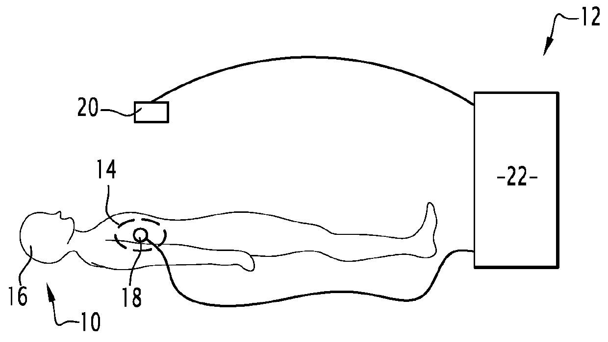
[0081]An object 10 and a device 12 are represented on FIG. 1.

[0082]In the specific example of FIG. 1, the object 10 is an area 14 of a subject 16.

[0083]The area 14 is delimited on FIG. 1 by a dotted line.

[0084]The area 14 is, for instance, a portion of the lung.

[0085]The subject 16 of FIG. 1 is a human being.

[0086]Alternatively, the subject 16 is an animal.

[0087]The device 12 is device adapted to determine at least one property P of the object 10.

[0088]For instance, in the specific example of FIG. 1, the device 12 is adapted to determine if a tumorous cell is present in the area 14. In such case, the determined property P is the presence of a tumorous cell.

[0089]The device 12 is also adapted to carry out a method for determining at least one property P of the object 10 as described in reference to FIG. 2.

[0090]The device 12 comprises a first imager 18, a second imager 20 and a controller 22.

[0091]The first imager 18 is adapted to image the object 10 according to a first imaging moda...


PUM


Abstract

The invention relates to a method for determining at least one property of an object, the method comprising a step of: a) obtaining first data relative to the object by an ultrasound imaging technique imaging the object at a frame rate superior to 300 Hz, characterized in that the method further comprises a step of: b) obtaining second data relative to the object by imaging the object with at least one of a X-ray and a γ-ray, and c) determining the at least one property of the object based on the first data and the second data.

Description

TECHNICAL FIELD OF THE INVENTION[0001]The present invention concerns a method for determining at least one property of an object. The present invention also relates to an associated device.BACKGROUND OF THE INVENTION[0002]Conventional Ultrasound Imaging (CUI) is the most widely used imaging modality in the clinic as it is portable, real-time, low-cost, and fully non-invasive.[0003]CUI is used in multiple modes that provide different kinds of contrast sources. The B-mode is the standard anatomical imaging mode and relies on the acoustic impedance of tissue, which, while providing limited specificity, has the notable advantage of allowing for the visualization of rapidly moving structures such as the heart. A distinctive feature of these B-mode ultrasound images is the presence of speckle of which the signature can be tracked from one frame to the other and can be used to quantify blood flow and tissue motion in ‘Doppler’ modes, as long as high frame rates are achieved.[0004]Unfortuna...

Claims


Application Information

Patent Timeline
IPC IPC(8): A61B6/00A61B6/03A61B8/06A61B8/08A61N7/00
CPCA61B6/5247A61B6/032A61B6/037A61B8/06A61B8/488A61B8/5261A61N7/00A61B6/4258A61B6/4417A61B8/4416
Inventor PROVOST, JEANGAROFALAKIS, ANIKITOSPERNOT, MATHIEUTANTER, MICKAËLTAVITIAN, BERTRANDVIEL, THOMAS
Owner INST NAT DE LA SANTE & DE LA RECHERCHE MEDICALE (INSERM)
Why Eureka
Try Eureka