
Methods and devices for connecting nerves

Active Publication Date: 2019-02-07
UNIV OF UTAH RES FOUND
View PDF0 Cites 1 Cited by
  • Summary
  • Abstract
  • Description
  • Claims
  • Application Information

AI Technical Summary

Benefits of technology

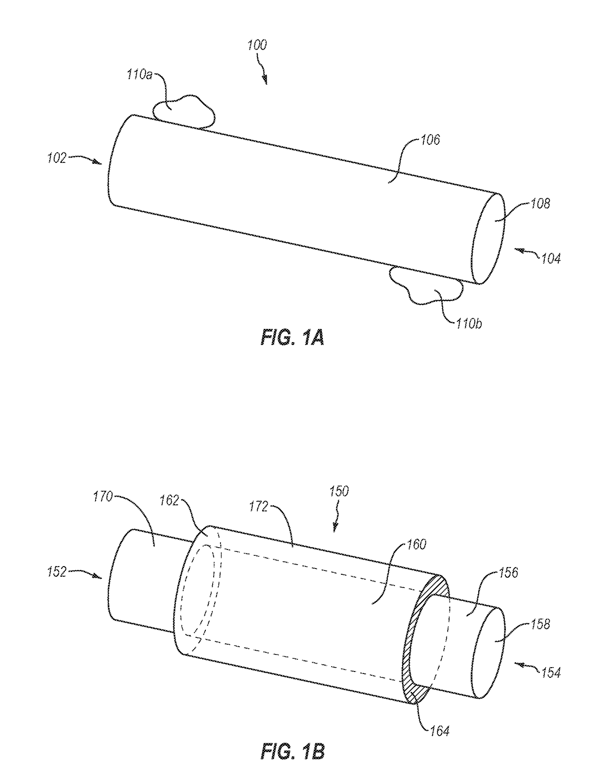
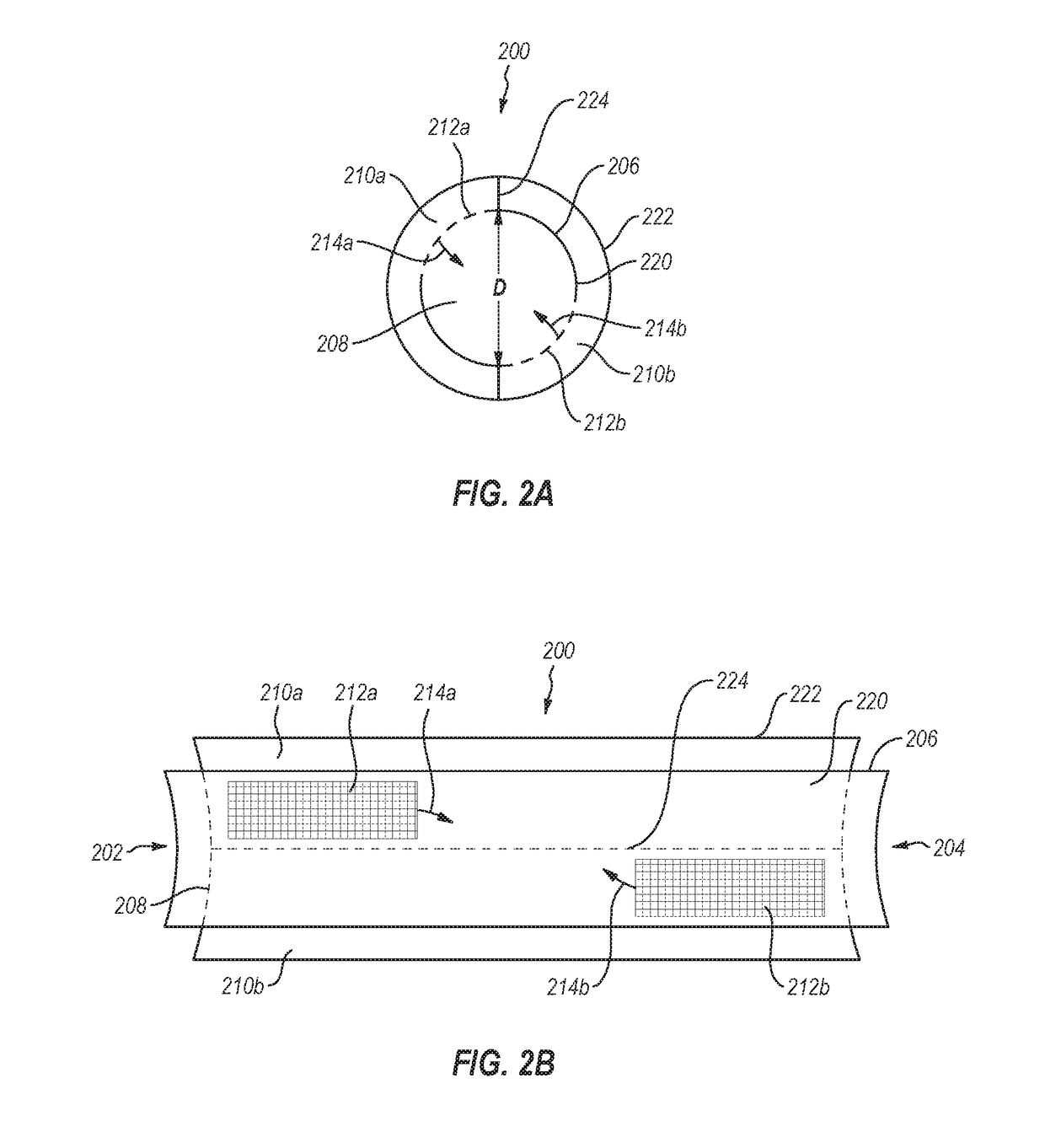
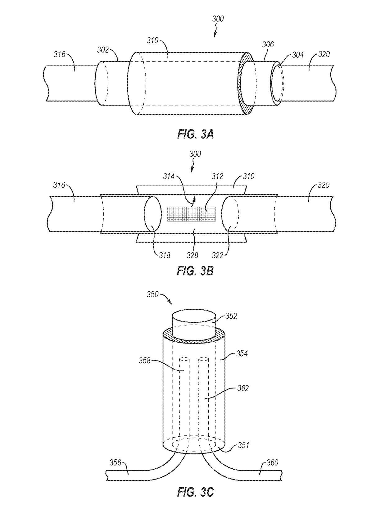
The present patent relates to a new type of nerve repair conduit that can cover a damaged portion of a nerve and deliver drugs to increase the rate of functional recovery. These conduits can be positioned around crushed, partially transected, or fully transected nerves. The conduits have drug reservoirs that continuously deliver small molecules or growth factors to the adjoined nerves at a controlled rate and improve the degree of axon regeneration and functional recovery. This delivery configuration allows for more controlled and uniform delivery of drugs, which may have inconsistent delivery kinetics and significant burst effects with other methods.

Problems solved by technology

These injuries can result in chronic debilitating pain from crush or neuroma formation.
Patients often suffer from life-long loss or functional disturbances mediated by the injured nerve, which can severely diminish their quality of life.
Nerve injuries have a tremendous socioeconomic impact from loss of work and associated healthcare costs.
Inadequate or untimely repair can result in lifelong deficits in muscle function or sensation.
These grafts are harvested from donor sensory nerves (sural nerve or posterior interosseous nerve) and have the disadvantage of loss of sensation at the donor site as well as the need for an additional surgical site.
While autologous nerve grafts serve as the state-of-the-art in repairing nerve gaps, numerous challenges associated with this approach results in functional benefits to less than about 50% of patients (e.g., only about 40-50%).
These challenges include morbidity at the site of harvesting of donor nerve(s), limitations to the amount of nerve that can be harvested, mismatches in size and fascicular patterns between the nerve stumps and the graft.
Acellular conduits and scaffolds have limited regenerative capacity and depend on the migration of supporting cells into the local environment.

Method used


Image

  • Methods and devices for connecting nerves
  • Methods and devices for connecting nerves
  • Methods and devices for connecting nerves

Examples


example 1

Release / Leakage Test

[0075]A 7-day release / leakage test was performed in order to explore the capacity of the PLGA device to deliver drugs of interest. Bovine serum albumin (BSA) was chosen to serve as the drug because its size is bigger than most of the growth factors. Two samples filled with 30 μL 300 mg / mL BSA were tested with two negative controls, two leakage test samples and a positive control, as shown in Table 1.

TABLE 1List of samples involved in the PLGA-BSA release / leakage testProtein in theReceiverSample NamePurposeReservoirReservoirchamberPositive ControlThe maximum possiblen / a42 uL 300 mg / mL7.5 mL PBS(PC) est. conc.conc. in the receiverBSA directly(tween free)1800 ug / mLchamber.injected into the*Note: useUse design's dimension:PBS7 mL Ambermax 42 uL drug reservoirGlass Vialvolume.Leakage Test 1To test if no BSA is#130 uL 300 mg / ml7.5 mL PBS(LT1)detected when using aBSAdevice filled with BSAand without window &filter.Leakage Test 1Duplicate LT1#230 uL 300 mg / ml7.5 mL PBS(L...

example 2

Placement of PLGA Repair Conduits

[0080]PLGA conduits with drug reservoirs were used to repair transected nerves in an end-to-end and a side-to-side fashion in a mouse sciatic nerve model in order to determine whether biodegradation of PLGA causes an adverse reaction. Walking track analysis was performed every two weeks until animals were sacrificed at day 90. It was found that long-term placement of PLGA conduits and biodegradation of PLGA produced no adverse reaction. In general, side-to-side repair seemed to show some advantages, but side-to-side repair is not always an option if there is a nerve gap. However, it is expected that all repair outcomes (e.g., side-to-side, end-to-end, side-to-end, etc.) can be improved with the drug-delivering conduits described herein.

example 3

Animal Testing

[0081]The drug delivery conduits described herein are tested in a mouse sciatic nerve model. NGF and GDNF are two of many growth factors implicated in improved peripheral nerve regeneration and are the test drugs for these experiments as they have been shown to enhance nerve growth both in vitro and in vivo. The mouse sciatic nerve has been established as a viable model for evaluation of peripheral nerve injury because of its easy accessibility and reasonable size for testing. It is hypothesized that the novel strategy described herein will provide results superior to nerve conduit alone (without drug) and will approach the results of autologous nerve graft. The device is used in combination with an autologous nerve graft and it is hypothesized that this combination will provide superior results to autologous nerve graft alone.

[0082]Experimental design: This efficacy study utilizes a mouse sciatic nerve gap (10 mm) model using transgenic mice that express GFP (green fl...


PUM

PropertyMeasurementUnit
Fractionaaaaaaaaaa
Fractionaaaaaaaaaa
Fractionaaaaaaaaaa

Abstract

A nerve repair conduit configured to be secured on first and second portions of a selected nerve. The nerve repair conduit includes a polymeric body having a proximal end, a distal end, an exterior surface and an interior surface defining an interior lumen. In addition, the nerve conduit includes at least one drug reservoir to hold agent(s) that may facilitate nerve regeneration. The drugs diffuse from the drug reservoir(s) into the nerve repair conduit through an outlet (such as a hole or a semipermeable membrane) in proximity to the first and second portions of a selected nerve. The nerve repair conduit may be configured to deliver the agent(s) at a rate having substantially zero-order kinetics and / or at a constant rate over a selected period of time.

Description

CROSS-REFERENCE TO RELATED APPLICATIONS[0001]This application is a continuation-in-part of U.S. patent application Ser. No. 15 / 942,965 filed 2 Apr. 2018 and titled “METHODS AND DEVICES FOR CONNECTING NERVES,” which is a continuation of U.S. patent application Ser. No. 14 / 352,024 filed 15 Apr. 2014 and titled “METHODS AND DEVICES FOR CONNECTING NERVES,” which application claims the benefit of and priority to International Application No. PCT / US12 / 60439, filed 16 Oct. 2012 and titled “METHODS AND DEVICES FOR CONNECTING NERVES,” which claims the benefit of and priority to U.S. Prov. Pat. App. Ser. No. 61 / 689,263 filed 1 Jun. 2012 and titled “METHODS AND DEVICES FOR CONNECTING NERVES”, U.S. Prov. Pat. App. Ser. No. 61 / 634,362 filed 28 Feb. 2012 and titled “METHODS AND DEVICES FOR CONNECTING NERVES,” and U.S. Prov. Pat. App. Ser. No. 61 / 547,978 filed 17 Oct. 2011 and titled “METHODS AND DEVICES FOR CONNECTING NERVES,” the entireties of each of which are incorporated herein by this refere...

Claims


Application Information

Patent Timeline
IPC IPC(8): A61B17/11C08L67/04A61L27/38A61F2/00A61L27/18A61L27/36A61L27/54
CPCA61L27/383A61L27/3675A61B2017/1132A61L2430/32A61L2300/64A61L27/54A61B17/1128A61L27/18A61L2300/414A61F2250/0067A61F2/0063A61F2/00C08L67/04A61B2017/00893
Inventor AGARWAL, JAYANT P.GALE, BRUCE KENTSANT, HIMANSHU JAYANTLABROO, PRATIMASHEA, JILL
Owner UNIV OF UTAH RES FOUND
Learn More