
Automated segmentation of histological sections for vasculature quantification

a technology of vascular morphology and quantification, applied in image data processing, instruments, computing, etc., can solve the problems of introducing errors, varying the thickness of the arteriole wall, and challenging manual measurement of morphological characteristics, and achieve high accuracy

Active Publication Date: 2019-02-07
LONDON HEALTH SCI CENT RES
View PDF15 Cites 3 Cited by
  • Summary
  • Abstract
  • Description
  • Claims
  • Application Information

AI Technical Summary

Benefits of technology

The present invention is about a way to segment things up after color deconvolution. It can detect borders and other objects using a 3D reconstruction of the actual section. This helps to identify staining artifacts and make things easier to analyze.

Problems solved by technology

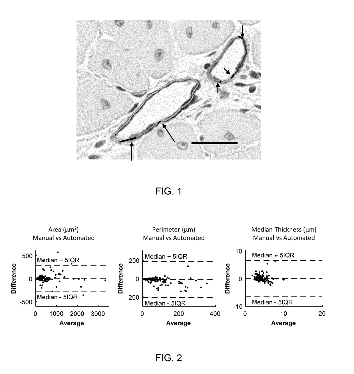
However, morphological characteristics can be challenging to measure manually, and some measurements, such as the varying thickness of the arteriole wall, approach the limits of practicality of manual measurement.
Manual measurements are generally taken in a small number of fields of view and extrapolated to the whole slide, potentially introducing error.
However, global approaches on 2D sections are challenged by within-section staining variability and lack of 3D context to distinguish true artefacts from small features of interest; for microvasculature analysis, the latter limitation is particularly important.

Method used


Image

  • Automated segmentation of histological sections for vasculature quantification
  • Automated segmentation of histological sections for vasculature quantification
  • Automated segmentation of histological sections for vasculature quantification

Examples


example

Materials

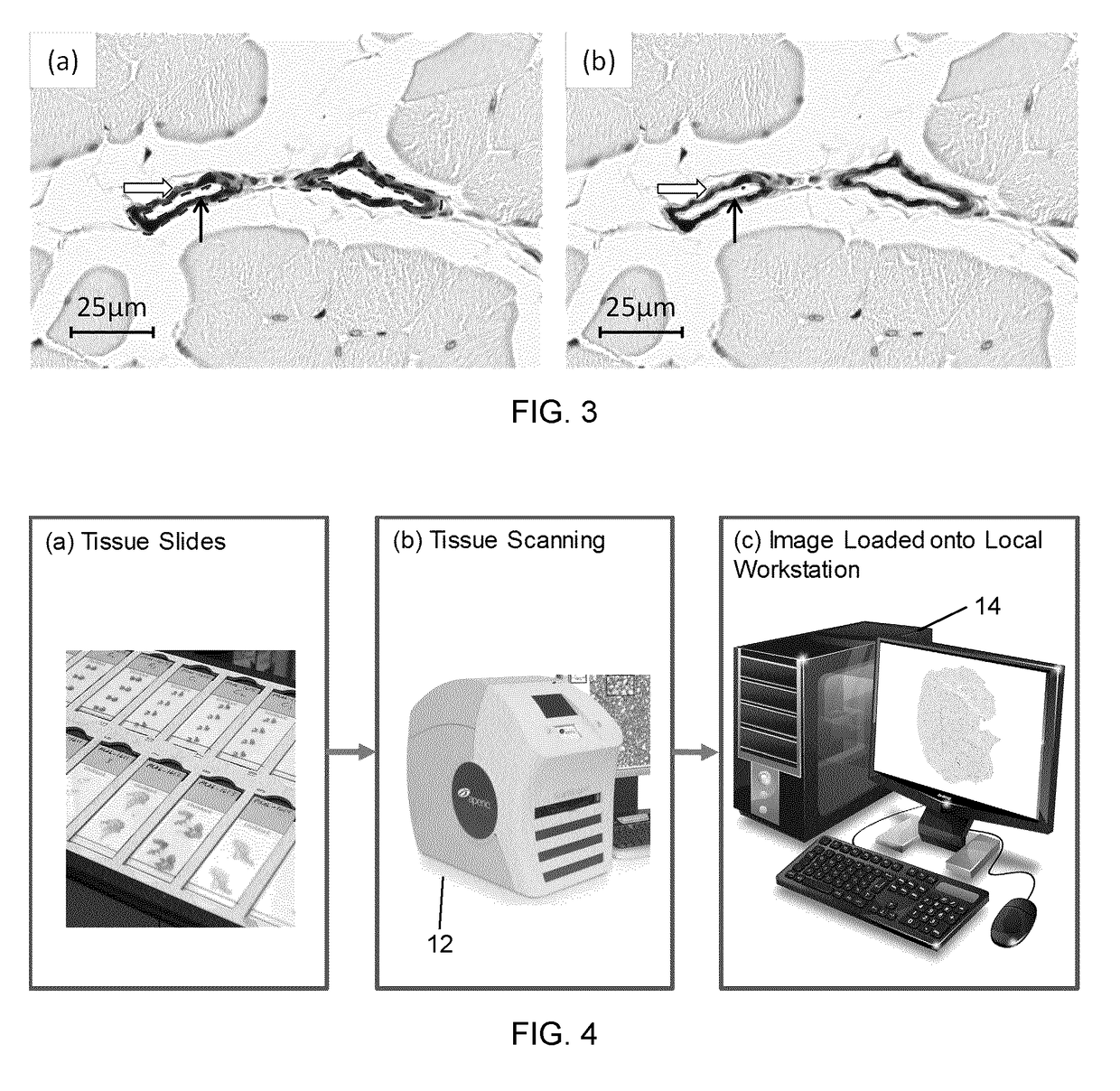
[0042]The experiments were conducted on normal and regenerated vasculature of the mouse hind limb. The tibialis anterior (TA) muscle bundle was used from a wild type C57BL / J6 mouse (Sample 1 and Sample 2), and a mouse of the same strain two weeks after induction of hind limb ischemia by femoral artery excision (Sample 3). Sample 1, Sample 2 and Sample 3 comprised 10, 9, and 12 serial sections, respectively. 3 normal and 3 regenerated separate C57BL / J6 mouse whole hind limb was used for validation of the segmentation and vessel measurements (n=110 manual delineated vessels). The mice were perfused with saline postmortem to remove red blood cells from vessel lumina and then perfusion-fixed at physiological pressure with 4% paraformaldehyde. The tissues were processed and paraffin-embedded, and then cut into 7×5 mm blocks and sectioned at 5 μm.

[0043]The tissues can be sectioned from about 2 to about 10 μm for bright field microscopy (2, 3, 4, 5, 6, 7, 8, 9 or 10 μm). However, ...


PUM


Abstract

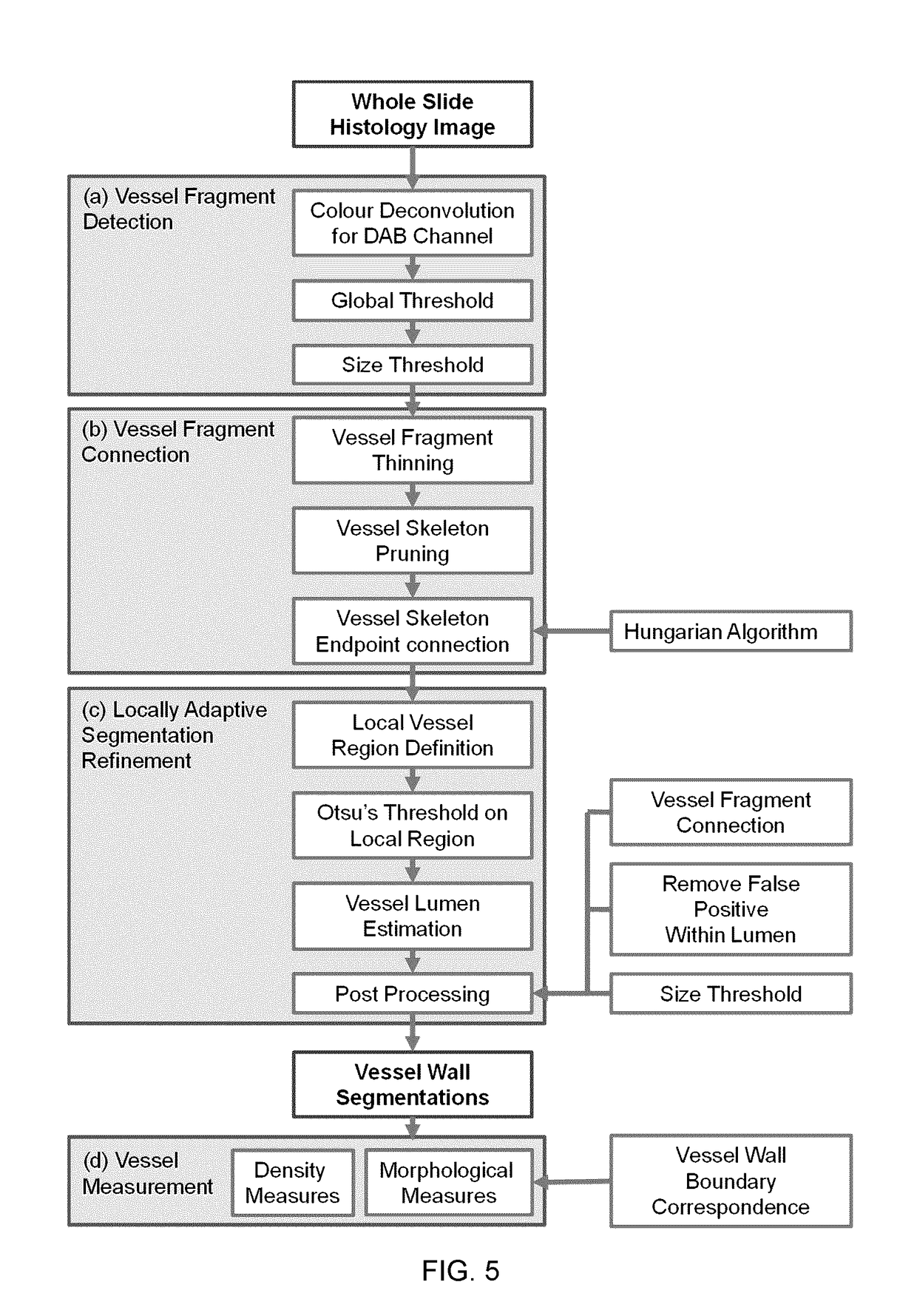
A fully automated method for detecting and measuring a target of interest such as vasculature, capable of processing whole slide images and extracting large number of targets of interest per slide. The method includes the steps of: (a) obtaining a digital image of a tissue specimen; (b) using a first set of mathematical algorithms based on objectively-defined criteria to isolate the one or more targets of interest from the slide, thereby detecting the one or more targets of interest; and (c) using a second set of mathematical algorithms based on objectively-defined criteria to construct boundaries around the detected targets of interest and obtain quantitative attributes of these one or more targets throughout the slide, thereby measuring the one or more targets.

Description

FIELD OF THE INVENTION[0001]The present invention relates to automatic measurement of vascular morphology and density.BACKGROUND OF THE INVENTION[0002]Vascular restructuring through angiogenesis in the microvasculature is a prominent feature of disease repair in tissues, which can impact tissue perfusion and organ function. To visualize and identify the features of interest, histology is used to depict localized protein expression of vascular components, with staining for specific vessel wall components of interest. Immunohistochemistry is a routine method for the evaluation of localized antigen distributions at high resolutions. An important feature which varies throughout the vasculature is the vessel medial layer comprised of smooth muscle, which varies in thickness along the circulatory system, and can be visualized when staining for α-actin. This distribution of smooth muscle may modulate vascular dilation, and thus the flow rate and tissue perfusion, which is imperative to org...

Claims


Application Information

Patent Timeline
Patent Type & Authority Applications(United States)
IPC IPC(8): G06T7/11G06T7/12G06T7/136G06T7/181
CPCG06T7/11G06T7/12G06T7/136G06T7/181G06T2207/10056G06T2207/20044G06T2207/30101G06T2207/30024
Inventor WARD, AARON DEVINXU, YI WENPICKERING, JOHN GEOFFREY
Owner LONDON HEALTH SCI CENT RES
Learn More