(1997), as described in Prior Document 8, in which the imaging probe was referred to as an “optoacoustic
endoscope”, a number of PAE probes have been developed to address technical requirements, such as “probe
miniaturization” and “specifying a device configuration or operation principle for
endoscopy.” However, no commercially successful or clinically applicable PAE
system has yet been developed that satisfies both of these technical requirements due to many underlying technical challenges.
The most well-known and difficult challenge is that, in order to successfully create a working PAE probe, all the optical and acoustical elements should be effectively integrated and arranged in a small and restricted space; an adequate scanning mechanism, through which a
tomographic image can be produced, should also be developed and integrated into the device.
Moreover, the costs for implementing the related instruments are relatively low.
However, the main drawback of the mechanical scanning mechanism is that, since a single-element ultrasonic
transducer that can receive the signals that are bounced back only from the aiming direction of the
transducer surface is mounted on the scanning tip of an endoscopic probe, in order to obtain a 2D image or a 3D image, a series of processes that emit a
laser pulse, and then detect the generated photoacoustic
waves, should be repeatedly performed by changing the physical position or the aiming direction of the ultrasonic
transducer (e.g., rotational scanning in general).
First, its major drawback is that it is relatively more difficult to reduce the size of the related endoscopic probe than the size of the mechanical scanning mechanism because it employs multiple transducer elements to detect
ultrasound waves.
Thus, problems, such as
crosstalk or
signal interference between channels, may occur and the cost of implementing the
system may also be high.
However, Prior Document 9 only mentioned the possibility of applying this array sensor to PAE or
intravascular imaging; it does not disclose any detailed shape or structure of a PAE probe or the associated implementation methods.
However, the endoscopic
system as is may not be applicable to a clinical
endoscopic procedure because its probe is still too big (9 cm in length and 3 cm in
diameter), and the
glass material utilized for the probe encapsulation is not suitable for actual clinical use in terms of safety.
Moreover, the endoscopic system of Prior Document 10 has a drawback in that a light emitting area (LEA) and an
ultrasonic sensor area (USA) are spaced apart from each other (the problem caused by such an arrangement will be explained later).
However, the proposed structure has a
disadvantage in that, since a limited number of optical fibers are simply added to the outer surface of an existing array transducer-based IVUS
catheter, the intensity of light illumination may not be uniform over the 360° scan area, and the probe's flexibility may not be good enough.
However, in this case, the probe flexibility may be seriously decreased in proportion to the total number of optical fibers that are used.
However, the endoscopic probe presented in Prior Document 13 has a
disadvantage in that, since the probe has a structure that obtains a photoacoustic
signal by using only an 8-element-based array transducer, which is placed facing the distal end of the probe, a parabolic mirror, which has a 45°-tilted reflection surface and faces that array transducer, has to mechanically rotate in order to obtain a photoacoustic image over a desired scan range.
Thus, the suggested scanning mechanism may not be a desirable direction for actual clinical use because the main purpose of using this type of array transducer is to avoid any mechanically moving component inside an endoscopic probe.
However, as mentioned above (Prior Document 13), the core benefit of employing an array transducer was not fully utilized, and the mismatch issue between an LEA and a USA still exists in this endoscopic system.
However, in any case, the endoscopic structures presented in Prior Document 15 have the limitation that the LEA of an endoscopic probe is limited to the surrounding area of an array transducer or several spots that are discontinuously distributed around a
linear array transducer.
This limitation was probably caused because the authors of Prior Document 15 only focused on a PAE probe embodiment by using an existing commercial ultrasonic array transducer, rather than focusing on working out an ideal probe structure that enables a uniform light illumination to an object to be examined over the entire area where the transducer elements are distributed.
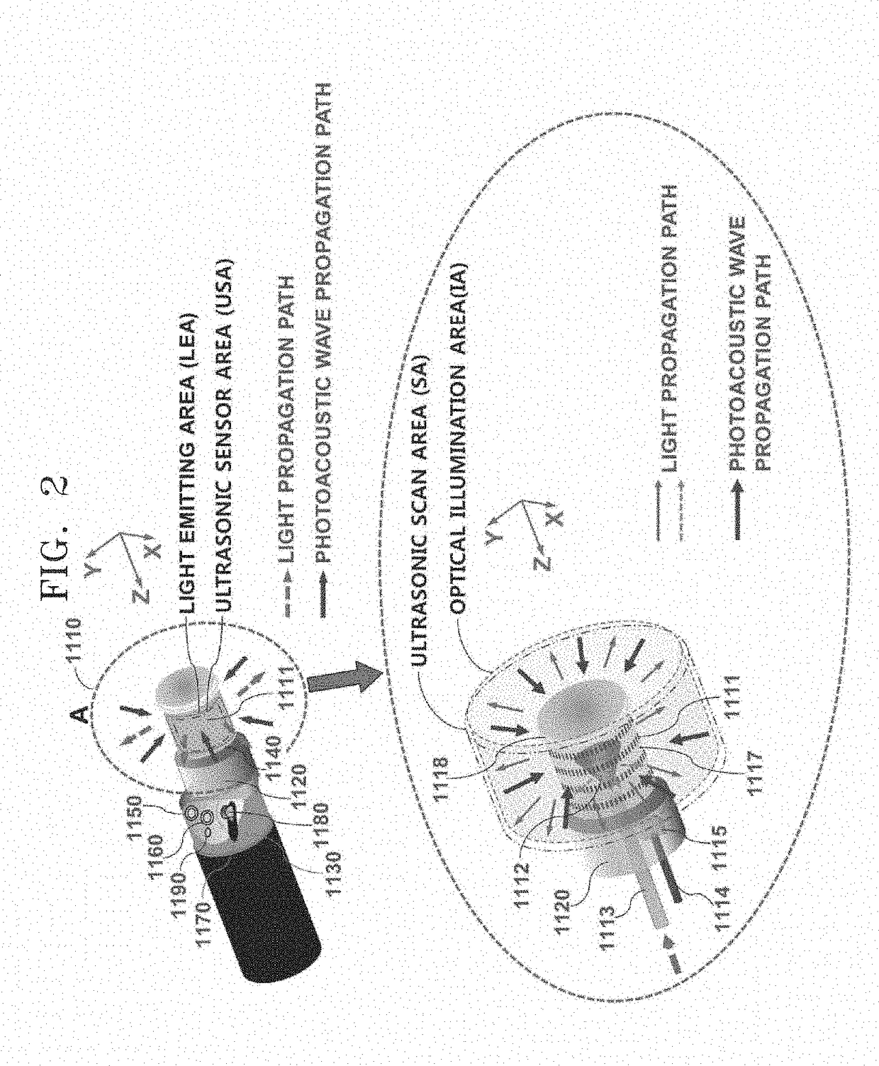
When any of the previous light illumination methods is utilized for a PAE probe, one
biggest problem is that an optical illumination area (IA) formed inside an object and an ultrasonic scan area (SA) formed inside the object by the collective effect of the sensor elements that constitute an array transducer do not perfectly coincide.
This leads to the following problem:
light energy is not uniformly delivered over the entire scan area (i.e., SA) of an array transducer during its scanning process, so a dead zone occurs in an acquired photoacoustic image.
However, if the size of a scanning head is reduced while maintaining any of the mentioned arrangement structures, one serious effect is that the maximum imaging depth of the PAE probe may be greatly reduced because the total available space for the scan head still has to be divided into the two compartments, i.e., an LEA and a USA, as the arrangement structure is pre-assumed.
It is true that, in prior inventions, an LEA is limited only to specific positions so the total amount of
light energy that is actually delivered to an object to be examined may be significantly limited.
Therefore, reducing the size of the probe may not be a desirable way to solve the aforementioned discrepancy issue.

