3D image compounding for ultrasound fetal imaging

a 3d image and ultrasound technology, applied in the field of ultrasound fetal imaging, can solve the problems of no longer feasible or plausible mosaicking scenarios, not providing a good quality compounded image, etc., and achieve the effect of reducing the overall confidence value and improving the visualization of the volumetric region

Active Publication Date: 2019-04-25
KONINKLJIJKE PHILIPS NV
View PDF0 Cites 14 Cited by
  • Summary
  • Abstract
  • Description
  • Claims
  • Application Information

AI Technical Summary

Benefits of technology

[0012]The ultrasound system of the present invention allows evaluating the overall quality of the 3D images of the fetus acquired for the given volumetric region. This evaluation is done by determining an overall confidence value of the plurality of the 3D images based on the incorporated knowledge of an articulated position taken by the fetus during each 3D frame acquisition. While segmenting each ultrasound 3D frame a variation of the common fetal structure in a given 3D frame compared to another 3D image frames can be detected, in the case the ultrasound imaging system allows adapting the articulated fetal model applied to the given 3D image frame. In some cases the relative orientation of the fetus may vary substantially between opposite directions from one 3D image to another. For such cases it may better to reacquire some 3D image frames than giving a user a low quality compounded image. Therefore, the image quality analyser of the system is arranged to compare the determined overall confidence value with an image compounding threshold, above which a spatially compounded 3D image of the fetus would provide an improved visualization of the volumetric region. The system of the present invention may further include a user feedback capability informing the user, when the overall confidence value is below the image compounding threshold. This provides user with an additional flexibility of either performing an additional 3D image frames acquisition or continue with the rest of the selected diagnostic protocol, if time is a limiting factor.
[0013]The benefit of the present ultrasound system lies in using the articulated fetal model, which includes the common fetal structure and at least one joint-limb relationship, in combination with the model's subsequent adaptation during the segmentation process of the 3D image frames. This permits the ultrasound system to automatically assess an extent of a fetal pose change, which might have occurred during the acquisition of the plurality of three dimensional (3D) ultrasound image frames. If the common fetal structure in between the subsequently acquired frames varies substantially such that the model's adaptation causes a reduction of the overall confidence value of the plurality of the 3D images the user may be notified.

Problems solved by technology

This does not only hamper image compounding (image stitching), but can also create scenarios where mosaicking is no longer feasible or plausible (especially in case of a large motion of the extremities).
In this situation a standard image compounding by spatially stitching images acquired at different orientations will not provide a good quality compounded image.

Method used


Image

  • 3D image compounding for ultrasound fetal imaging
  • 3D image compounding for ultrasound fetal imaging
  • 3D image compounding for ultrasound fetal imaging

Examples


Embodiment Construction

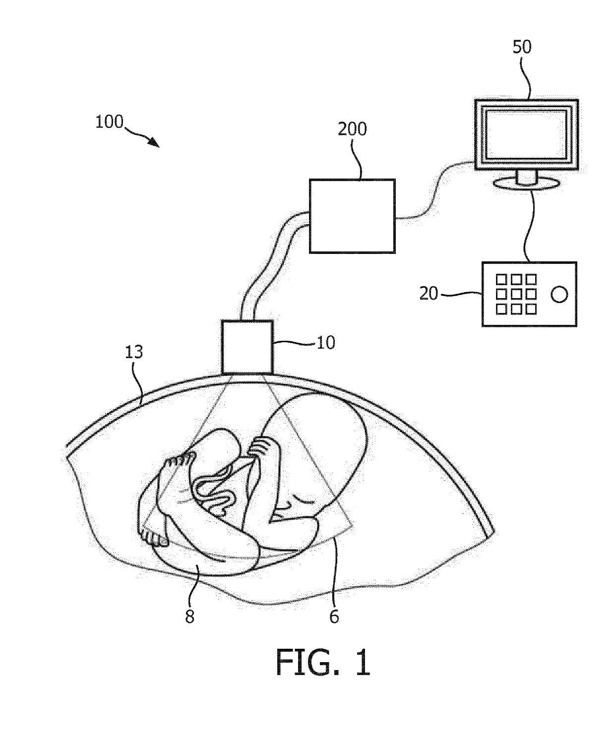
[0049]FIG. 1 shows a schematic illustration of an ultrasound imaging system according to an embodiment generally denoted by 100. The ultrasound imaging system 100 is applied to inspect a volumetric region of an anatomical site, in particular an anatomical site of a patient 13 including a fetus 8. The ultrasound imaging system 100 comprises an ultrasound probe 10 having at least one transducer array having a multitude of transducer elements for transmitting and / or receiving ultrasound waves. The transducer elements are preferably arranged in a two-dimensional (2D) array, which is constructed to electronically steer ultrasound beams within the volumetric region such that a three-dimensional ultrasound image frame of said region is provided. Alternatively, the array may be a one-dimensional array (1D) constructed to be mechanically steered through the volumetric region in order to provide a three-dimensional ultrasound image frame. The probe 10 is adapted to transmit ultrasound waves i...


PUM


Abstract

The present invention provides an improved ultrasound imaging system arranged to evaluate a set of acquired 3D image data in order to provide a compounded 3D image of a fetus irrespective of its position and movement. This is achieved by providing an ultrasound imaging system comprising: an ultrasound probe having an ultrasound transducer array operable to acquire at different look directions a plurality of three dimensional (3D) ultrasound image frames of a volumetric region comprising a fetus; a compound image memory for storing the acquired plurality of the 3D ultrasound image frames and an articulated fetal model with a common fetal structure; an ultrasound image processor responsive to the plurality of 3D ultrasound image frames, said processor comprising a fetal segmentation unit arranged to segment each 3D image frame based on the articulated fetal model thereby providing a plurality of spatially related 3D images of the volumetric region; and an image quality analyzer coupled to the segmentation unit and arranged to determine, based on the articulated fetal model, an overall confidence value of the plurality of the 3D images, said image quality analyzer is further arranged to compare the overall confidence value with an image compounding threshold.

Description

FIELD OF THE INVENTION[0001]The present invention relates to an ultrasound imaging apparatus for inspecting a volume of the subject comprising a fetus, in particular to an ultrasound imaging apparatus arranged combine three dimensional (3D) ultrasound image data into a spatially compounded 3D image of the fetus. The present invention further relates to a method for combining 3D ultrasound image data of the fetus into a spatially compounded 3D image of the fetus.BACKGROUND OF THE INVENTION[0002]Fetal imaging is currently developing towards 3D ultrasound imaging. In the later gestational ages of fetus a field of view of the ultrasound transducer is no longer covering the full fetus as illustrated in FIG. 1. As a result, different portions of the body are acquired separately (e.g., brain, abdomen, femurs, see FIG. 3). In order to get one compounded view image stitching or mosaicking is known technique extensively used for 2D imaging (e.g., panorama viewing for camera pictures).[0003]On...

Claims


Application Information

Patent Timeline
Patent Type & Authority Applications(United States)
IPC IPC(8): A61B8/08A61B8/00G06T7/149G06T7/174A61B5/107
CPCA61B8/0866A61B8/483A61B8/5253A61B8/466G06T7/149G06T7/174A61B5/1071A61B8/467G06T2207/10132G06T2207/20124G06T2207/30044A61B8/5223A61B8/5276G06T2207/10136G06T7/344G01S7/52098G01S15/8993G01S15/8995G06T2207/30196
Inventor KLINDER, TOBIASLORENZ, CHRISTIANWAECHTER-STEHLE, IRINA
Owner KONINKLJIJKE PHILIPS NV
Why Eureka
Try Eureka