3D cell culture and ex vivo drug testing methods

a cell culture and ex vivo technology, applied in the field of 3d cell culture technology in methods for ex vivo drug testing, can solve the problems of poor reproducibility and stability of 3d cell culture techniques, inability to accurately represent the real 3d world in disease progression, drug testing and/or biochemical and physiological research, and the current limitations of conventional 2d (2-dimensional) cell culture methods, etc., to achieve the effect of optimizing the labeling

Inactive Publication Date: 2019-05-02
MOLECULAR RESPONSE
View PDF0 Cites 0 Cited by
  • Summary
  • Abstract
  • Description
  • Claims
  • Application Information

AI Technical Summary

Problems solved by technology

Moreover, it is now known that conventional 2D (2-dimensional) cell culture methods do not accurately represent the real 3D world in disease progression, drug testing and / or biochemical and physiological research.
However, 3D cell culture techniques have current limitations including poor reproducibility and stability, complexity of components, difficulty with scaling up or down, and / or a need for improvement of physiological substrates.
In particular, anti-proliferative predictive assays of oncology drug candidates using patient-derived tumor cells, tested in a 3D growth environment, produce unpredictable results due to high heterogeneity, low proliferation, and / or low availability of well-characterized samples.

Method used


Image

  • 3D cell culture and ex vivo drug testing methods
  • 3D cell culture and ex vivo drug testing methods
  • 3D cell culture and ex vivo drug testing methods

Examples


example 1

Ex Vivo Proliferation Assay

[0068]Patient-derived tumor cells were allowed to thaw in 37° C. water bath until frozen cell suspension melts enough to form a slushy solution. The cell suspension was slowly transferred through a cell strainer to a 50 ml conical tube filled with conditioning media warmed to 37° C. Cells were pelleted by spinning the tubes at 800 RPM for 5 minutes at 4° C. The supernatant was carefully removed without disturbing the pellet by aspiration of the supernatant. The pellet was resuspended in 1-2 ml warmed conditioning medium. Cells were counted using TC10. 10 μl of the cell suspension was removed and transferred into a tube for Trypan Blue exclusion assay to assess the cell density and overall health of the cells in the vial. LIVE-DEAD cells were counted.

[0069]6-well plates were coated with Cultrex® (ECM from Trevigen). ECM was diluted 1:20 with ice cold conditioning medium without growth factors and supplements. 1 ml per well of 6-well plate or 3 ml per 100 mm...

example 2

Use of Patient-Derived Xenografts (PDX)

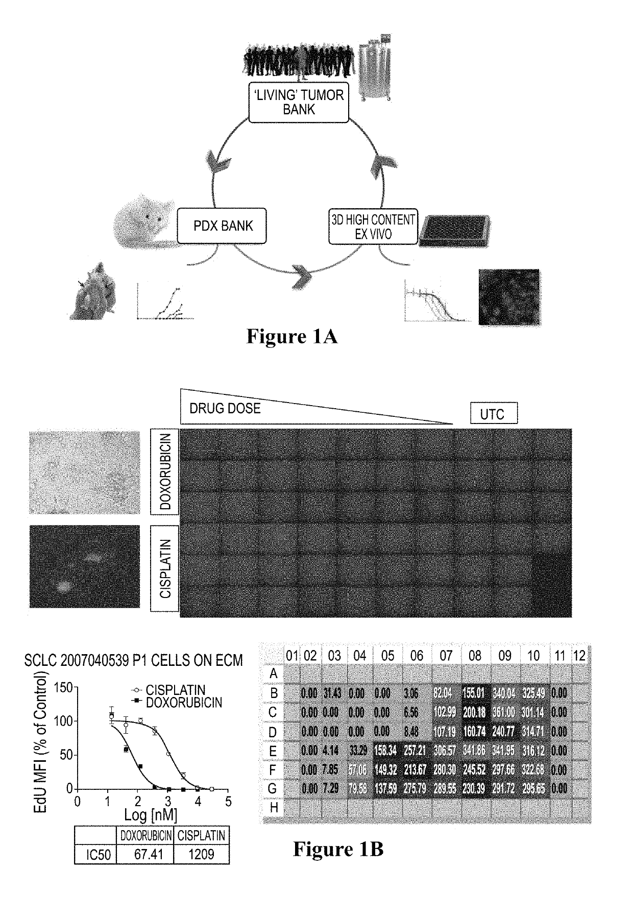
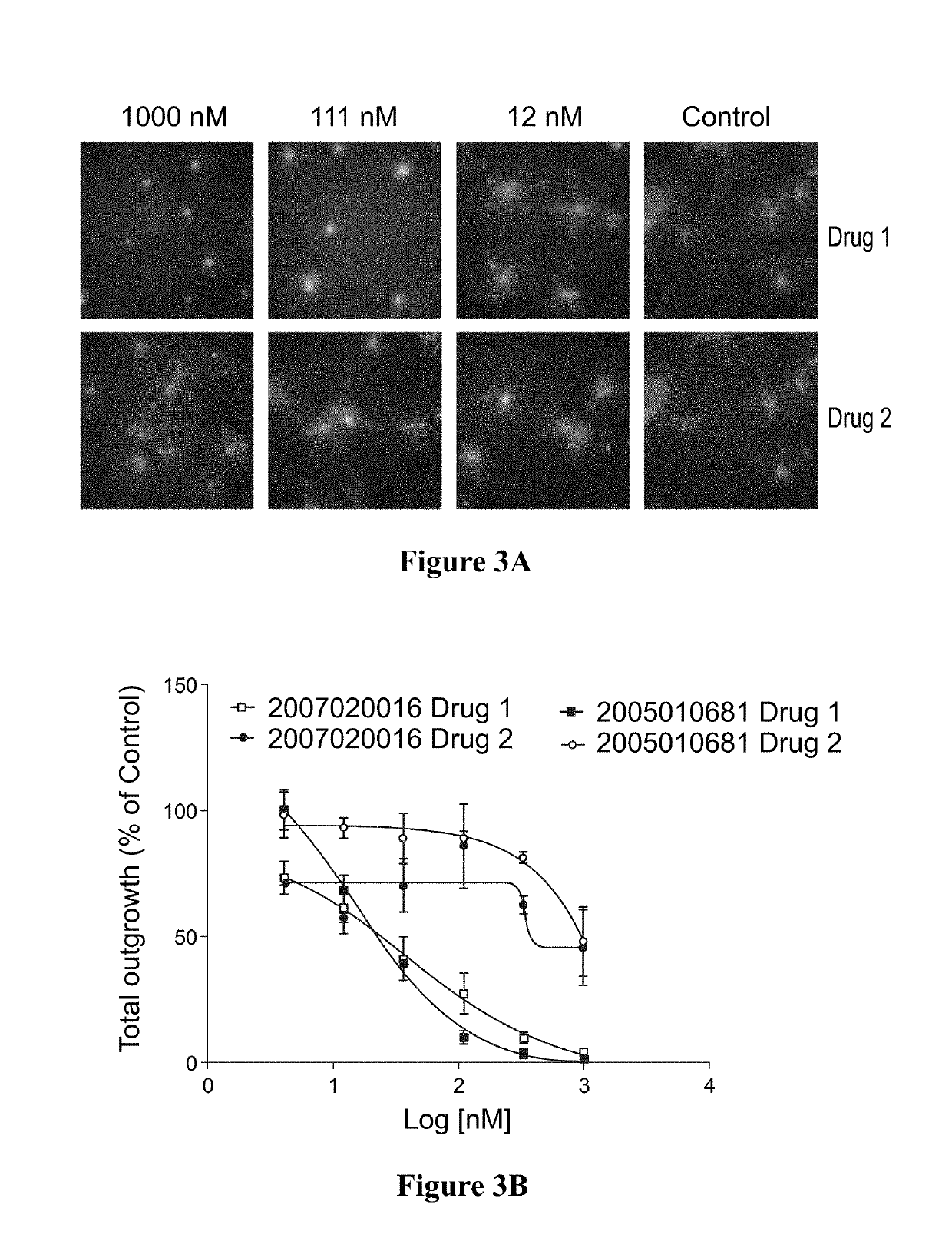
[0080]Fresh or banked patient-derived tumor cells were injected into immunocompromised mice. Cells were harvested and plated to form spheroids. Spheroids were subjected to ex vivo drug treatment using chemotherapy drug candidates, cisplatin and doxorubicin. Cell proliferation was measured by HC imaging by EdU incorporation assay. (FIG. 1A-B)

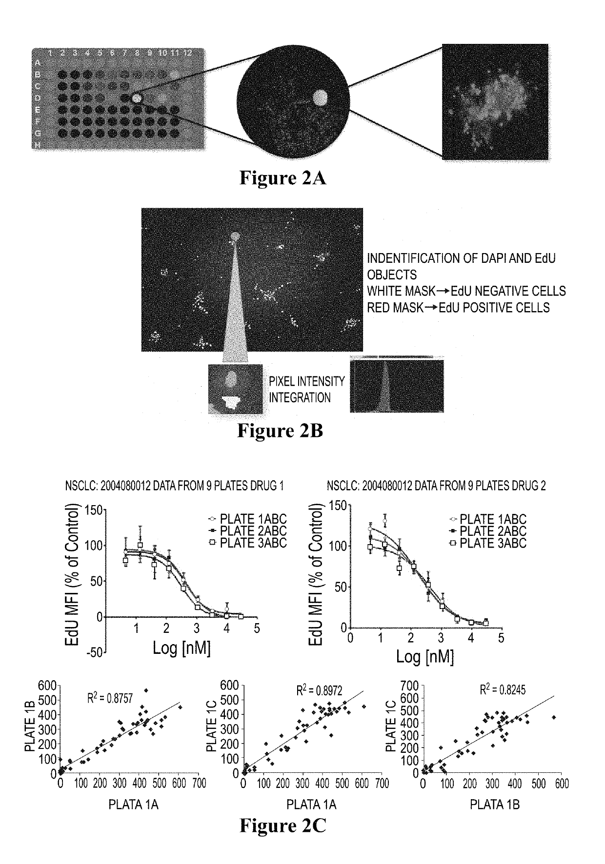
[0081]Cell images are captured and processed using multiple algorithms corresponding to the experiment. DAPI was selected on channel 1 to identify valid objects. EdU was selected on channel 2 to identify a population of cells incorporating DNA. Intensity for each pixel for each channel was recorded and integrated to obtain total intensity. Total intensity was divided by total pixels to give mean fluorescent intensity (MFI) for that channel. Integrated mean fluorescent intensity of multiple cells divided by total cells per well gave mean of MFI. To normalize the variability of cell numbers between wells, the ...


PUM

PropertyMeasurementUnit
molecular weightaaaaaaaaaa

Abstract

Provided herein are methods for testing proliferative responses of a drug on patient-derived tumor cells; the method comprising obtaining cells from biopsy or tumor resection material; culturing the cells on a 3D extracellular matrix (ECM); treating the cells in ECM with a drug; subjecting the treated cells to high-content (HC) imaging; and evaluating the HC imaging of the treated cells; thereby testing the proliferative responses of the drug on the patient-derived tumor cells. In some embodiments, the methods disclosed herein comprise obtaining cells from biopsy or tumor resection material; xenografting the cells into an animal model (patient-derived xenograft; PDX) for tumor formation; and obtaining tumor cells from the animal.

Description

[0001]The present application is a continuation of U.S. patent application Ser. No. 15 / 036,808, filed May 14, 2016, which is the U.S. national phase of International Patent Application No. PCT / US2014 / 065549, filed Nov. 13, 2014, which designated the U.S. and claims the benefit of priority to U.S. Provisional Application No. 61 / 905,040, filed Nov. 15, 2013, each of which is hereby incorporated in its entirety including all tables, figures, and claims.FIELD OF THE INVENTION[0002]The present invention relates to the use of 3D (3-dimensional) cell culture technology in methods for ex vivo drug testing, wherein cells acquire a natural 3D phenotype resulting in the capability for increased cell proliferation, differentiation, and function.BACKGROUND OF THE DISCLOSURE[0003]The current trend in drug discovery and disease-related research is moving away from the use of non-human primate animal models. This trend was underscored recently by the significant reduction of the use of chimpanzees ...

Claims


Application Information

Patent Timeline
Patent Type & Authority Applications(United States)
IPC IPC(8): G01N33/50
CPCG01N33/5011G01N33/5088G01N2510/00
Inventor NAIR, PRAVEENMIRSAIDI, CYRUSBROUDY, TOMMY
Owner MOLECULAR RESPONSE
Why Eureka
Try Eureka