Method for automatically generating a volume model of correction data for an x-ray based medical imaging device

a technology of medical imaging and volume model, applied in the field of automatic generation of volume model of correction data for x-ray based medical imaging device, can solve the problems of no longer being able to make meaningful statements, occurrence of zones with apparently higher or even lower absorption, and inhomogeneous tissue, so as to reduce the required storage capacity and reduce the effect of quality loss

Pending Publication Date: 2019-06-27
SIEMENS HEALTHCARE GMBH
View PDF4 Cites 0 Cited by
  • Summary
  • Abstract
  • Description
  • Claims
  • Application Information

AI Technical Summary

Benefits of technology

The patent describes a method for improving the accuracy of planning radiotherapy by using a three-dimensional image data model of the treatment area. This model is generated from a plurality of X-ray images and is used to identify areas where image artifacts may be present. These areas are then corrected to improve the accuracy of the image data. The resulting corrected image data is then used to plan the radiotherapy treatment. This method simplifies the planning process and avoids the need for manual corrections. Additionally, the patent describes an X-ray based medical imaging device that automatically generates a volume model of correction data using X-ray images, which can be used for planning without any loss of quality.

Problems solved by technology

However, if the body tissue to be mapped by the CT scanner for radiotherapy planning contains a foreign body that absorbs X-rays to a significantly different degree than the surrounding body tissue, the image data output by the CT scanner might contain artifacts that do not correspond to the real situation in the mapped body tissue.
Such foreign bodies may include a significantly higher density than the surrounding body tissue so that, when recording an individual X-ray image, it is no longer possible to make meaningful statements relating to the region that is shaded by the foreign body from the X-ray source of the CT scanner due to the much higher absorption by the foreign body.
The reconstruction of the volume model of the body region to be examined from a plurality of such X-ray images in which a large region no longer supplies any useful absorption information results in the occurrence of regions in the volume model corresponding to an apparently high degree of absorption not only at the site of the actual foreign body.
The faulty absorption information may also result in the occurrence of zones with apparently higher or even lower absorption or apparently inhomogeneous tissue in the environment of the foreign body in the volume model.
However, this is extremely complicated.
Herein, in the worst case, human errors may result in scenarios where highly sensitive tissue covered by an artifact is not detected correctly and hence receives an excessive dose of irradiation.
There is also a residual risk of critical body tissue being covered by the artifacts in the original image data so that the critical body tissue may no longer be identifiable as such in the corrected image data after correction of the artifacts.

Method used


Image

  • Method for automatically generating a volume model of correction data for an x-ray based medical imaging device
  • Method for automatically generating a volume model of correction data for an x-ray based medical imaging device

Examples


Embodiment Construction

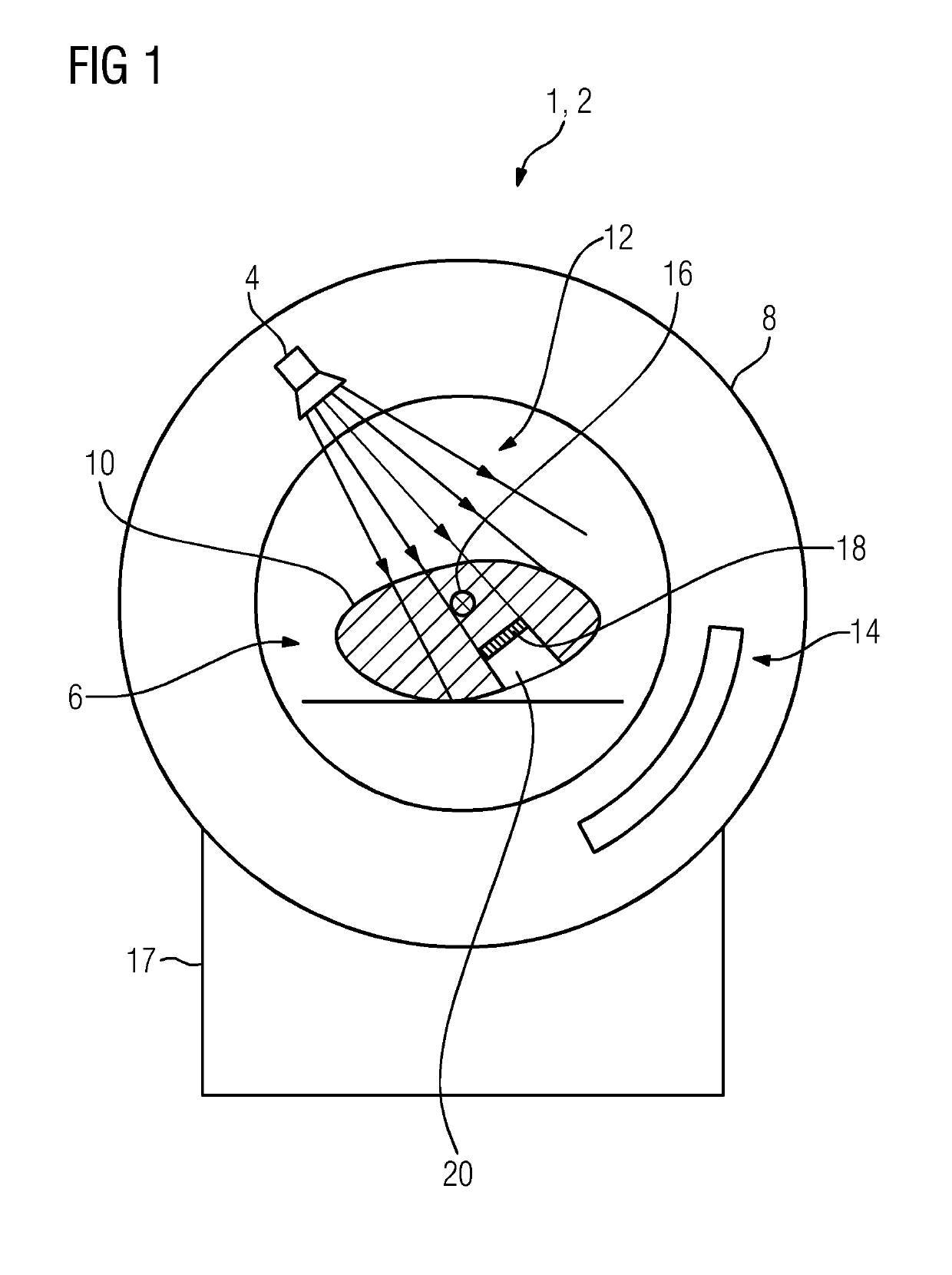
[0028]FIG. 1 depicts a schematic cross-sectional view of an X-ray based medical imaging device 1 that is configured as a CT scanner 2. In the CT scanner 2, an X-ray source 4 irradiates a body region 10 of a patient positioned in the interior 6 of the rotating ring 8 of the CT with X-rays 12. The portions of the X-rays 12 that are not absorbed by the body region 10 of the patient are measured on the opposite side relative to the interior 6 of the X-ray source 4 by an X-ray detector 14 and processed to form an individual X-ray image. For complete imaging, different X-ray images are recorded. For the individual recordings, both the X-ray source 4 and the X-ray detector 14 rotate around an axis 16 perpendicular to the image plane. There may be an axial displacement of the X-ray source 4 and X-ray detector 14 along the axis 16. Both the X-ray source 4 and the X-ray detector 14 perform the movement of discretized coverage of a cylinder surface. The individual X-ray images are then transfe...


PUM


Abstract

Method and systems are provided for automatically generating a volume model of correction data for an X-ray based medical imaging device. A plurality of X-ray images is recorded of a body region of a patient to be examined from different positions in each case. The plurality of X-ray images is used to generate a first volume model of the body region. Image artifacts are corrected in the first volume model using the plurality of X-ray images and thus a corrected volume model is generated. The corrected volume model is used to determine a contour of an artifact volume affected by image artifacts in the first volume model and the contour of the artifact volume is defined as a volume model of correction data. The volume model of correction data is stored on a data medium and / or output via an interface.

Description

CROSS REFERENCE TO RELATED APPLICATIONS[0001]This application claims the benefit of DE 10 2017 223 604.3 filed on Dec. 21, 2017, which is hereby incorporated by reference in its entirety.FIELD[0002]Embodiments relate to a method for automatically generating a volume model of correction data for an X-ray based medical imaging device.BACKGROUND[0003]When planning radiotherapy, as used, for example, to control a tumor, it is usual to ascertain physical parameters of the irradiation, for example angle of incidence, radiation dose and profile, based on medical image data, that is generated by computed tomography (CT). For the planning, individual regions that each correspond to different tissue structures and also include the tumor tissue are identified in the image data. Knowledge of the spatial distribution of the different tissue structures will provide the optimal dose distribution to be calculated, e.g. the maximum possible irradiation dose in the tumor tissue in conjunction with th...

Claims


Application Information

Patent Timeline
Patent Type & Authority Applications(United States)
IPC IPC(8): A61B6/00A61B6/12G06T7/13G06T7/11G06T15/08
CPCA61B6/5282A61B6/12G06T7/13G06T7/11G06T15/08G06T2207/30052G06T2207/10081G06T2207/10116G06T11/008A61B6/032A61B6/5258
Inventor RITTER, ANDREHOFMANN, CHRISTIANDYBAN, PAVLOGEORGI, JENS-CHRISTOPHSCHUBERT, KAIOETZEL, DIETERTONNDORF-MARTINI, ERIC
Owner SIEMENS HEALTHCARE GMBH
Why Eureka
Try Eureka