Method for measuring immunogenicity of protein agent

Inactive Publication Date: 2019-06-27
MOGAM INST FOR BIOMEDICAL RES
View PDF0 Cites 0 Cited by
  • Summary
  • Abstract
  • Description
  • Claims
  • Application Information

AI Technical Summary

Benefits of technology

The present invention provides a method for accurately predicting the immunogenicity of protein agents before they enter clinical phase. This method can be performed using in vitro experiments, increasing the efficiency of protein agent development while reducing costs. It allows for the relative immunogenicity of protein agents to be predicted with high accuracy.

Problems solved by technology

Further, the induced immune response to the drug may cause side effects such as fever and inflammatory reaction.
However, this test method cannot assess immunogenicity unless the protein drug of interest is under clinical application, thereby having a disadvantage in that the immunogenicity cannot be predicted in a development phase of the drug.
In consideration of huge costs and time involved in the development of a drug till the entry on the clinical phase, the fact that the immunogenicity is not determined till the entry on the clinical phase is the major obstacle in development of protein drugs.

Method used


Image

  • Method for measuring immunogenicity of protein agent
  • Method for measuring immunogenicity of protein agent
  • Method for measuring immunogenicity of protein agent

Examples


example 1-2

DC:T Cell Assay

[0060]1. Procedures for Preparation of Dendritic Cells

[0061]1.1. PBMC Thawing

[0062]After quickly unfreezing cryopreserved PBC in a water bath at 37° C., the solution was moved to a 50 mL conical tube. While whirling the tube, a thawing medium (RPMI supplemented with 10% (vol / vol) FBS) was added dropwise and mixed well (15 mL / vial). After centrifugation at 1200 rpm for 10 minutes, a supernatant was discarded and the product was suspended in 30 mL of MACS buffer (PBS supplemented with 0.5% (vol / vol) FBS and 2 mM EDTA). After measuring the number of cells to determine cell viability, a supernatant was discarded by centrifugation at 1200 rpm for 10 minutes.

[0063]1.2. Monocyte Isolation

[0064]CD14 microbeads (Miltenyi Biotech) were added to the thawed PBMC and stained in ice for 15 minutes. After adding 30 mL of MACS buffer to the above product and centrifuging the same at 1350 rpm for 8 minutes, a supernatant was discarded. After suspending the product in 500 μL MACS buffe...

example 2

Verification of Immunogenicity Determining Method

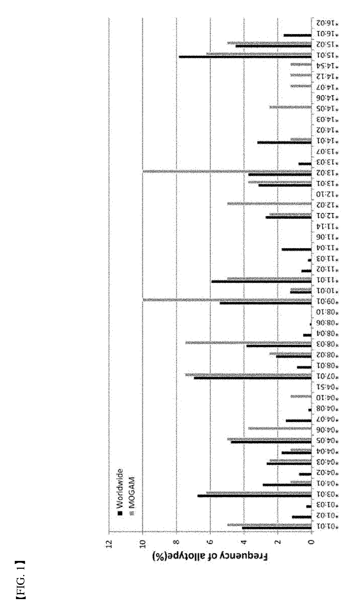
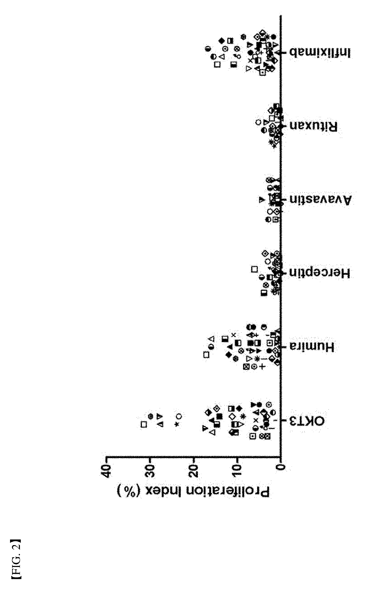
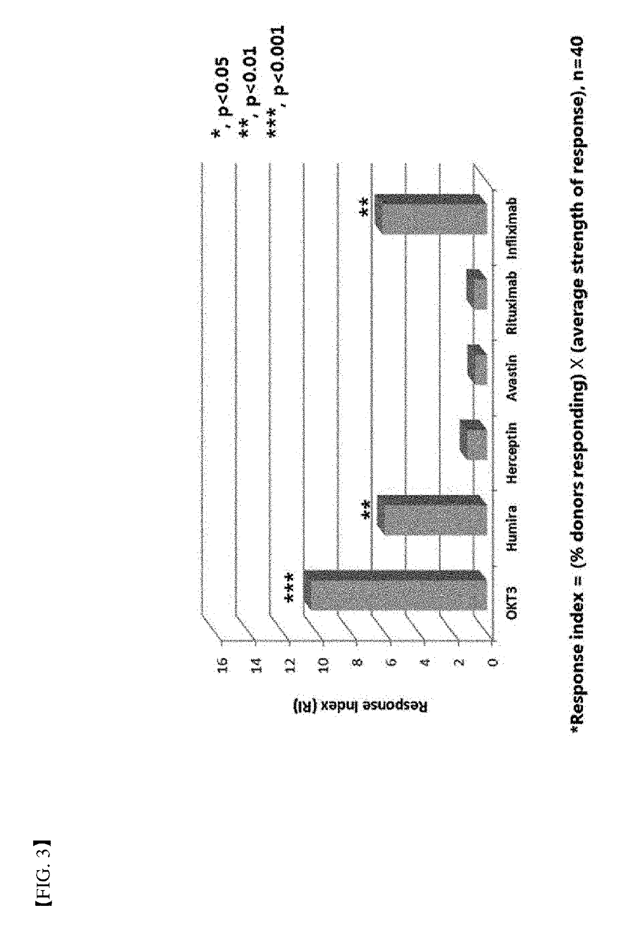
[0084]In order to verify the method for determination of immunogenicity according to the present invention, this method was compared to a world population distribution with reference to HLA-DRB1 allotype. Following this, PBMC in 40 or more different donors corresponding to about 80% coverage thereof was used as a target (FIG. 1). Six (6) types of protein agents (Table 2) with ADA generation extent identified in a clinical phase were assayed by the method for determination of immunogenicity according to the present invention. FDA-approved antibody therapeutic agents with immunogenicity reported in the art were summarized in Table 2 below. Further, assay results of immunogenicity according to the present invention were briefly illustrated in FIGS. 2 and 3.

TABLE 2AntibodyReportednameCompanyTypeTargetIndication(s)immunogenicity*MuromanabOrtho BiotechMurineCD3Allograft rejection47%(50)(OKT3)AdalimumabAbbottHumanTNFαRA / Crohn / PsA / JIA / 2.6%-26...


PUM


Abstract

A method for determining immunogenicity of a protein agent. The method includes constructing a library of peripheral blood mononuclear cells having various HLA-DRB1 genotypes; culturing peripheral blood mononuclear cell CD14+ monocyte-derived immature dendritic cells for each genotype in a medium containing a protein to be measured, GM-CSF, IL-4, TNF-α, IL-1β, IL-6 and PGF2 to prepare mature dendritic cells; removing CD8+ T cells from the peripheral blood mononuclear cells for each genotype to prepare CD8+ T cell-free peripheral blood mononuclear cells; co-culturing the mature dendritic cells and the CD8+ T cell-free peripheral blood mononuclear cells at a cell count ratio of approximately 1:5 to 1:20; and quantifying the CD4+ T cells proliferated by co-cultivation per genotype.

Description

TECHNICAL FIELD[0001]The present invention relates to a method for determining immunogenicity of a protein agent.BACKGROUND ART[0002]For purpose of treating a variety of diseases, diverse protein formulations are under research and development and used, in particular, there are a number of clinical cases with very successful effects for therapeutic antibodies. Representative examples of such therapeutic antibodies may include adalimumab (Humira®), etanercept (Enbrel®), etc. targeting inflammatory diseases such as rheumatoid arthritis, rituximab (Mabthera®) targeting lymphoma, trastuzumab (Herceptin®) targeting the breast cancer, or the like.[0003]A number of cases of causing immune response not intended in clinical examples with application of antibody therapeutics have been reported. In a case of first generation antibody therapeutics, an immune response to an antibody derived from mice, in particular, an immune response against a therapeutic antibody administered, that is, an anti...

Claims


Application Information

Patent Timeline
IPC IPC(8): C12N5/0783C12N5/0784C12N5/00
CPCC12N5/0636C12N5/0639C12N5/0018C12N2501/22C12N2501/2304C12N2501/25C12N2501/2301C12N2501/2306C40B40/02C12N5/064C12N2502/1114C12N5/0087G01N33/15G01N33/5008G01N33/5047G01N33/505G01N2333/70514G01N2500/10
Inventor LIM, OK JAESHIN, DUCK HYANG
Owner MOGAM INST FOR BIOMEDICAL RES
Why Eureka
Try Eureka