
Detection of microcalcifications in anatomy using quantitative transmission ultrasound tomography

a technology of quantitative transmission ultrasound and microcalcification, applied in the field of anatomy microcalcification detection using quantitative transmission ultrasound tomography, can solve the problems of not being able to apply the effort to imaging modalities, unable to support the clinical use of hhus for detection of microcalcification, and unable to achieve the effect of detecting microcalcifications in the clinical field

Inactive Publication Date: 2019-08-15
QT ULTRASOUND
View PDF0 Cites 1 Cited by
  • Summary
  • Abstract
  • Description
  • Claims
  • Application Information

AI Technical Summary

Benefits of technology

The patent describes a method for detecting small amounts of calcium in anatomical structures using ultrasound tomography. The method involves analyzing the refraction-corrected images generated by the ultrasound system and using image filters to remove unwanted brightness points. The technique can be used to identify microcalcifications and has potential use in the diagnosis and treatment of breast and other medical conditions.

Problems solved by technology

A challenge with using microcalcifications as a biomarker for breast cancer detection or screening is that microcalcifications do not generate any special symptoms and are not something that can be felt during a routine breast exam.
However, these efforts are not applicable to imaging modalities such as ultrasound due, for example, to available resolution of the image.
Indeed, while conventional handheld ultrasound (HHUS) has been a mainstay of diagnostic breast imaging, the ability of HHUS for evaluation of breast microcalcifications is somewhat limited and the results to date do not support the clinical use of HHUS for detection of microcalcifications.

Method used


Image

  • Detection of microcalcifications in anatomy using quantitative transmission ultrasound tomography
  • Detection of microcalcifications in anatomy using quantitative transmission ultrasound tomography
  • Detection of microcalcifications in anatomy using quantitative transmission ultrasound tomography

Examples


Embodiment Construction

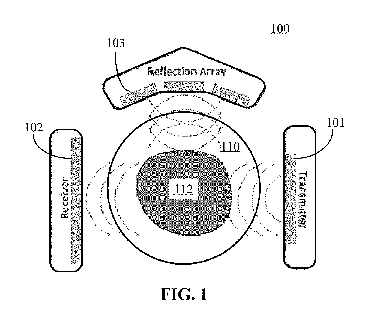
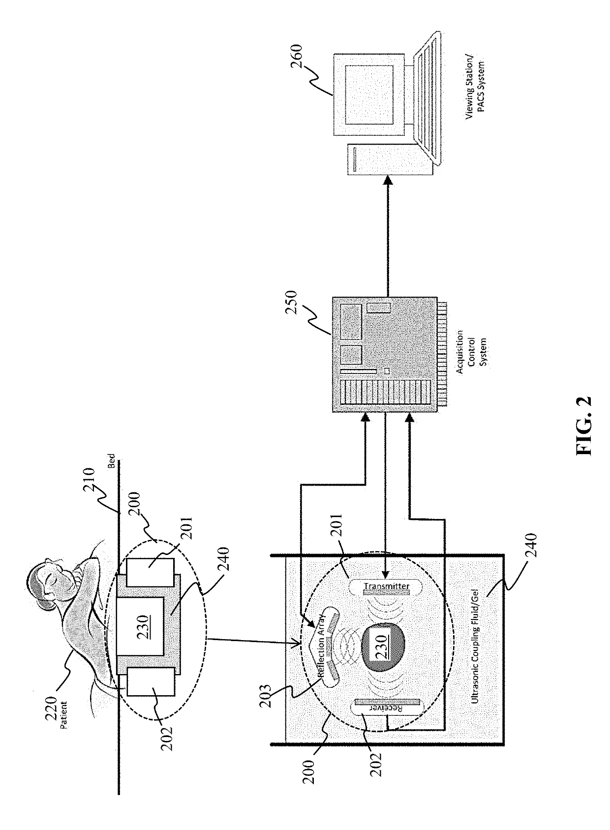
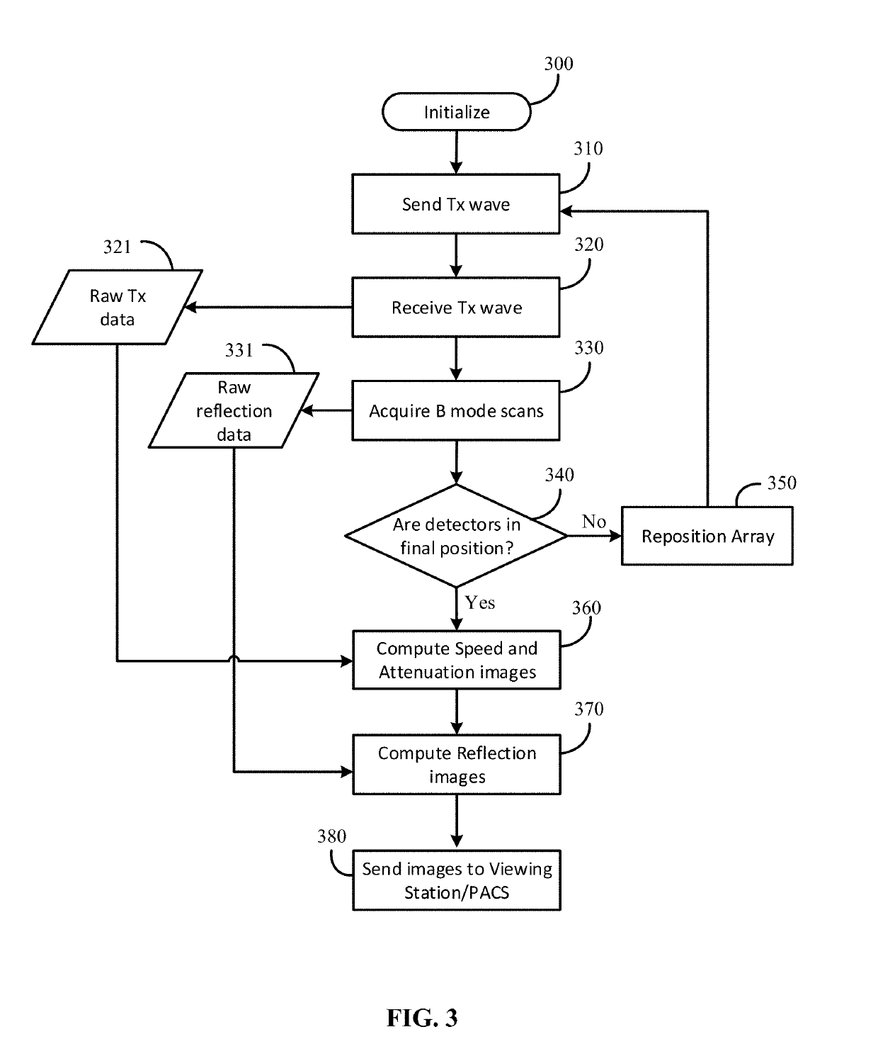
[0024]Detection of microcalcifications in anatomy using quantitative transmission ultrasound tomography is described. Refraction-corrected reflection images (e.g., reflection images corrected by using speed-of-sound data) can be processed to extract microcalcifications from background or other anatomy.

[0025]Quantitative Transmission Ultrasound (QTUS) performs both reflection and transmission ultrasound methods to gather data. The reflection portion directs pulses of sound wave energy into tissues and receives the reflected energy from those pulses—hence it is referred to as “reflection ultrasound.” Detection of the sound pulse energies on the opposite side of a tissue after it has passed through the tissue is referred to as “transmission ultrasound.” QTUS enables evaluation of tissue in clinical ultrasound by offering high spatial and contrast resolution, with absolute spatial registration (no image warping or stretching) quantitative imaging.

[0026]In particular, QTUS uses inverse s...


PUM


Abstract

Microcalcifications can be detected using quantitative ultrasound tomography. Refraction-corrected reflection images generated from a quantitative ultrasound system can be thresholded, and morphologically analyzed to isolate voxels corresponding to microcalcifications.

Description

CROSS-REFERENCE TO RELATED APPLICATION[0001]This application claims the benefit of U.S. Provisional Application Ser. No. 62 / 630,420, filed Feb. 14, 2018.BACKGROUND[0002]In breast cancer detection, one possible early indicator for breast cancer or precancerous changes to breast tissue involves certain types of breast calcifications. Breast calcifications are small calcium deposits that can develop in a woman's breast tissue (and may even line blood vessel walls). Calcifications are categorized by form, size, density, and distribution. Many microcalcifications are benign; however, certain shapes and distributions can indicate possible cancer or precancerous cells.[0003]For example, suspicious microcalcifications can be those that are clustered together, exhibit a branching or linear pattern, have variable density of distribution, or are distributed in a segmental or haphazard way. Other features that may be suspicious include the form of the microcalcifications that are either round, ...

Claims


Application Information

Patent Timeline
Patent Type & Authority Applications(United States)
IPC IPC(8): A61B8/08A61B8/14G06T7/00
CPCA61B8/0825A61B8/5207A61B8/5269A61B8/085A61B8/14G06T7/0014G06T2207/20036G06T2207/10132G06T2207/20032G06T2207/20182G06T2207/30068G06T7/0012G06T7/11G06T7/136G06T7/155A61B8/15A61B8/4477A61B8/5253A61B8/4281A61B8/406
Inventor MALIK, BILAL HAMEEDLENOX, MARK WAYNE
Owner QT ULTRASOUND
Learn More