Ultrasensitive electrochemical biosensors

a biosensor and ultra-sensitive technology, applied in the field of biosensors, can solve the problems that the technological and commercial success of other electrochemical biosensors has not been paralleled, and achieve the effects of high specificity, easy production, and easy detection of the presence or activity of target molecules

Inactive Publication Date: 2019-08-29
MOLECULAR WAREHOUSE LTD
View PDF0 Cites 0 Cited by
  • Summary
  • Abstract
  • Description
  • Claims
  • Application Information

AI Technical Summary

Benefits of technology

[0005]The present invention addresses a need to develop quantitative, relatively inexpensive and easily produced molecular biosensors that readily detect the presence or the activity of target molecules (e.g analytes) on short time scales that are compatible with treatment regimes. Such biosensors can either be applied singly or in multiplex to validate and / or diagnose molecular phenotypes with high specificity and great statistical confidence irrespective of the genetic background and natural variations in unrelated physiological processes. Such biosensors may be used in other testing procedures such as where the target molecule or analyte is an illicit drug or performance-enhancing sub stance.
[0006]The biosensors of the present invention are further particularly suited to incorporation into electrical devices such as point-of-care devices for analysis and transmission of diagnostic results. The biosensors of the invention typically have specificity for a target molecule and produce an electrical response to detection of the target molecule. Preferred biosensors of the present invention provide high sensitivity to a target molecule through allosteric peptide-regulated reversible changes to catalytic activity which are further linked to binding of the target molecule to a binding moiety. The peptide-regulated change may be couplable to a range of different binding interactions thus allowing for detection of different target molecules based on the same common peptide-regulated architecture. The biosensors of the invention may thus be suitable for engineering for use in detection of more than one target molecule when configured with appropriate binding moieties. The biosensors of the invention typically comprise an oxidoreductase enzyme or a variant thereof.
[0007]The present invention provides an oxidoreductase enzyme comprising a heterologous amino acid sequence which is responsive to a peptide, wherein binding of the peptide to the heterologous amino acid sequence reversibly regulates catalytic activity of the enzyme. The binding of the peptide may cause a reduction in the catalytic activity of the enzyme, or may enhance the catalytic activity of the enzyme.

Problems solved by technology

Remarkably, this technological and commercial success has not been paralleled by other electrochemical biosensors despite the need for better and cheaper diagnostics and analytics in many industries.
While this is a common strategy it creates potential problems when binding of two protein modules to a single small molecule is required.

Method used


Image

  • Ultrasensitive electrochemical biosensors
  • Ultrasensitive electrochemical biosensors
  • Ultrasensitive electrochemical biosensors

Examples


example 1

for Construction of Insertion Peptide-Responsive Mutant of PQQ-GDH

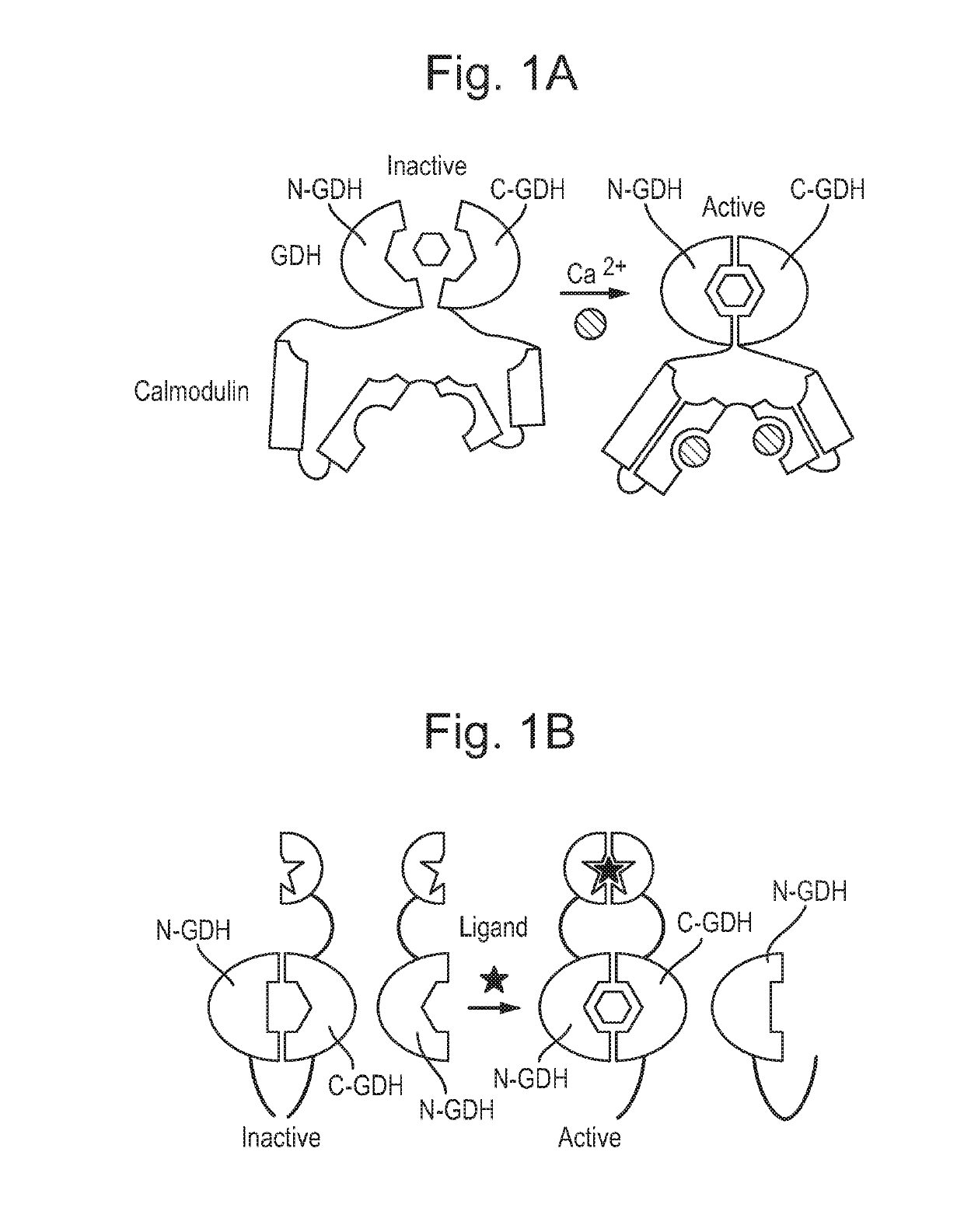
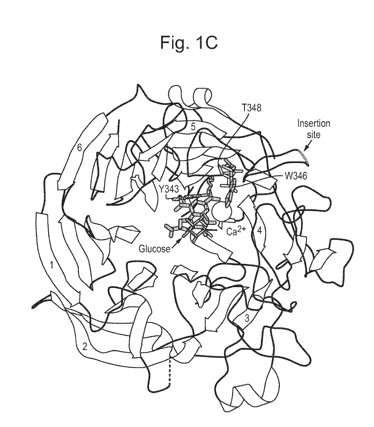
[0173]We previously identified the loop connecting strands A and B of beta-sheet 3 of PQQ-GDH as a site able to tolerate insertion of a calmodulin protein. The resulting calmodulin-GDH chimera (FIG. 1A) demonstrated calcium-binding activity and acted solely as a calcium sensor. The same location was also amenable to splitting GDH into two inactive fragments that could then be reconstituted into an active enzyme via scaffolding interactions with an analyte (FIG. 1B, SEQ ID NOs 43 and 44). The splitting approach allowed for coupling of GDH activity to detection of various analytes, but required proteolytic cleavage and purification of components, and also had a response rate governed by the rate of reconstitution of components. Analyte-drived dimerisation was also required for activity.

[0174]We sought an approach that would allow conversion of GDH into a allosteric peptide regulated ON or OFF switch that can be subseque...

example 2 -

Example 2-Construction of Two Component Biosensors Based on the Peptide-Activated GDH Chimera

[0176]We next decided to test if the developed allosteric module can be used to construct a generic biosensor architecture. To this end we fused the developed CaM-GDH chimera C-terminally with rapamycin-binding FKBP domain (SEQ ID NO: 11) and produced the protein in recombinant form. As expected the protein displayed minimal GDH activity in the absence of the CaM-BP. We then constructed a fusion between FKBP binding partner FRB and CaM-BP that would associate with the former reporter molecule in the rapamycin dependent fashion. We reasoned that such a unit should operate cooperatively and its overall affinity in the absence of the ligand should be at least an order of magnitude lower than in its presence.

[0177]Therefore we analyzed the structure of CaM:CaM-BP complex (PDB;2BBM) to design a mutation of CaM-BP that would on one hand significantly reduce the affinity of the CaM-BP. We concluded...

example 3

ion of a Ligand-Activated Sterically Auto-Inhibited Cam-GDH Module

[0181]Next we attempted to aggregate the developed biosensor architecture into a single sensory unit. We conjectured that if the activating peptide could be kept away from the Cam-GDH in the ligand controlled fashion it would allow both parts of the biosensor to reside in the same molecule. To this end we constructed a fusion protein consisting of Cam-GDH chimera flanked by the PDZ domain and a fusion of CaM-BP fused to PDZ domain binding peptide via thrombin cleavage site (FIG. 4A, SEQ ID NO: 29). The resulting fusion protein displayed reduced GDH activity that could be induced by the exposure to the PDZ peptide or thrombin protease (FIG. 4B).

[0182]Further improvements of the biosensors could include additional binding sites for steric inhibitor that would shift the equilibrium towards the sterically auto inhibited state (FIG. 4C). While the presented example is based on detection of PDZ peptide any other binder liga...


PUM

PropertyMeasurementUnit
pHaaaaaaaaaa
catalytic activityaaaaaaaaaa
affinityaaaaaaaaaa

Abstract

The invention relates to biosensors. More particularly, this invention relates to an electrochemical biosensor and to electrochemically active enzymes or variants thereof that are suitable for detection of one or more target molecules in a sample.

Description

TECHNICAL FIELD[0001]THIS INVENTION relates to biosensors. More particularly, this invention relates to an electrochemical biosensor and to electrochemically active enzymes or variants thereof that are suitable for detection of one or more target molecules in a sample. The biosensor molecule may also relate to the field of synthetic biology such as for constructing artificial cellular or extracellular signalling networks.BACKGROUND[0002]Detection of target molecules or analytes in biological samples is central to diagnostic monitoring of health and disease. Key requirements of analyte detection are specificity and sensitivity, particularly when the target molecule or analyte is in a limiting amount or concentration in a biological sample. Previous approaches include use of monoclonal antibodies which specifically bind the analyte. This type of diagnostic approach has become well known and widely used in the enzyme-linked immunosorbent sandwich assay (ELISA) format which is the gold ...

Claims


Application Information

Patent Timeline
Patent Type & Authority Applications(United States)
IPC IPC(8): C12Q1/26C12N15/52G01N33/68C12Q1/00
CPCC12Q1/26C12N15/52C12Y101/01C12Q1/005G01N33/6845C12N9/0006
Inventor GUO, ZHONGALEXANDROV, KIRILL
Owner MOLECULAR WAREHOUSE LTD
Why Eureka
Try Eureka