Screenable liver disease models and methods

a liver disease and model technology, applied in the field of 3d tissue culture, can solve the problems of limited preclinical application of hsc into myofibroblasts, limited use of preclinical applications of hsc, and inability to describe the reconstitution of liver, and achieve the effect of superior physiological behavior

Pending Publication Date: 2019-10-17
INSPHERO AG
View PDF0 Cites 1 Cited by
  • Summary
  • Abstract
  • Description
  • Claims
  • Application Information

AI Technical Summary

Benefits of technology

The present invention provides a liver disease model that has the ability to mimic the natural behavior of the liver. This will be useful for researchers in studying the causes and effects of liver disease.

Problems solved by technology

The common limitation of these culture systems are that the HSC activate in culture into myofibroblasts, due to the interaction of HSC with the 2D culture dish.
In addition, these models are just an incomplete representation of the liver's morphology and physiology, which limits their use in preclinical applications, e.g., assaying agents for their risk to cause fibrosis.
However, current state of the art does not describe the reconstitution of liver organoids through the assembly of the four major liver cell types, i.e. hepatocytes, Kupffer cells, stellate cells and endothelial cells, and its use for the study of human liver disease, in particular but not exclusively the progression from healthy liver to NASH / fibrosis.

Method used


Image

  • Screenable liver disease models and methods
  • Screenable liver disease models and methods
  • Screenable liver disease models and methods

Examples


example 1

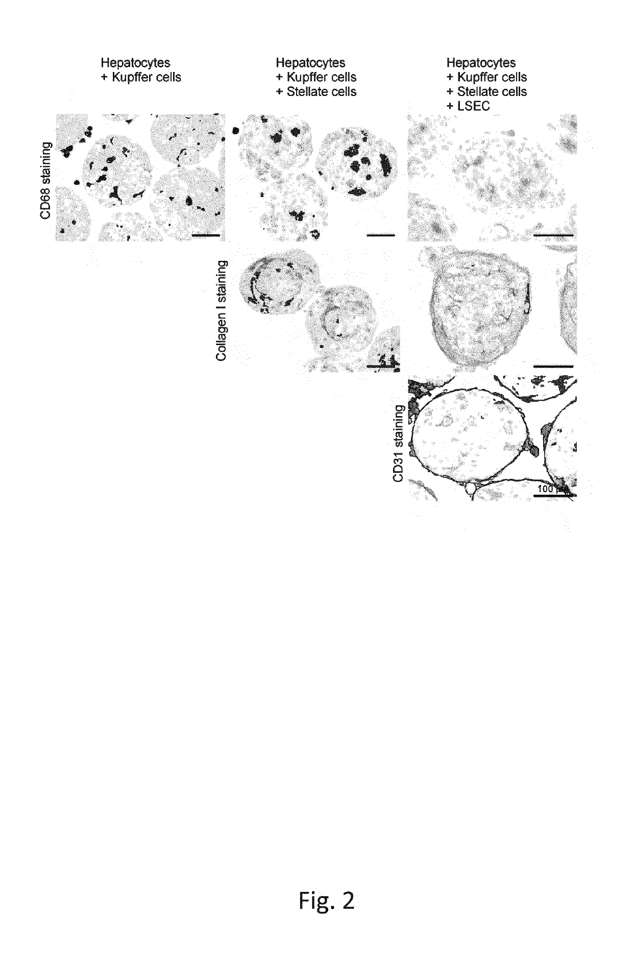
Production Of Triple Cell Type Spheroidal Microtissues (Hepatocytes, Kupffer Cells, Stellate Cells)

[0197]After growth in 2D culture homo- and heterotypic microtissues were produced in hanging drops using InSphero's GravityPLUS™ (InSphero) platform. For protocols see WO2015158777A1, the content of which is incorporated by reference herein[0198]First, hepatocytes and hepatic stellate cells were detached from cell culture flasks[0199]Therefore, medium was aspirated and the cell monolayer was washed with phosphate buffered saline (PBS)[0200]Detaching of hepatocytes and HSCs was by Trypsin treatent[0201]After optical confirmation of dissociation, thaw / wash medium was added, stopping the enzymatic reaction[0202]Kupffer cells were thawed and directly used for MT production[0203]Hepatocytes, HSCs and KCs were separately washed in thaw / wash medium by centrifugation[0204]After the centrifugation step, the cells were resuspended in thaw / wash medium[0205]Their cell number and viability was asse...

example 2

Coating Of Spheroidal Microtissues With Liver Endothelial Cells (LEC)

[0207]After The artificial spheroidal microtissue formation (Day 4), LEC have been thawed, washed with LEC Coating Medium—and centrifuged[0208]In addition to the centrifugation step, the cells were resuspended in LEC Coating Medium and their cell number and viability has been assessed[0209]For LEC seeding and coating of the spheroidal microtissues a cell suspension of the desired composition was prepared in LEC Coating Medium[0210]spheroidal microtissues are being provided in suitable wells or vessel[0211]5 μ of the LEC suspension are dispensed to each well or vessel.[0212]LEC sink down and get in contact with the microtissues and the extracellular matrix surrounding them[0213]After 2 days of incubation, microtissues are fully coated with LECs

example 3

Induction Of Steatotic Phenotype by Lipid Loading of Liver Microtissues

[0214]Dissolve FFA-free BSA at RT for 1 h in H2O for desired stock solution concentration[0215]Dissolve desired amount of OA-Na in 1 mL 0.1 M NaOH→at 70° C.[0216]Dissolve desired amount of PA-Na 1 mL 0.1 M NaOH→at 70°[0217]Keep the FFAs on 70° C., while the BSA is placed on 37° C.[0218]Mix FFA with BSA with a magnetic stirrer (for 30 min)[0219]Dilute BSA with 0.1 M NaOH→vehicle control[0220]Cool down to RT, sterile filter, aliquot and freeze.[0221]For a final fatty acid (FA) solution dilute master stock in medium.[0222]Replace medium of liver microtissues with FA-containing medium and incubate 2-7 days.

[0223]Oleate (OA), a monounsaturated omega-9 fatty acid, incorporates in hepatocytes within small lipid droplets (microvesicular steatosis), while palmitic acid (PA), saturated hexadecanoic acid, shows a rather macrovesicular phenotype (FIG. 3A1). Lipid accumulation is the first step for recapitulating NASH (Non-Al...


PUM


Abstract

The present invention relates to an in vitro testing platform for the study of metabolic liver disease and related therapeutic strategies, composed of an artificial spheroidal microtissue comprising at least hepatocytes and hepatic stellate cells, and at least one type of hepatic inflammatory cells and further medium and reagents capable to establish and simulate different stages of metabolic liver disease and their progression and testing preventive and therapeutic strategies by pharmacological and / or dietary interventions.

Description

FIELD OF THE INVENTION[0001]The present invention relates to the field of 3D tissue cultures and its applications in drug testing.INTRODUCTION[0002]Liver fibrosis is a common outcome of virtually all chronic hepatic insults including viral hepatitis (i.e., hepatitis B and C), alcoholic or obesity-associated steatohepatitis (i.e., nonalcoholic steatohepatitis (NASH)), parasitic disease (i.e., schistosomiasis), metabolic disorders (i.e., Wilson's), hemochromatosis and other storage diseases, congenital abnormalities, autoimmune and chronic inflammatory conditions (i.e., sarcoidosis), and drug toxicity, among others (Puche, Saiman, & Friedman, 2013).[0003]Liver Fibrosis is defined by the World Health Organisation as the presence of excess collagen due to new fiber formation. The fibers result from production of extracellular matrix (ECM) due to activation of quiescent hepatic stellate cells into myofibroblasts.[0004]Liver fibrosis is currently mainly studied in animal experiments, but ...

Claims


Application Information

Patent Timeline
IPC IPC(8): C12N5/071G01N33/50C12N5/00
CPCG01N33/5088G01N2800/52G01N33/5067C12N2503/04C12N5/0697C12N2513/00C12N5/0062C12N5/0671C12N2502/1157C12N2502/28
Inventor MESSNER, SIMONKELM, JENS M.KOSTADINOVA-ANGELOVA, RADINAMORITZ, WOLFGANG
Owner INSPHERO AG
Why Eureka
Try Eureka