Therapy for frontotemporal dementia
a frontotemporal dementia and therapy technology, applied in the field of frontotemporal dementia therapy, can solve the problems of predisposing neurons to degeneration, ad and parkinson's disease, and ad risk, and achieve the effects of preventing, reducing, or increasing the degeneration or death of cortical neurons, preserving or rescuing neuron function, or viability
- Summary
- Abstract
- Description
- Claims
- Application Information
AI Technical Summary
Benefits of technology
Problems solved by technology
Method used
Image
Examples
example 1
[0194]FTD is the second-most common cause of dementia in individuals younger than 65 years of age, and mutations in the gene encoding progranulin (GRN) are a common Mendelian cause of FTD. To date, all mutations (>50 different mutations) in GRN that cause FTD have been shown to do so by haploinsufficiency—the vast majority are nonsense mutations causing the affected individual to express only 50% of normal transcript levels. As a consequence, replacement of progranulin should provide a therapy to ameliorate, reverse, or even prevent FTD due to GRN mutations.
[0195]AAV viral vectors will be used to express the progranulin gene (GRN) in the central nervous system (CNS) as a therapy for frontotemporal dementia (FTD). Multiple AAV vectors are developed that can deliver human progranulin. As disclosed herein, AAV vectors can deliver measurable amounts of progranulin to the CNS by administration routes such as intraparenchymal or intraventricular injection in studies involving mice. A seri...
example 2
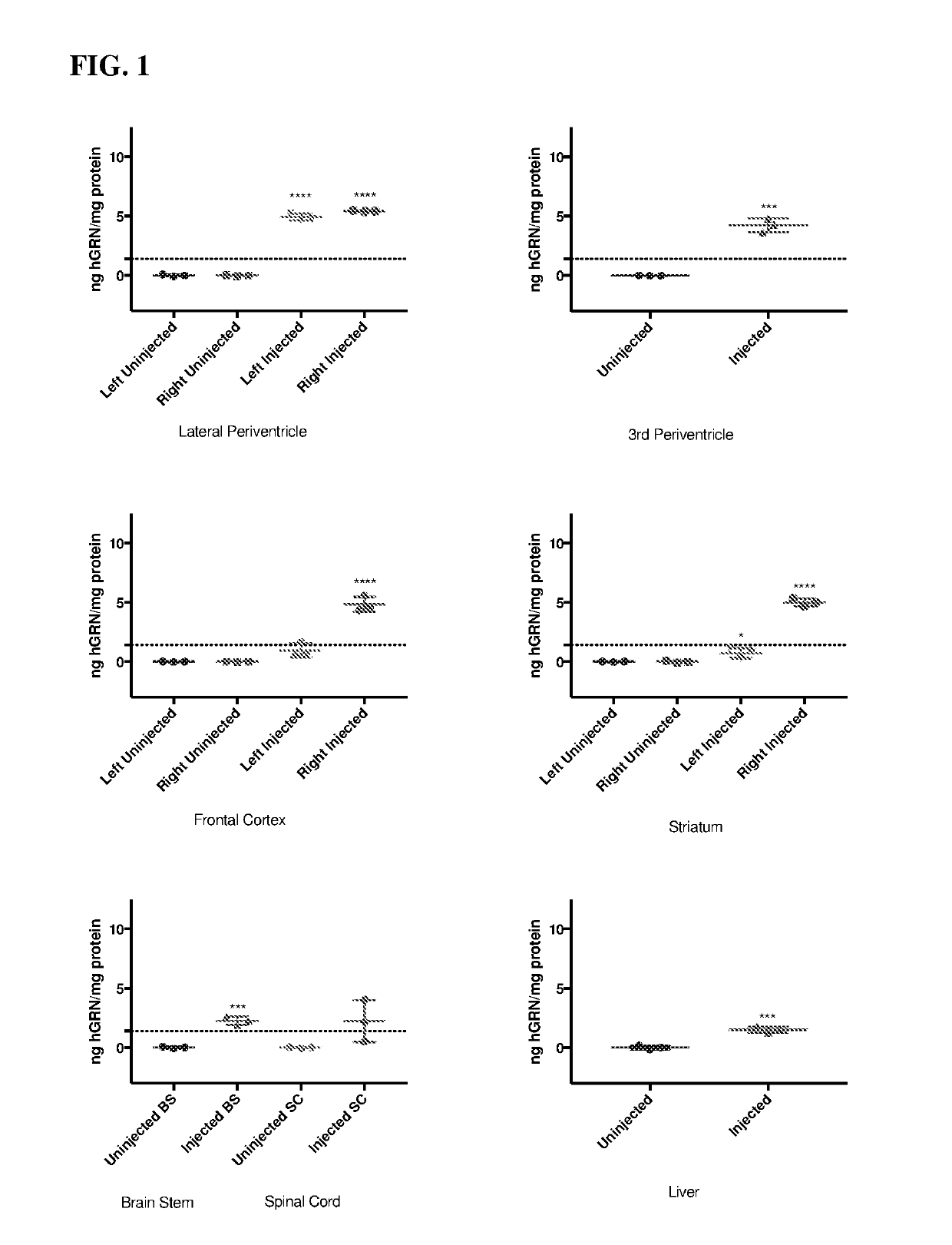
[0197]Human progranulin (hGRN) overexpression in the lateral periventricle, 3rd periventricle, frontal cortex, striatum, brain stem, spinal cord, and liver of GRN null mice 1 month post unilateral injection of 5E10 vg of AAV9.CMV.hGRN.bGHpA, compared to uninjected littermates, as measured by ELISA (FIG. 1). Dotted line indicates normal levels of hGRN in human frontal cortex as measured by ELISA. For lateral periventricle, frontal cortex, and striatum left and right indicate hemispheres of the brain, all mice were injected in the caudal right lateral ventricle.
example 3
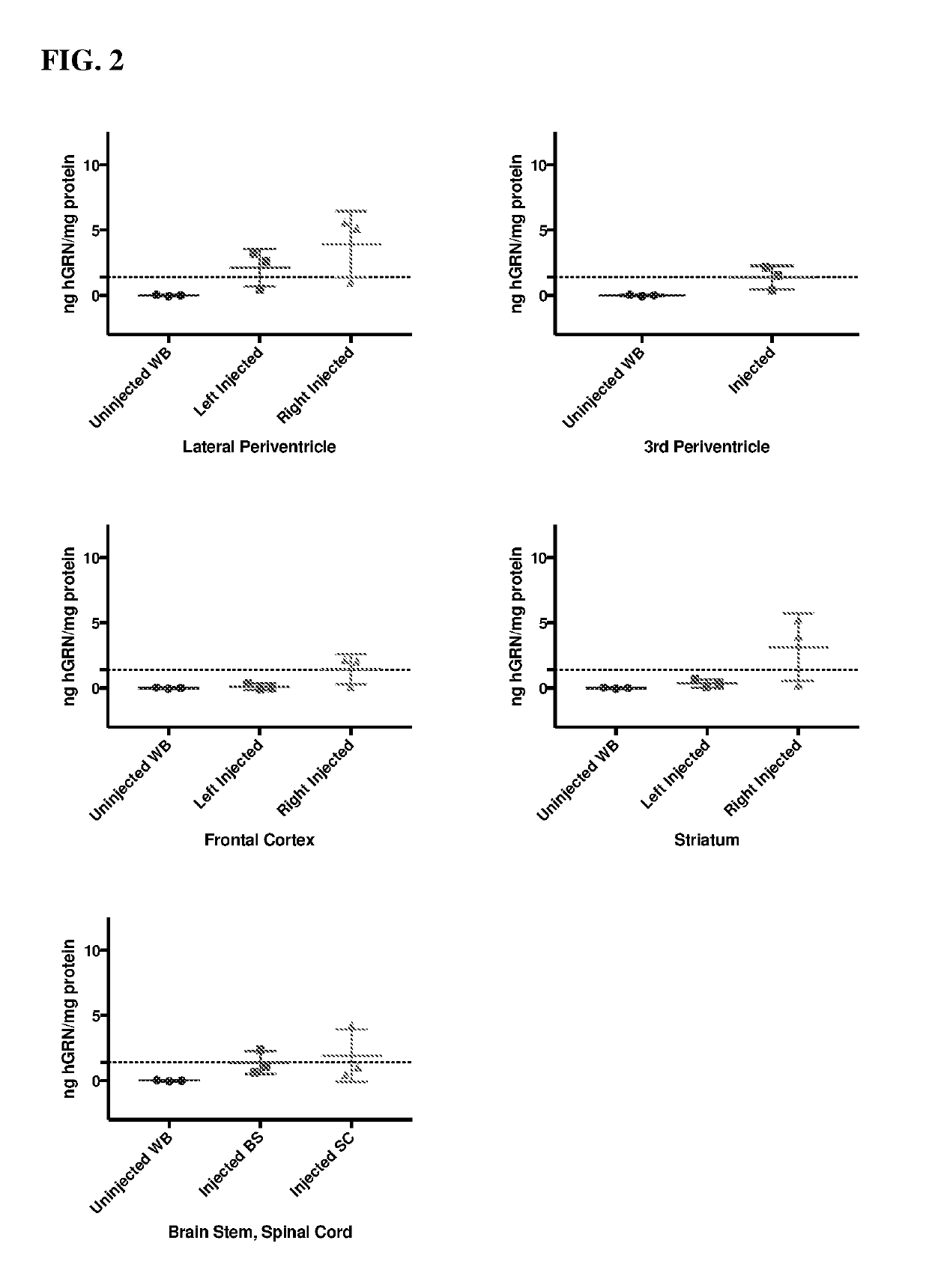
[0198]hGRN overexpression in the lateral periventricle, 3rd periventricle, frontal cortex, striatum, brain stem, and spinal cord of GRN null mice 3 month post unilateral injection of 5E10 vg of AAV9.CMV.hGRN.bGHpA compared to uninjected GRN-null whole brain (WB), as measured by ELISA (FIG. 2). Dotted line indicates normal levels of hGRN in human frontal cortex as measured by ELISA. For lateral periventricle, frontal cortex, and striatum left and right indicate hemispheres of the brain, all mice were injected in the caudal right lateral ventricle.
PUM
| Property | Measurement | Unit |
|---|---|---|
| Fraction | aaaaa | aaaaa |
Abstract
Description
Claims
Application Information
 - R&D
- Intellectual Property
- Life Sciences
- Materials
- Tech Scout
- Unparalleled Data Quality
- Higher Quality Content
- 60% Fewer Hallucinations
Browse by: Latest US Patents, China's latest patents, Technical Efficacy Thesaurus, Application Domain, Technology Topic, Popular Technical Reports.
© 2025 PatSnap. All rights reserved.Legal|Privacy policy|Modern Slavery Act Transparency Statement|Sitemap|About US| Contact US: help@patsnap.com


