

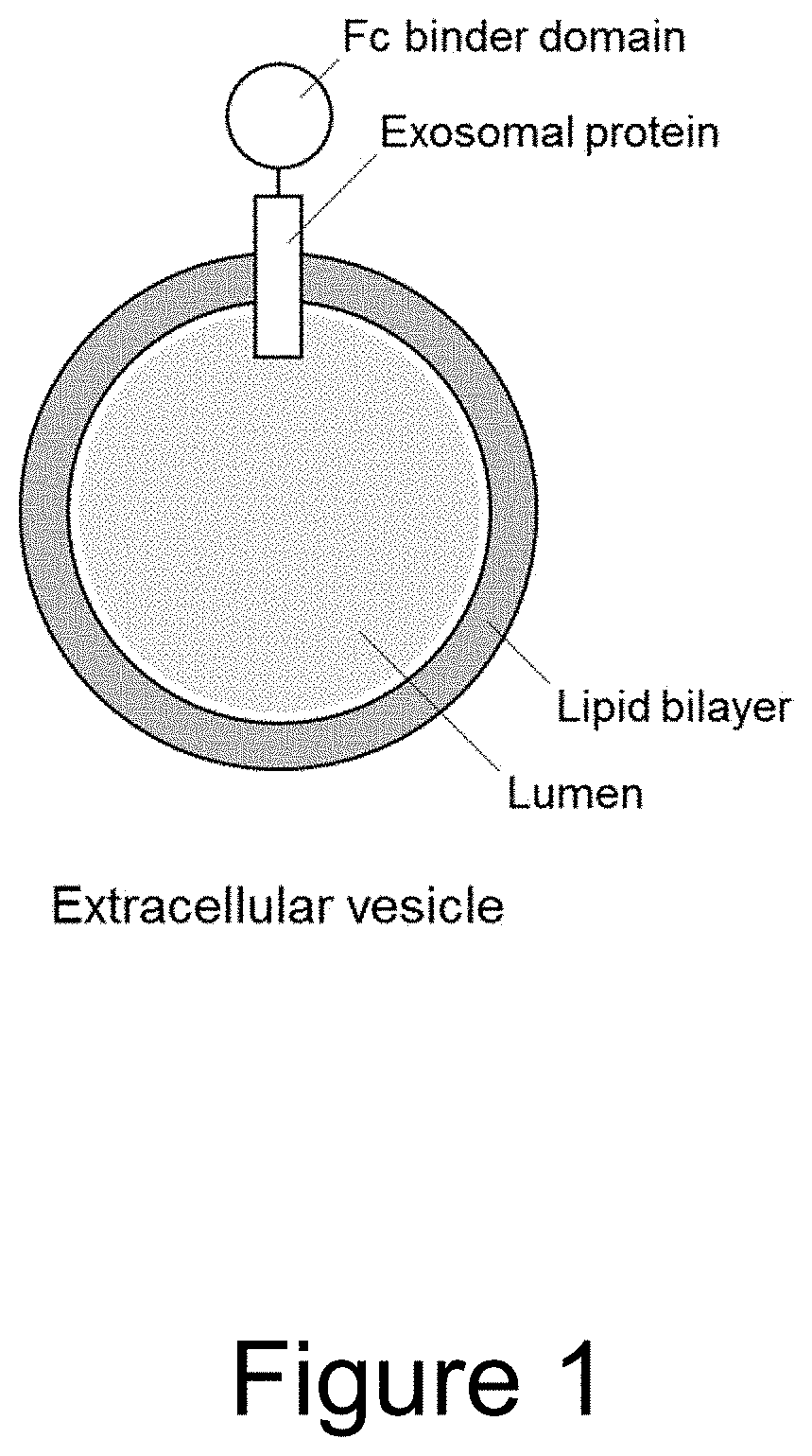
Extracellular vesicle comprising a fusion protein having fc binding capacity
- Summary
- Abstract
- Description
- Claims
- Application Information
AI Technical Summary
Benefits of technology
Problems solved by technology
Method used
Image

Examples
example 1
f IgG to EVs Comprising Fc Binding Polypeptides (Fc-Binding EVs)
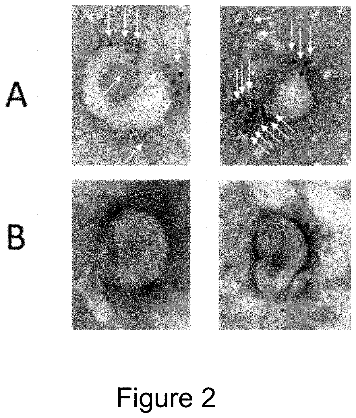
[0107]EVs were isolated from the conditioned medium from engineered HEK293T cells (control versus Fc-binding construct that stably express Gp130 Extracellular domain-2XGGGGS linker-Z domain-Gp130 transmembrane domain-Leucine Zipper-N terminal syntenin-His tag) using tangential flow filtration with 300 kd hollow fiber columns, followed by ultrafiltration using 10 kd spin filters for concentration. The binding capacity for IgG by the Fc-binding EVs were then assessed using electron microscopy and flow cytometry.
[0108]For electron microscopy, 1×10{circumflex over ( )}9 EVs were incubated with Rabbit anti-goat 10 nm antibody conjugated with gold Nanoparticles for 2 h at 37° C. As shown in FIG. 2, Fc-binding EVs (A) are decorated with nanogold labeled antibodies (i.e. Fc containing proteins), whereas control EVs (B) do not have any antibodies bound.
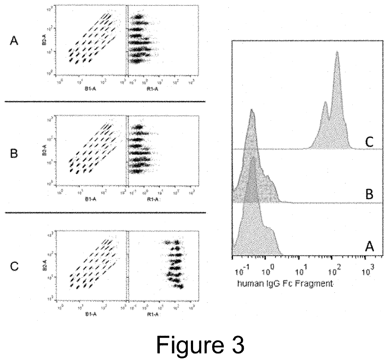
[0109]For flow cytometry, 1×10{circumflex over ( )}8 EVs were incubated overn...
example 2
mp2B EVs for Delivery of anti-HER2 Antibody
[0110]EVs were isolated from HEK293T cells (either stably expressing FCGR1A Extracellular domain-4XGSlinker-Lamp2b or their wild type controls) using ultrafiltration and size exclusion chromatography. EVs were labelled with PKH26 red fluorescent dye, and decorated with anti-HER2 antibody or its isotype control by co-incubating EVs and antibody for 1 h at 37° C. Unbound antibody was removed by size exclusion chromatography. Uptake of antibody decorated EVs was characterized in HER2 low-expressing cell line MDA-MB-231 and in HER2 high-expressing cell line MDA-MB-361 using flow cytometry. FIG. 4 shows that anti-HER2 antibody increases uptake of decorated EVs as compared to isotype control decorated and wild type EVs only in HER2 high-expressing cell line MDA-MB-361, but not in HER2 low-expressing cell line MDA-MB-231. Similar results were obtained with EVs expressing CD63-ZZ fusion proteins.
example 3
t Delivery by EVs Comprising CD81-Protein A / G Fusion Proteins
[0111]EVs were isolated from HEK293T cells (either stably expressing CD81-ProteinA / G CD81 Second loop fusion protein or their wild type control) using ultrafiltration and size exclusion chromatography. EVs were decorated with etanercept or a control antibody by co-incubating EVs and etanercept for 1 h at 37° C. Unbound etanercept was removed by size exclusion chromatography. To study anti-inflammatory effect of the etanercept-decorated EVs, the well-studied TNBS-induced colitis mouse model was used. This model simulates the gut inflammation, cytokine storm and weight decrease associated with IBD patients. 24 mice were divided into four treatment groups, with 6 mice per group. The mice were pre-sensitized by applying 150 μl of a olive oil-acetate solution with 2% TNBS, on the skin, 1 week prior to colitis induction. Colitis was then induced by giving a rectal infusion of 100 μl solution containing 1.5% TNBS in 40% ethanol. ...
PUM
 Abstract
Description
Claims
Application Information