Devices and Methods for Detection and Quantification of Immunological Proteins, Pathogenic and Microbial Agents and Cells

a technology of immunological proteins and microorganisms, applied in the field of immunoassays and microfluidic devices, can solve the problems of increasing the cost and complexity of assays, the manual counting of microbeads using microscopes, and the inability to detect and quantify immunological proteins, so as to reduce the cost and complexity, increase the sensitivity of lateral flow tests, and maintain the specificity and accuracy.

Pending Publication Date: 2020-04-09
CARNEGIE MELLON UNIV
View PDF3 Cites 5 Cited by
  • Summary
  • Abstract
  • Description
  • Claims
  • Application Information

AI Technical Summary

Benefits of technology

[0011]The pScreen™ microfluidic immunoassay device, based on the inventions disclosed herein, fulfills all of the above-described needs in a single device. The detection and quantification of an unknown concentration of analyte in a liquid sample is obtained by exploiting the fluid-dynamic properties of magnetic-responsive micro-beads in liquid solution rather than using optical effects or magnetic field sensing as in current technologies. The unknown concentration of the target analyte is derived by measuring the differential flow rate between the sample flow in two micro-channels, one of which is under the influence of an applied magnetic field gradient. The present invention significantly reduces the cost and complexity of current laboratory-based immunoassay diagnostic tests, and greatly increases one thousand-fold the sensitivity of lateral flow tests, while maintaining the specificity and accuracy of laboratory-based tests, and the ability to detect targeted antigen concentration over a predefined range.

Problems solved by technology

POC tests are designed to be used in the field and require limited training, but they are far less sensitive and accurate with most POC tests providing only binary positive / negative or semi-quantitative results.
Thus, although sensitivity of the assay may increase, the cost and complexity of the assay also increases.
Typically, micro-bead counting using a microscope involves the manual, and often tedious, counting of beads through a microscope objective.
This method requires skilled technicians in a laboratory setting, is time consuming and is subject to a technician's interpretation.
Cell counters, while accurate, are expensive and also require skilled technicians in sophisticated laboratory settings.
In addition, these devices typically include handset and consumable components, resulting in increased manufacturing, calibration and maintenance costs.
Thus, these devices have limited applications in the field of POC immunoassay diagnostics.

Method used


Image

  • Devices and Methods for Detection and Quantification of Immunological Proteins, Pathogenic and Microbial Agents and Cells
  • Devices and Methods for Detection and Quantification of Immunological Proteins, Pathogenic and Microbial Agents and Cells
  • Devices and Methods for Detection and Quantification of Immunological Proteins, Pathogenic and Microbial Agents and Cells

Examples


example 1

c Basis and Technology Feasibility of the Present Invention

(A) Introduction

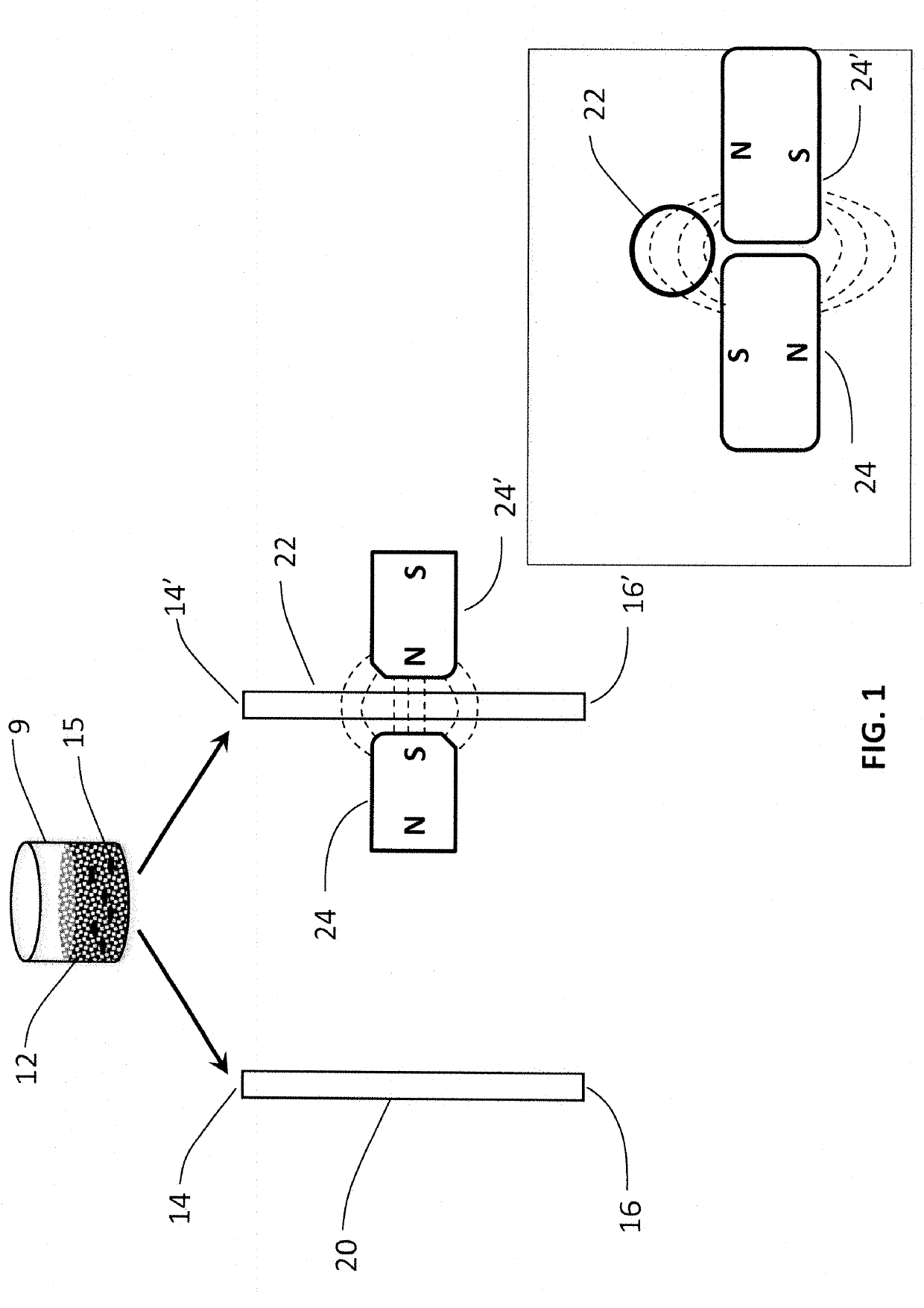
[0144]The data presented herein describe the effect that flocculation of magnetic-responsive micro-beads in a micro-channel has on the flow resistance of liquid in the micro-channel. A liquid seeded with magnetic-responsive micro-beads in a micro-channel that is exposed to a magnetic field gradient produces a localized micro-bead flocculation. This localized micro-bead flocculation results in a localized reduction of the cross section of the micro-channel, and thus in a localized increase of the flow resistance across the flocculation region. The increase in resistance, in turn, results in an increased pressure drop across the flocculation region due to the energy loss in maintaining the flow across the reduced cross-section of the micro-channel. If the external forces responsible for the formation of the micro-bead flocculation are stronger than the flow shear-stress on the micro-beads and their aggregates, ...

example 2

Prototype Fabrication and Testing

(A) Fabrication

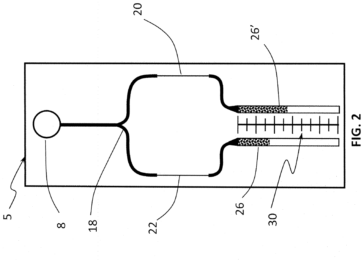
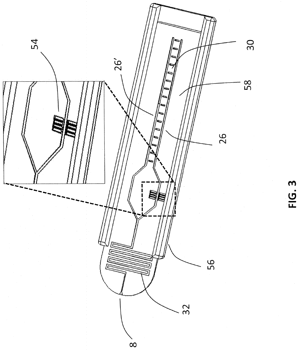
[0151]Two sets of pScreen™ prototypes were fabricated: (1) a bench top prototype with multiple micro-channels for simultaneous testing of various samples; and (2) a single-use, portable, lab-on-card device. The pScreen™ bench-top prototype was described in the previous section. The pScreen™ lab-on-card prototype was realized using standard microfluidics fabrication techniques. In brief, the micro-channels were etched using a laser etcher system on a poly(ethylene terephthalate) glycol (Petg) substrate. The channels then were sealed using a matching poly(lactic-co-glycolic acid) (PLGA) top by thermal bonding using a hot press. The magnetic field gradient was obtained by placing two small magnets in an N-S configuration underneath the test channel. The pScreen™ lab-on-card prototype also was fabricated using injection cast molding in which the prototype was fabricated in poly(methyl methacrylate) (PMMA).

(B) Experimental Data for a Microf...

example 3

ationships between Flow Rate and Micro-Bead Concentration

[0155]The present invention provides a method of detecting and quantifying the concentration of magnetic-responsive micro-beads in a liquid sample, by calculating the ratio {tilde under (O)}m / {tilde under (O)}o, the difference {tilde under (O)}o−{tilde under (O)}m, and the ratio ({tilde under (O)}o−{tilde under (O)}m)p / ({tilde under (O)}m)q, wherein p and q are derived through a calibration process, wherein the ratios {tilde under (O)}m / {tilde under (O)}o and ({tilde under (O)}o−{tilde under (O)}m)p / ({tilde under (O)}m)q are a proxy for the number of magnetic-responsive micro-beads in the liquid sample, and then quantifying the concentration of magnetic-responsive micro-beads in the liquid sample.

[0156]Three methods are provided to show how to use these proxy relationships and how to derive in practice the number of magnetic-responsive micro-beads in a liquid sample. The first method analytically derives these relationships fr...


PUM

PropertyMeasurementUnit
concentrationaaaaaaaaaa

Abstract

The present invention provides microfluidic pScreen™ devices for quantifying the concentration of DNA fragments in a liquid sample by using magnetic-responsive silica micro-beads and nonmagnetic-responsive silica micro-beads. The devices of the present invention allow for rapid, simple and inexpensive quantification of DNA fragment concentration in a sample. The devices do not require complex instrumentation and can be performed in less than three minutes. Moreover, they are compatible with complex samples including, without limitation, unpurified PCR amplification products, and thus can be expected to seamlessly integrate into various common molecular biology techniques and workflows.

Description

CROSS REFERENCE TO RELATED APPLICATIONS[0001]The present application is a continuation-in-part of U.S. patent application Ser. No. 15 / 666,918, filed Aug. 2, 2017, which is a continuation of U.S. patent application Ser. No. 14 / 148,010, filed Jan. 6, 2014, which is a continuation-in-part of U.S. patent application Ser. No. 13 / 862,899, filed Apr. 15, 2013, now U.S. Pat. No. 8,623,596, which is a continuation application of U.S. patent application Ser. No. 13 / 684,618, filed Nov. 26, 2012, now U.S. Pat. No. 8,445,192, issued May 21, 2013, which is a continuation-in-part application of U.S. patent application Ser. No. 13 / 590,859, filed Aug. 21, 2012, which claims priority to U.S. Provisional Patent Application No. 61 / 539,210, filed Sep. 26, 2011, all of which are incorporated by reference herein in their entirety.FIELD OF THE INVENTION[0002]The present invention relates to the field of immunoassay and microfluidic devices and, in particular, to a point-of-care diagnostic method and device...

Claims


Application Information

Patent Timeline
Patent Type & Authority Applications(United States)
IPC IPC(8): G01N33/543B01L3/00
CPCB01L3/5027G01N33/54326Y02A50/58B01L3/502746B01L2200/0647B01L2300/0816B01L2300/028B01L2300/0864B01L2200/148B01L2400/043B01L2400/0406B01L2400/084B01L2300/0867G01N33/54333Y02A50/30
Inventor GANDINI, ALBERTOANTAKI, JAMES F.CHUAN, BYRON WANGMARHEFKA, JOIE N.MATTILA, JUHA-PEKKASALAS, ELISEO
Owner CARNEGIE MELLON UNIV
Why Eureka
Try Eureka