
Ultrasound, photoacoustic, and viscoelastic imaging systems and methods for cervical analysis to assess risk of preterm delivery

a technology of ultrasound and ultrasound, applied in the field of ultrasound, can solve the problems of increasing the risk of a myriad of birth defects, the difficulty of accurate detection of expectant mothers at risk of preterm birth of a fetus, and the difficulty of sensitivity and sensitivity required

Pending Publication Date: 2020-04-23
WAYNE STATE UNIV
View PDF5 Cites 2 Cited by
  • Summary
  • Abstract
  • Description
  • Claims
  • Application Information

AI Technical Summary

Benefits of technology

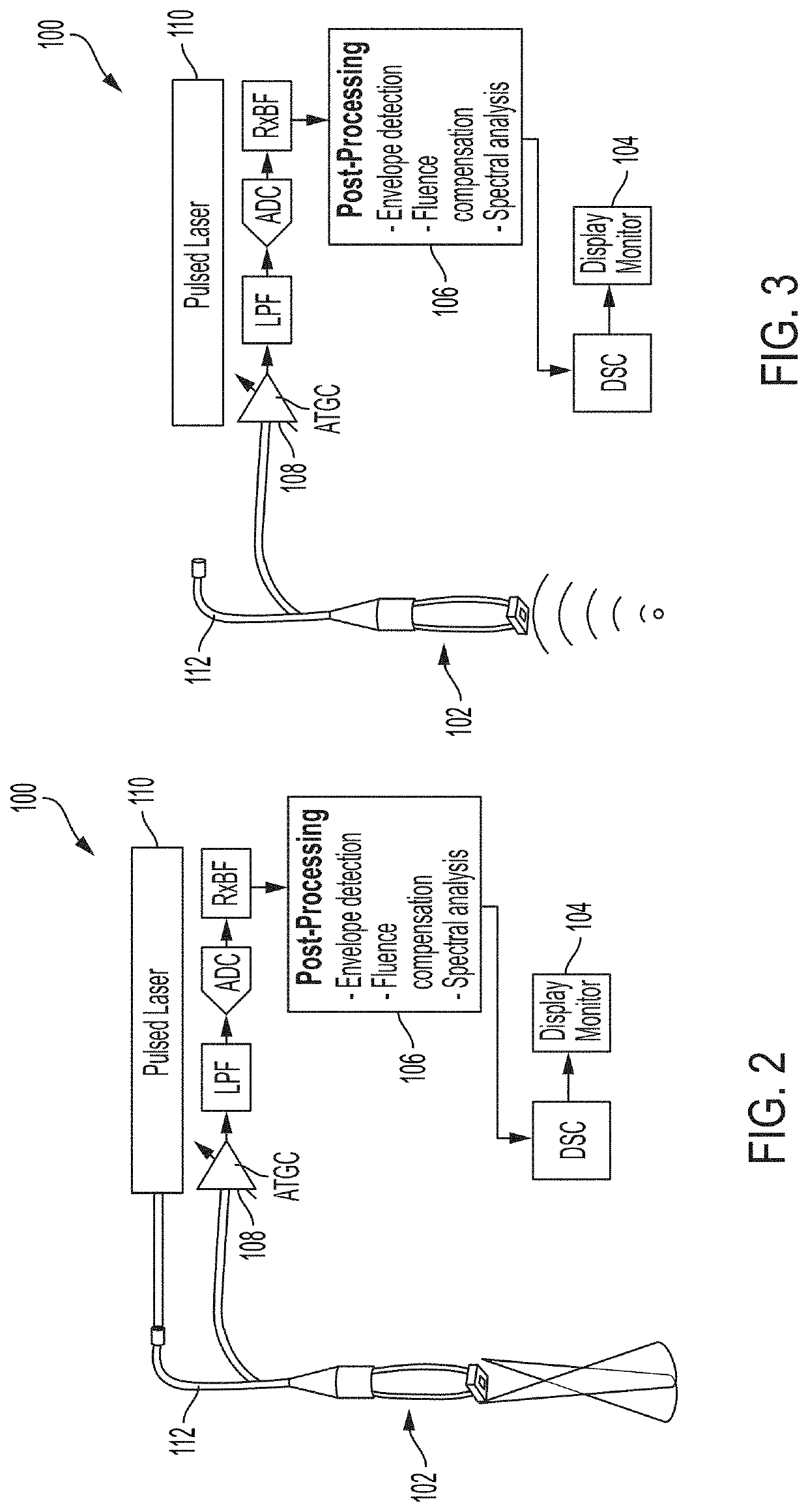
The patent describes a system and method for detecting a risk of preterm delivery during delivery. The system includes an ultrasound machine and a probe device that sends and receives signals to and from the cervical tissue of the mother. The signals are transmitted as ultrasound waves and photoacoustic waves, which are converted into electrical signals by the probe device. The system can generate images and biomarker parameters of the cervical tissue in real-time, and can also generate a risk parameter based on the biomarker parameters to alert the medical professionals of a potential preterm delivery. The technical effects of this system and method include improved accuracy in predicting preterm delivery and earlier intervention to prevent preterm labor.

Problems solved by technology

Accurate detection of expectant mothers that are at risk of preterm birth of a fetus is a challenging clinical problem.
Complications from such preterm birth is a leading cause of death of children less than five years of age and is a major cause of an increased risk of a myriad of illnesses and conditions ranging from, and not limited to, chronic respiratory disease, blindness, and neurological disorders.
However, less than 40% of mothers delivering preterm are diagnosed with such a shortened cervix biomarker through such US imaging, which reflects a lack of sensitivity and sensitivity required for more accurate detection of those expectant mothers at risk of preterm birth.

Method used


Image

  • Ultrasound, photoacoustic, and viscoelastic imaging systems and methods for cervical analysis to assess risk of preterm delivery
  • Ultrasound, photoacoustic, and viscoelastic imaging systems and methods for cervical analysis to assess risk of preterm delivery
  • Ultrasound, photoacoustic, and viscoelastic imaging systems and methods for cervical analysis to assess risk of preterm delivery

Examples


Embodiment Construction

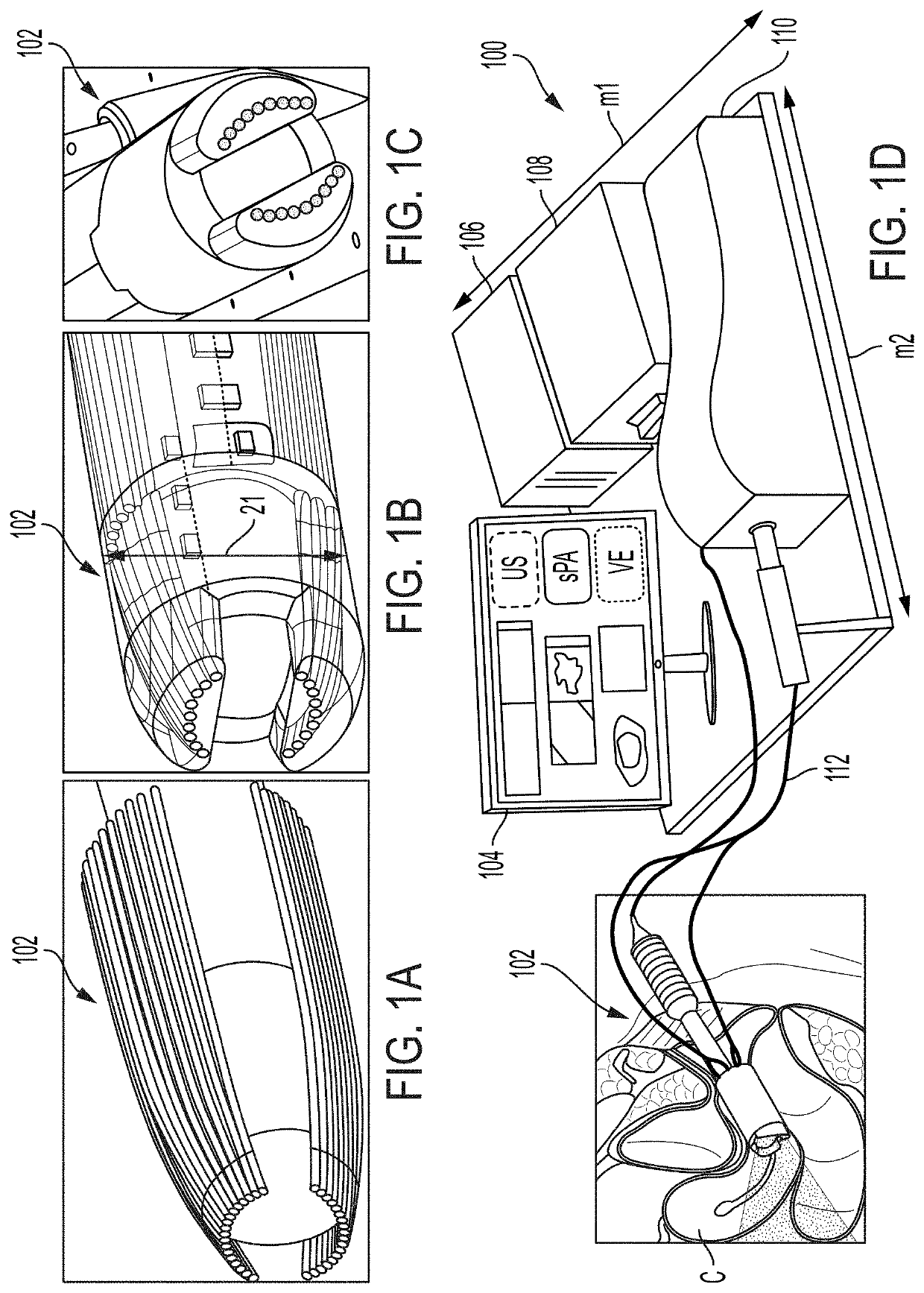
[0033]The present disclosure relates to systems and methods to optimize clinical care of a fetus and mother to assess a risk of preterm delivery through use of a multi-model probe to image a cervix through a vaginal canal and generate multi-modal imaging providing a variety of cervix tissue characteristic data. The systems and methods described herein further permit a visualization of cervix tissue to determine oxygen saturation with respect to the cervix tissue.

[0034]Due to a lack of a highly sensitive and accurate diagnostic modality to predict the risk of preterm delivery, preterm delivery is a main cause of perinatal morbidity and mortality worldwide, which is associated with a significant healthcare cost spent on prematurity. Although measurement of cervical length by transvaginal ultrasound has assisted to guide diagnosis and management of preterm delivery, such technology alone fails to capture a majority of preterm deliveries that occur. The multi-modal imaging tool as descr...


PUM


Abstract

Methods and system are described for multi-modal, multi-parametric, non-invasive, and real-time assessment of cervical tissue through a multi-modal probe device for use within a vaginal canal and an associated imaging system to assess a risk of preterm delivery of an expectant mother. The multi-modal system may include ultrasound (US) imaging, viscoelastic (VE) imaging, and / or photoacoustic (PA) imaging of the cervical issue to determine cervical biomarker information indicative of parameters including, but not limited to, a collagen to water ratio such that a more water dominant ratio is indicative of a risk of preterm delivery.

Description

CROSS-REFERENCE TO RELATED APPLICATIONS[0001]The present disclosure claims the benefit of U.S. Provisional App. No. 62 / 748,613, filed Oct. 22, 2018, and entitled “ULTRASOUND, PHOTOACOUSTIC, AND VISCOELASTIC IMAGING SYSTEMS AND METHODS FOR CERVICAL ANALYSIS TO ASSESS RISK OF PRETERM DELIVERY,” the entirety of which is incorporated by reference herein.TECHNICAL FIELD[0002]The present disclosure generally relates to multi-modal imaging systems and methods to assess risk of preterm delivery and, more specifically, to ultrasound (US), viscoelastic (VE), and photoacoustic (PA) systems and methods for cervical analysis of an expectant mother to assess risk of preterm delivery of the fetus.BACKGROUND[0003]Accurate detection of expectant mothers that are at risk of preterm birth of a fetus is a challenging clinical problem. A fetus that is born preterm is born before 37 completed weeks of gestation. Complications from such preterm birth is a leading cause of death of children less than five ...

Claims


Application Information

Patent Timeline
IPC IPC(8): A61B5/00A61B8/08
CPCA61B8/461A61B5/4331A61B8/5223A61B8/0866A61B5/7275A61B5/0095A61B8/4416A61B8/12A61B5/0035A61B5/435A61B5/4343
Inventor MEHRMOHAMMADI, MOHAMMADHASSAN, SONIA S.HERNANDEZ-ANDRADE, EDGARYAN, YANBASIJ, MARYAMGOMEZ-LOPEZ, NARDHY
Owner WAYNE STATE UNIV
Learn More