
Blood pathology image analysis and diagnosis using machine learning and data analytics

a blood pathology and image analysis technology, applied in healthcare informatics, instruments, computing models, etc., can solve the problems of slow and subject to pathologist subjectivity, delay in diagnosis and appropriate patient care, and process that is both manually intensive and time-consuming, so as to achieve faster and less prone to errors

Inactive Publication Date: 2020-05-14
IBM CORP
View PDF0 Cites 11 Cited by
  • Summary
  • Abstract
  • Description
  • Claims
  • Application Information

AI Technical Summary

Benefits of technology

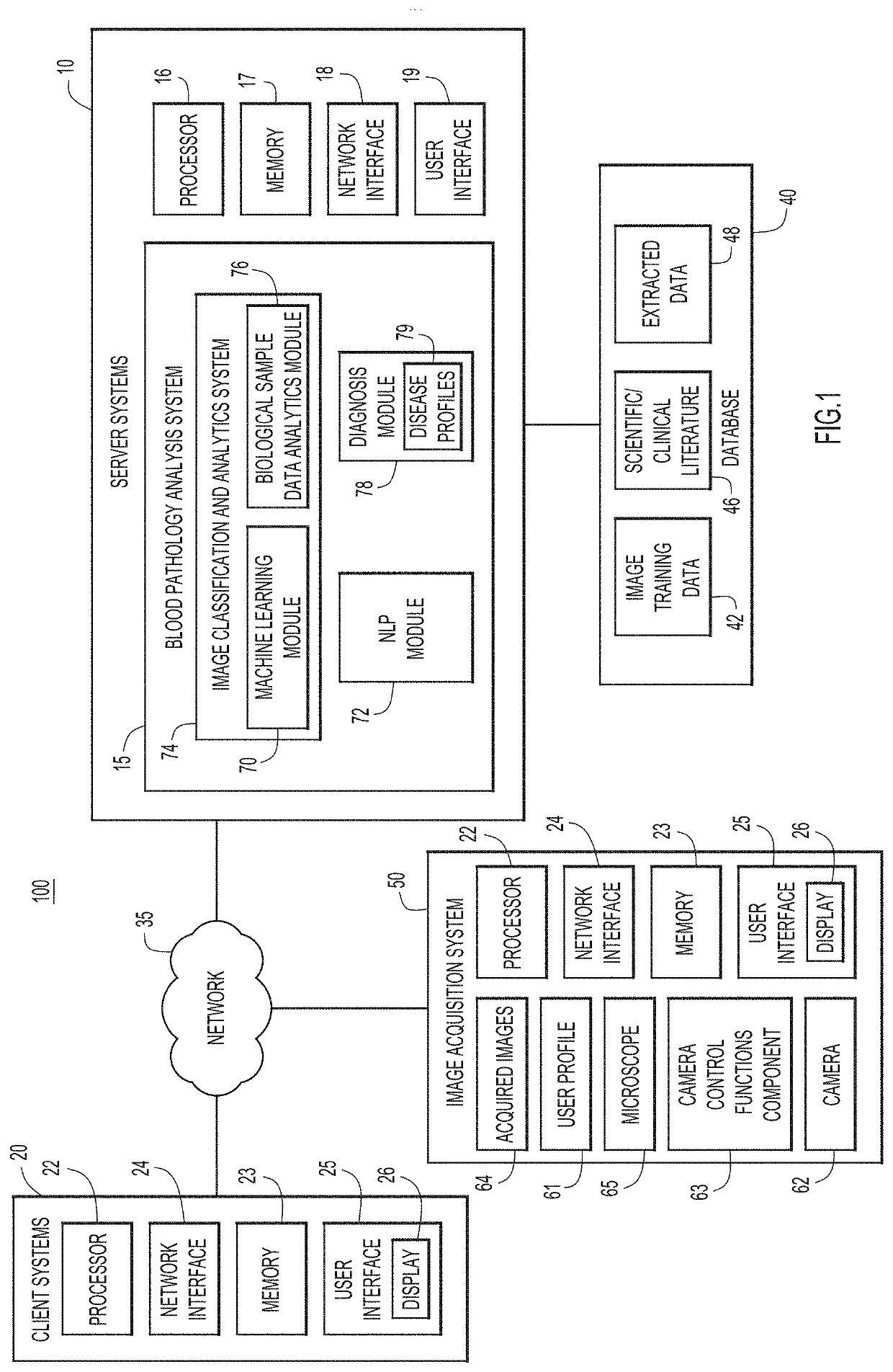
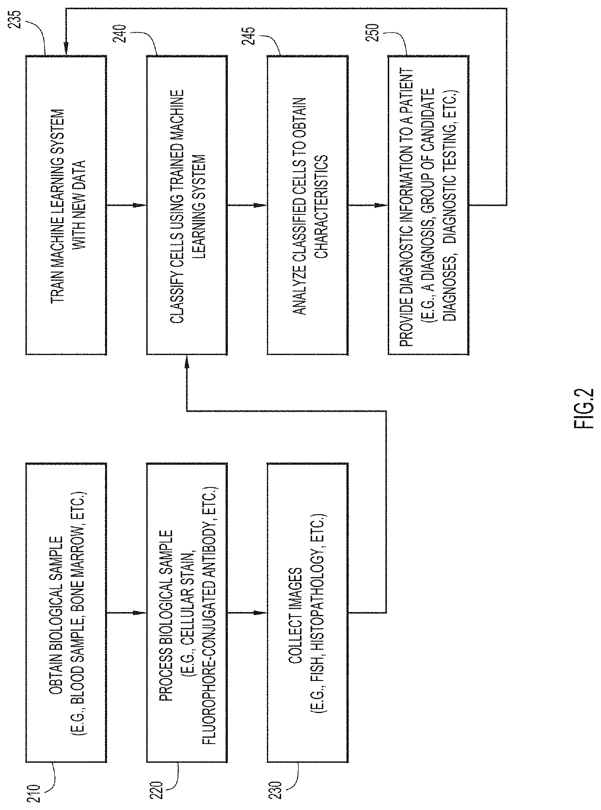
The present invention provides a computer-implemented method for analyzing microscopic images of a biological sample, such as a blood sample, using machine learning techniques and data analytics to generate a diagnosis for a patient. This approach allows for a standardized, uniform analysis of cells, resulting in faster and less prone to error results. The system can categorize cells into a variety of different categories and identify subcategories of cells, which can aid in the diagnosis of cancer / tumor cells and other non-tumoral diseases. The system can also update with current information and reduce physician error by providing a list of potential diagnoses and a recommendation for additional testing.

Problems solved by technology

Often, physicians or other medical personnel conduct and interpret the results of immunophenotyping and IHC assays, which typically involve generation of a series of images of the biological sample, a process that is both manually intensive and time consuming.
However, manual review is slow and subject to pathologist subjectivity, and different pathologists may reach different conclusions when analyzing the same slide.
This may lead to delays in diagnosis and in appropriate patient care, especially if the patient is misdiagnosed.
However, these automated techniques are often limited in scope and are not standardized.
Hospitals, clinics, and other laboratories which analyze biological samples for diagnosing blood cancers or other diseases do not typically use automated methods, instead relying on clinician judgement and pathologist analysis, a process that may be both time consuming and subject to human error.

Method used


Image

  • Blood pathology image analysis and diagnosis using machine learning and data analytics
  • Blood pathology image analysis and diagnosis using machine learning and data analytics
  • Blood pathology image analysis and diagnosis using machine learning and data analytics

Examples


Embodiment Construction

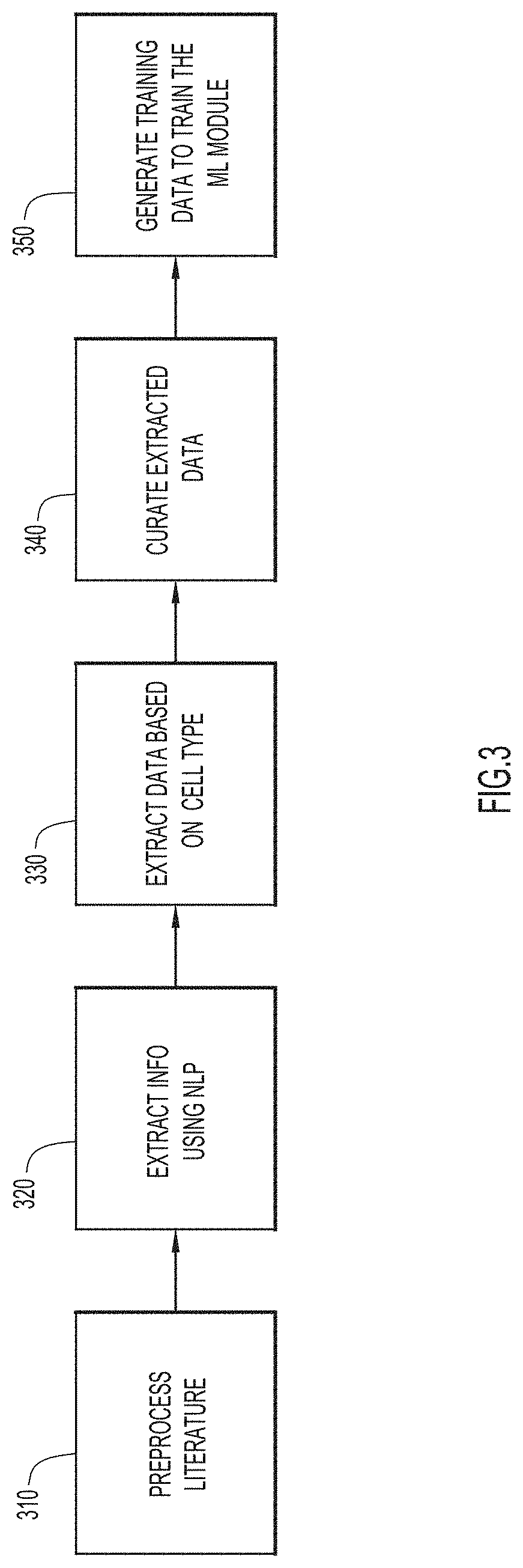
[0020]Methods, systems, and computer readable media are provided for analyzing images from blood smears using machine learning and data analytics to diagnose a patient. Present techniques allow for automated and consistent classification and analysis of cells of a blood sample to generate a diagnosis. These techniques use a machine learning module to classify cells, based on morphological characteristics of the cells and in some cases, the presence of a probe that identifies a nucleotide mutation (e.g., detectable by FISH) or the presence of a cell marker to identify the cell (e.g., detectable by fluorophore or fluorochrome-conjugated antibodies). As additional biological samples are processed, the machine learning module may be trained using this data, and classification of cells may improve over time. These techniques can be extended to a variety of cell types, including but not limited to basophils, eosinophils, neutrophils, red blood cells (erythrocytes), macrophages, mast cells...


PUM


Abstract

Methods, systems, and computer readable media are provided for processing microscopic images of a biological sample from a patient. One or more images of a blood sample from a microscope is obtained, each image comprising a plurality of different types of cells. The one or more images are processed by a machine learning system to classify individual cells into one of a plurality of cell categories. The cells in each cell category are analyzed to determine characteristics of the respective cell category. A diagnosis or list of possible diagnosis are determined based on the classification and characteristics of the cells for the patient in an automated manner.

Description

TECHNICAL FIELD[0001]Present invention embodiments relate to automated techniques for analysis of biological samples, and more specifically, to machine learning techniques and data analytics of images of blood samples for diagnosis of a disease.DISCUSSION OF THE RELATED ART[0002]Blood diseases are typically diagnosed by a combination of immunophenotyping and pathology imaging techniques such as microscopy. Immunophenotyping may involve applying various stains or other reagents to a biological sample, such as a bone marrow, blood or other tissue sample or any other suitable biological sample comprising a heterogeneous population of cells, in order to identify the presence of particular types of cells within the population of cells. Microscopy imaging, immunohistochemistry (IHC) or direct observation of a blood smear sample may also be performed as part of the diagnostic process. In some cases, the biological sample may be incubated under suitable conditions with an antibody that bind...

Claims


Application Information

Patent Timeline
IPC IPC(8): G16H50/20G06N99/00
CPCG16H50/20G06N20/00G16H30/40G06N3/08G06N20/20G06N5/01
Inventor SANCHEZ-MARTIN, MARTAHUETTNER, CLAUDIA S.XU, JIAEIFERT, CHERYLDEHAN, ELINORXUE, SHANGMICHELINI, VANESSA
Owner IBM CORP
Learn More