Methods of detection and treatment of urothelial cancer

a technology of urothelial cancer and detection method, which is applied in the field of diagnostic and treatment of cancer to achieve the effect of improving symptoms and increasing expression

Inactive Publication Date: 2020-06-11
THE JOHN HOPKINS UNIV SCHOOL OF MEDICINE
View PDF0 Cites 1 Cited by
  • Summary
  • Abstract
  • Description
  • Claims
  • Application Information

AI Technical Summary

Benefits of technology

This patent describes a method for treating cancer or symptoms associated with urothelial cancer in a patient. The method involves administering a substance that removes a layer of DNA called a methyl group from a specific gene called CCND2, which is turned on in cancer cells. This results in the gene being more active and helps to reduce the symptoms of cancer. It is believed that this method could be used in combination with other treatments for cancer and could help to improve the effectiveness of these treatments.

Problems solved by technology

As a result, typically the gene expression is down-regulated and in the case of a tumor suppressor gene, this is a direct cause for cancer cell growth.

Method used


Image

  • Methods of detection and treatment of urothelial cancer
  • Methods of detection and treatment of urothelial cancer
  • Methods of detection and treatment of urothelial cancer

Examples


example 1

Materials and Methods

[0079]Tissue and Urine Samples

[0080]A total of 36 formalin-fixed paraffin-embedded (FFPE) primary LGPUCC tissues were obtained from patients who underwent therapeutic surgery at The Johns Hopkins Hospital. The demographic and clinical information was obtained from the computerized tumor registry at The Johns Hopkins Healthcare System. Among the 36 LGPUCC samples, 17 were collected from patients who did not recur during any follow-up periods, and the remaining 19 were primary tumor samples that recurred within the follow-up periods after TURBT. We also performed analysis by considering the follow-up periods of 12, 18, and 24 months for recurrence to observe the association with promoter methylation of candidate markers.

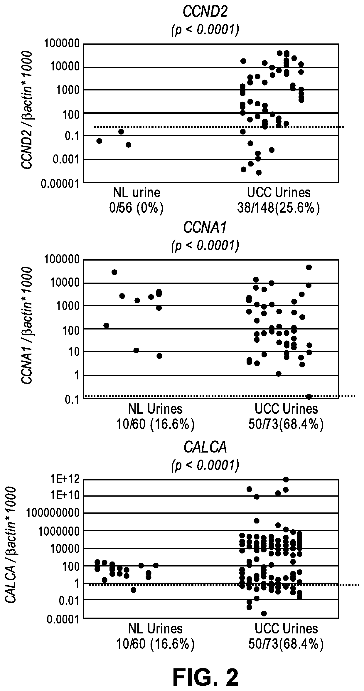
[0081]To be included in the cohort, an eligible patient had to have a confirmed diagnosis of LGPUCC and a sufficient amount of archived tumor material for DNA extraction.To determine the feasibility of detecting promoter methylation of genes in uri...

example 2

Detection and Quantitation of DNA Methylation by PCR

[0103]Methylation at cytosine residues is effectively detected and quantitated by quantitative fluorogenic methylation specific PCR (QMSP). The assay is performed as described (see Maldonado, L. et al., Oncotarget, 2014, 5(14): 5218-5233). Briefly, DNA is extracted from cells isolated from a biological sample by phenol-chloroform extraction protocol followed by ethanol precipitation as described previously (Hogue, M. et al., J. Clin Oncol. 2005, 23(27): 6569-6575). Next it is subjected to bisulfite treatment, which converts unmethylated cytosine residues to uracil residues, as specified in the manufacturer's instructions (EpiTect Bisulfite kit, Qiagen), as previously described (Herman J G. et al., Proc Natl Acad Sci USA. 1996, 93 (18): 9821-9826). Bisulfite-modified DNA was used for fluorescence-based real-time PCR, as previously described (Hogue, M. et al., J Natl Cancer Inst. 2006, 98(14): 996-1004) Amplification reactions are ca...

example 3

Promoter Methylation Detection in UCC Tumor Tissue Samples

[0107]DNA is extracted from formalin fixed paraffin embedded (FFPE) block containing tumor tissue. A representative FFPE block, reviewed and confirmed to contain the pathologic sample is sectioned to obtain multiple 10 micron slides, several of which are used for microdissection to obtain portions containing greater than 70% of neoplastic cells. The first and last slides of the representative block are stained with hematoxillin and eosin.

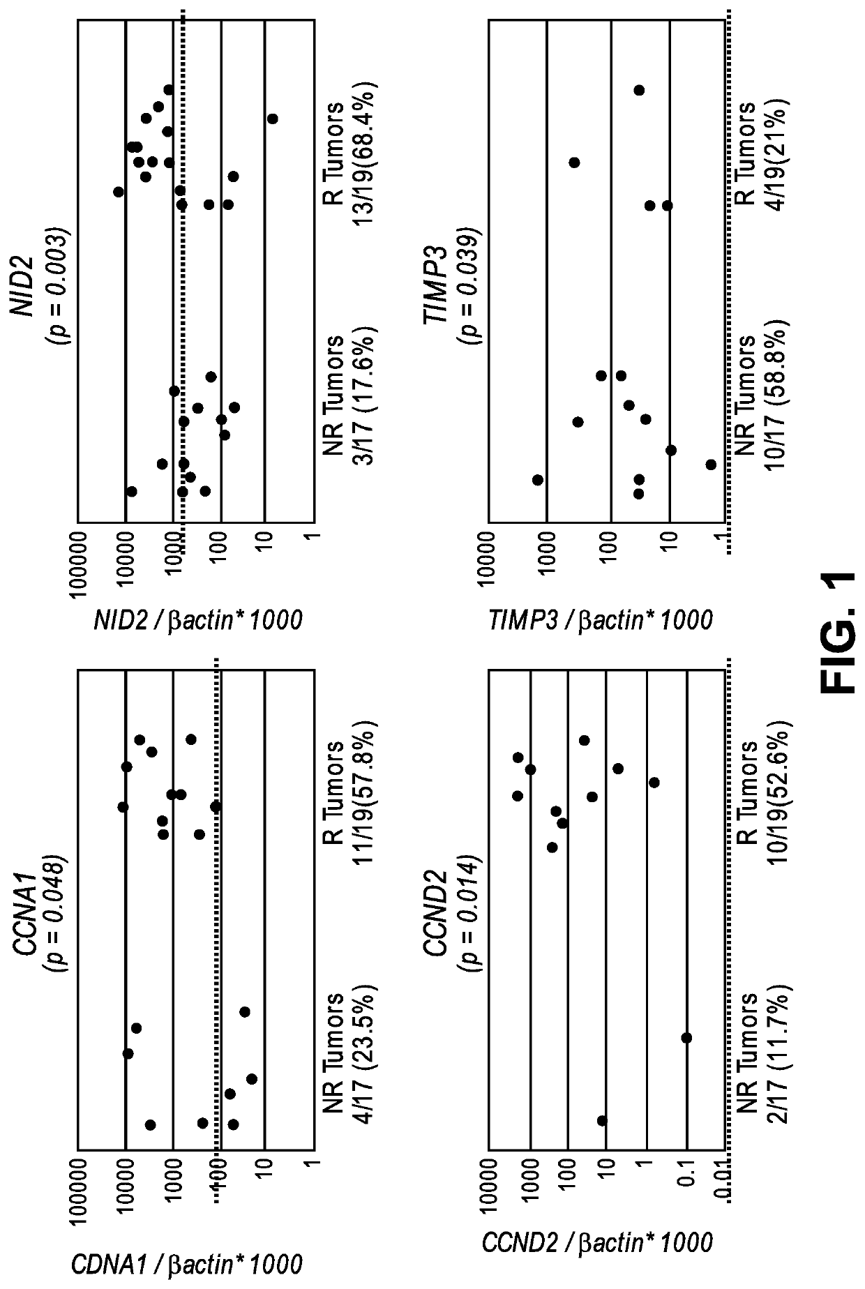
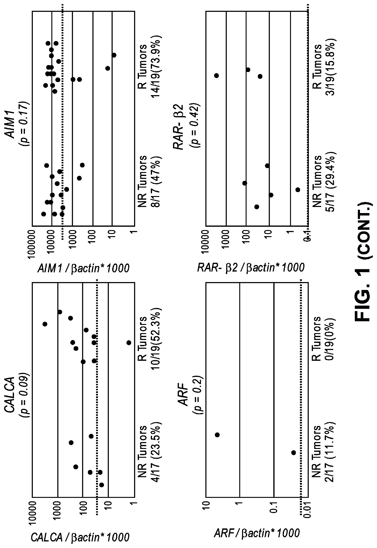
[0108]By a candidate gene approach, promoter methylation of 8 genes (ARF, TIMP3, RAR-β2, NID2, CCNA1, AIM1, CALCA and CCND2) were analyzed by quantitative methylation specific PCR (QMSP) in the DNA of 17 non-recurrent and 19 recurrent noninvasive low grade papillary urothelial cell carcinoma archival tissues. A total of 36 FFPE primary low-grade papillary urothelial cell carcinoma (LGPUCC) tissue samples were obtained from patients who underwent therapeutic surgery. Among them 17 samples were...


PUM

PropertyMeasurementUnit
Fractionaaaaaaaaaa
Fractionaaaaaaaaaa
Fractionaaaaaaaaaa

Abstract

The invention provides methods for detecting a cellular proliferative disorder (e.g., urothelial cancer) in a subject by assessing the methylation status of the CCND2, CCNA1 or CALCA promoter in a nucleic acid sample. The methods of the invention are useful for diagnostic, prognostic as well therapeutic regimen predictions.

Description

CROSS-REFERENCE TO RELATED APPLICATIONS[0001]This application claims benefit of priority under 35 U.S.C. § 119(e) of U.S. Ser. No. 62 / 175,897, filed Jun. 15, 2015, the entire contents of which is incorporated herein by reference in its entirety.FIELD OF THE INVENTION[0002]The invention relates generally to methods for diagnosing and treating cancer and more specifically to methods for detecting, diagnosing, providing prognosis for and treating urothelial cancers by detecting methylation changes in the regulatory region of specific nucleic acid sequences in a sample from a subject.BACKGROUND INFORMATION[0003]It has been shown that genetic and epigenetic changes contribute to the development and progression of tumor cells. Epigenetic alterations in promoter methylation and histone acetylation have been associated with cancer-specific expression differences in human malignancies. Methylation has been primarily considered as a mechanism of tumor suppressor gene (TSG) inactivation, and c...

Claims


Application Information

Patent Timeline
IPC IPC(8): C12Q1/6886A61K31/706A61K31/7068A61K31/164A61P35/00
CPCA61P35/00C12Q2600/106A61K31/164A61K31/7068C12Q1/6886C12Q2600/154A61K31/706
Inventor PARK, JONG CHULMALDONADO GONZALEZ, LEONEL FRANCISCOBRAIT RODRIGUES DE OLIVEIRA, MARIANAHOQUE, MOHAMMAD O.
Owner THE JOHN HOPKINS UNIV SCHOOL OF MEDICINE
Why Eureka
Try Eureka