
Capacitive micro-sensor for pathogen-specific antibody responses

a micro-sensor and antibody technology, applied in the field of capacitive micro-sensors for pathogen-specific antibody responses, can solve the problems of limiting the utility of elisas in low-resource settings, demonstrating interference from matrix components of unprocessed samples, and increasing complexity and cost, so as to achieve superior sensitivity and specificity, reduce cross-reactivity, and reduce the effect of cos

Pending Publication Date: 2020-07-30
COLORADO STATE UNIVERSITY
View PDF0 Cites 2 Cited by
  • Summary
  • Abstract
  • Description
  • Claims
  • Application Information

AI Technical Summary

Benefits of technology

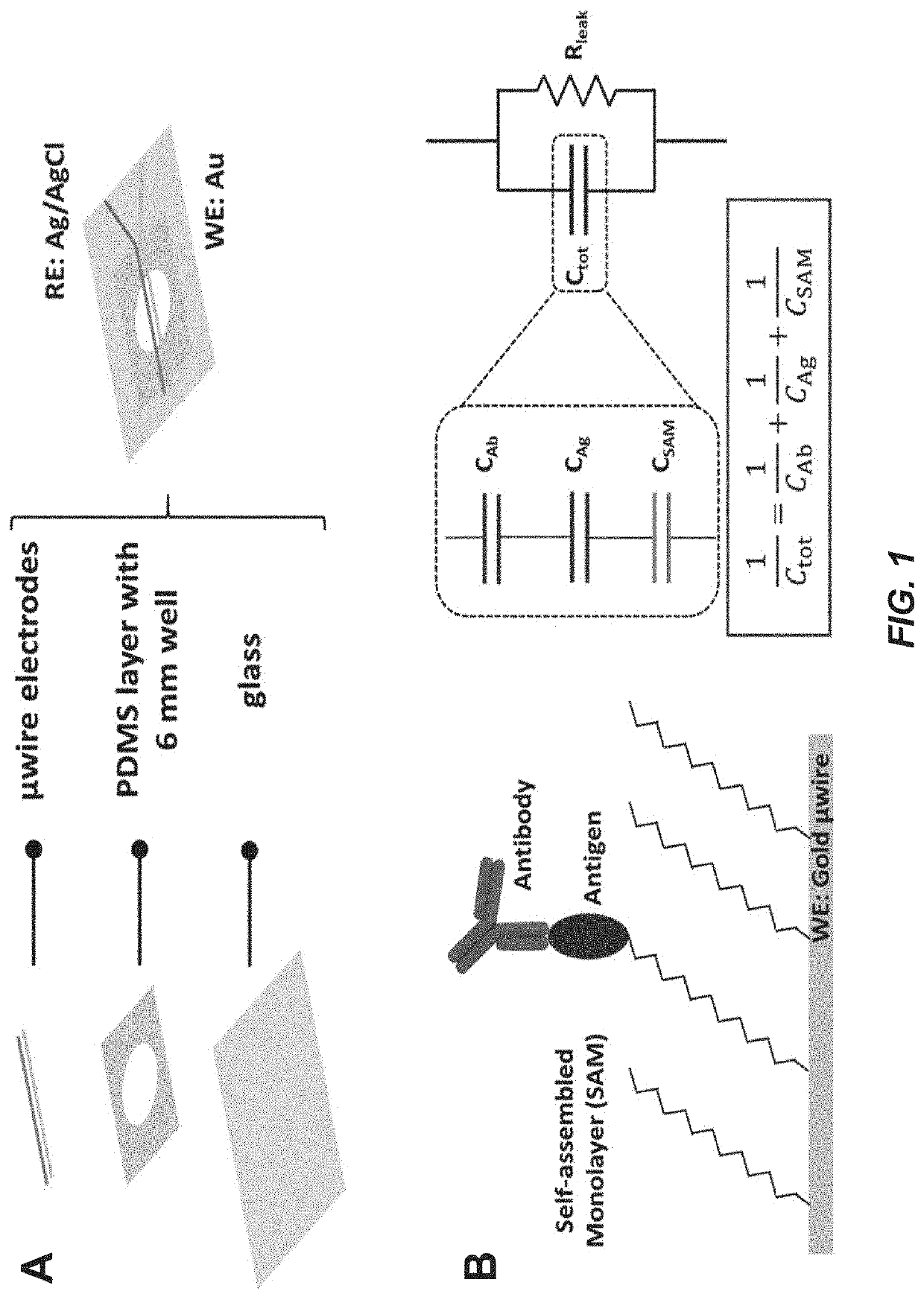
The patent describes a capacitive immunosensor that can detect specific antibodies in a sample. The sensor is made by modifying a sensor with the envelope protein of theZIKV or Chikungunya virus, and can detect very low levels of antibodies. The sensor is label-free and can distinguish between different types of antibodies. The method involves creating a self-assembled monolayer on a gold electrode and attaching pathogen-specific antigens to the monolayer. When an antibody binds to the antigen, it creates a detectable change in the electrical properties of the electrode. The patent also describes a method of detecting antibodies using a micro-sensor that is made by a similar process. Overall, the patent provides a simple and effective way to detect specific antibodies.

Problems solved by technology

However, ELISAs require large instrumentation in centralized laboratories and specialized training to execute and interpret the results which limits the utility of ELISAs in low-resource settings.
While lateral flow assays are promising candidates for POC applications, these assays often lack sensitivity and demonstrate interference from matrix components of unprocessed samples.
Other electrochemical antibody sensors have been developed for serological analysis, but these designs incorporate enzymatic labels or redox couples that increase complexity and cost.
Capacitive biosensors are thus an attractive sensing modality that has not yet been fully explored for specific antibody detection.
Resource intensive diagnostic tools limits their utility of for point of care service.

Method used


Image

  • Capacitive micro-sensor for pathogen-specific antibody responses
  • Capacitive micro-sensor for pathogen-specific antibody responses
  • Capacitive micro-sensor for pathogen-specific antibody responses

Examples


example 1

and Methods

[0108]Study Design.

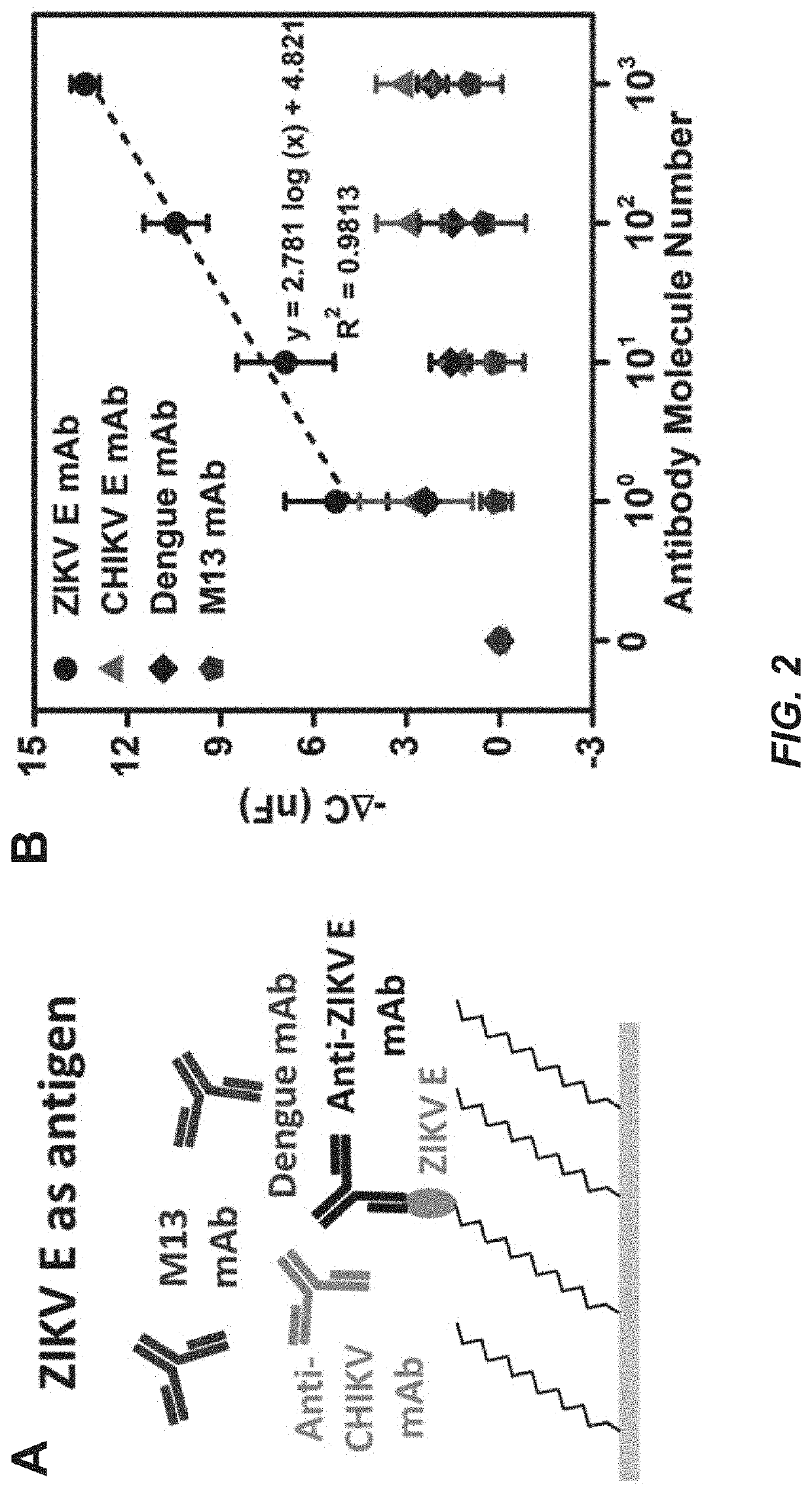
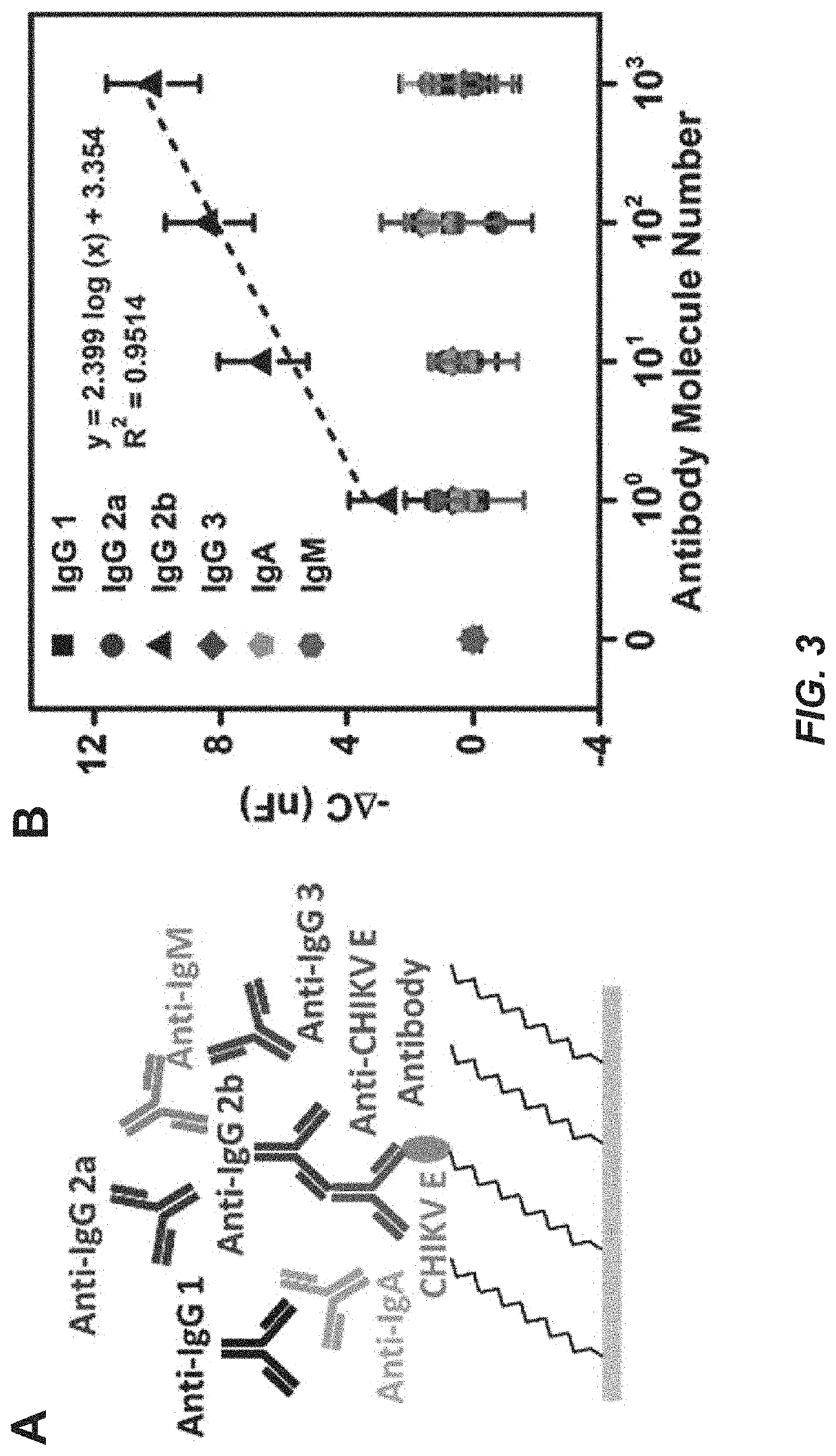
[0109]The working microwire surface was functionalized with E protein from either ZIKV (ZIKV E) or Chikungunya virus (CHIKV E). Lower dynamic range boundaries for the device were first determined with monoclonal antibody samples. Anti-ZIKV E antibody was employed as a specific target while anti-CHIKV E, anti-Dengue, and anti-M13 were used as nonspecific targets. The microwire biosensor was also used to isotype the monoclonal antibodies with anti-mouse IgG1, IgG2a, IgG2b, IgG3, IgA and IgM antibodies. The microwire sensor was then validated using pre-immune and immune mouse sera collected 4, 7, 14 and 21 days post-ZIKV immunization. Next, the sensor was used to isotype Day 4 and 21 mouse sera for IgM and IgG.

[0110]Information for immunization and sera characterization, electrode functionalization and sensor fabrication are described in the Examples. Representative serum samples positive for ZIKV IgG antibody by Western blot were included in the serum tes...

example 2

Functionalization and Sensor Fabrication

[0120]A 25 μm diameter Au microwire was used as the working electrode. To prepare the electrode surface, the Au microwire was immersed in a 20 mL solution of 50 mM KOH and 25% H2O2 for 10 min, and thoroughly rinsed in Milli-Q water to remove residual reagent. This widely used cleaning protocol removes debris that interferes with the stability of immobilized surface structures. The Au microwire was then plasma cleaned for 2 min in an 02 Plasma Etch PE-25 (Plasma Etch, Carson City, Nev., USA) at a pressure of 200 mTorr and with 150 W applied to the RF coil. A self-assembling monolayer (SAM) formation reaction was performed immediately after plasma cleaning which spontaneously forms an organized structure at the surface. Some SAM-forming molecules do not bind strongly to their substrate, like perylenetetracarboxylic dianhydride (PTCDA) on gold, and the resultant structures have poor stability. However, other molecules with stronger affinity such ...

example 3

on of Plasmids for Zika DNA Immunization

[0125]Genes for the Zika virus PRVABC59 strain (NCBI Accession: KX087101) capsid and prM-Env proteins were codon-optimized for mammalian expression and synthesized by Genescript Inc. The V5 epitope tagged capsid gene was cloned into the EcoRV site of pcDNA3.1 (plasmid pBG610), and a Japanese encephalitis virus prM signal sequence was added to the prM-Env gene and the construct was cloned into the EcoRV site of pcDNA3.1 (pBG611) as previous described (Virology 2006, 346, 53). Plasmid sequences will be provided upon request. Expression of capsid and prME proteins after transfection into Vero cells was verified by Western blot analysis using anti-V5 (Life Tech) and anti-Envelope (4G2 (ATCC HB-112 (D1-4G2-4-15))) antibodies, respectively.

[0126]DNA was prepared for immunization using the TempliPhi Rolling Circle Amplification Kit (GE HealthCare) according to the manufacturer's instructions. DNA was purified by phenol:chloroform extraction and ethan...


PUM

PropertyMeasurementUnit
mole percentaaaaaaaaaa
diametersaaaaaaaaaa
diametersaaaaaaaaaa

Abstract

A novel technique for label-free, rapid detection of ultra-low concentrations of virus specific antibodies is described. We have developed a simple, robust capacitive biosensor using microwires coated with Zika or Chikungunya virus envelope antigen. With little discernable nonspecific binding, the sensor can detect as few as 10 antibody molecules in a small volume (10 molecules / 30 μL) within minutes. It can also be used to rapidly, specifically, and accurately determine the isotype of antigen-specific antibodies. Finally, we demonstrate that anti-Zika virus antibody can be sensitively and specifically detected in dilute mouse serum and can be isotyped using the sensor. Overall, our findings indicate that our microwire sensor platform can be used as a reliable, sensitive, and inexpensive diagnostic tool to detect immune responses at the point of care.

Description

RELATED APPLICATIONS[0001]This application claims priority under 35 U.S.C. § 119(e) to U.S. Provisional Patent Application No. 62 / 796,647, filed Jan. 25, 2019, which is incorporated herein by reference.GOVERNMENT SUPPORT[0002]This invention was made with government support under Grant Nos. RO1 AI114675 and RO1 AI132668 awarded by the National Institutes of Health and 1332404 and 1450032 from the National Science Foundation. The government has certain rights in the invention.BACKGROUND OF THE INVENTION[0003]Analyzing the humoral antibody response in clinical samples is critical to diagnose infectious disease, understand pathogenesis and immune response kinetics, and develop vaccines, and the enzyme-linked immunosorbent assay (ELISA) is used as the gold standard clinical diagnostic tool for antibody detection. However, ELISAs require large instrumentation in centralized laboratories and specialized training to execute and interpret the results which limits the utility of ELISAs in low...

Claims


Application Information

Patent Timeline
Patent Type & Authority Applications(United States)
IPC IPC(8): G01N33/543G01N27/327G01N33/569
CPCG01N33/569G01N27/3275G01N33/5438G01N33/56983
Inventor WANG, LEIFILER, JESSICADANDY, DAVIDGEISS, BRIAN
Owner COLORADO STATE UNIVERSITY
Learn More