
Method and apparatus for ultrasound measurement and imaging of biological tissue elasticity in real time

a biological tissue and ultrasound technology, applied in the field of ultrasound measurement and imaging of biological tissue elasticity in real time, can solve the problems of low measurement accuracy, limited diagnostic value, and quantitative elastic properties, and achieve the effect of increasing measurement accuracy and reliability

Inactive Publication Date: 2020-08-13
AO NPF BIOSS
View PDF0 Cites 3 Cited by
  • Summary
  • Abstract
  • Description
  • Claims
  • Application Information

AI Technical Summary

Benefits of technology

The proposed invention aims to improve the accuracy and reliability of measuring biological tissue elasticity parameters in real-time ultrasound imaging. By adding additional elements and connections to the apparatus, the proposed method allows for more accurate and reliable measures, as well as higher resolution imaging.

Problems solved by technology

However, such evaluation of elastic properties is not quantitative and this limits its diagnostic value.
The disadvantage of this method is the low accuracy of measurements, since in real conditions it is impossible to create the homogeneous strain in biological objects, which have usually complex spatial geometry.
Therefore, at present this method is qualitative (see U.S. Pat. No. 9,220,479) and as a matter of fact, it does not allow quantitative measurements of the biological tissues elastic properties.
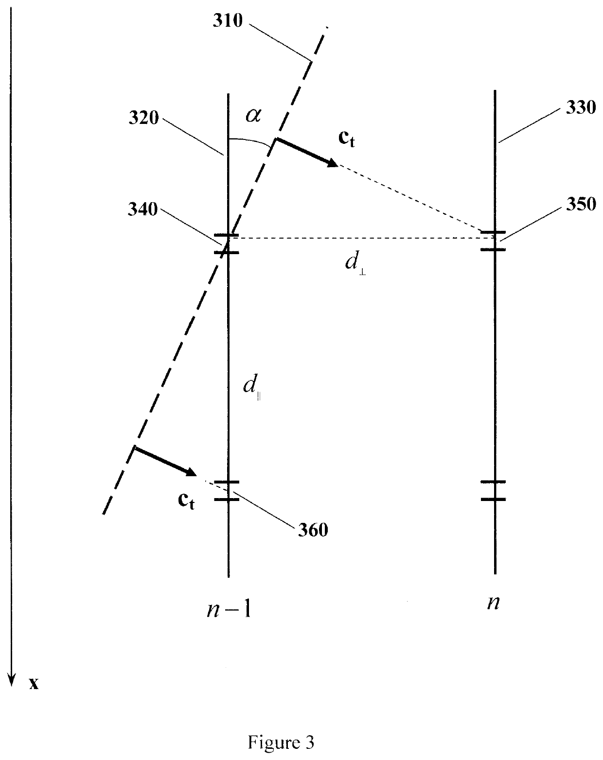
In practice, the same constant direction of propagation can not be achieved even for different parts of the same wave front, as discussed below.
The disadvantage of this method is the low accuracy of the determining the second derivatives and their spectral components that decreases the signal-to-noise ratio.
In addition, the spatial resolution of the method is substantially limited, since the determining the shear wave velocity requires the detecting the tissue displacement data from at least three probing lines and three sample volumes located at different depths.

Method used


Image

  • Method and apparatus for ultrasound measurement and imaging of biological tissue elasticity in real time
  • Method and apparatus for ultrasound measurement and imaging of biological tissue elasticity in real time
  • Method and apparatus for ultrasound measurement and imaging of biological tissue elasticity in real time

Examples


example

[0073]The claimed method and apparatus for ultrasound measurement of biological tissue elasticity in real time may be realized, for example, using the XILINX Spartan-6 XC6SLX45 field-programmable gate array (FPGA), static memory (SRAM) chips and a personal computer that can perform all required measurements and calculations in real time using appropriate program products.

[0074]A typical plurality of probing lines does not exceed 64, the maximum number of sample volumes on each probing line does not exceed 128 and therefore the total number of sample volumes on all probing lines does not exceed 8192.

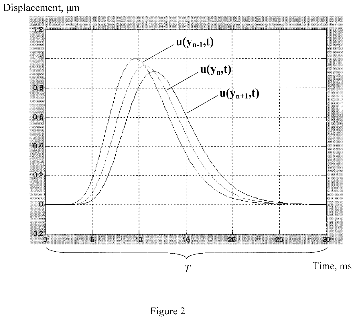
[0075]The maximum pulse repetition frequency of probing pulses does not exceed 10 kHz and a time of shear wave propagation in biological tissues does not exceed as rule 30 ms. Hence, the maximum number of displacement curve discrete values obtained for one sample volume does not exceed 300. Thus, the total number of discrete values for all sample volumes does not exceed 2457600.

[0076]The ...


PUM


Abstract

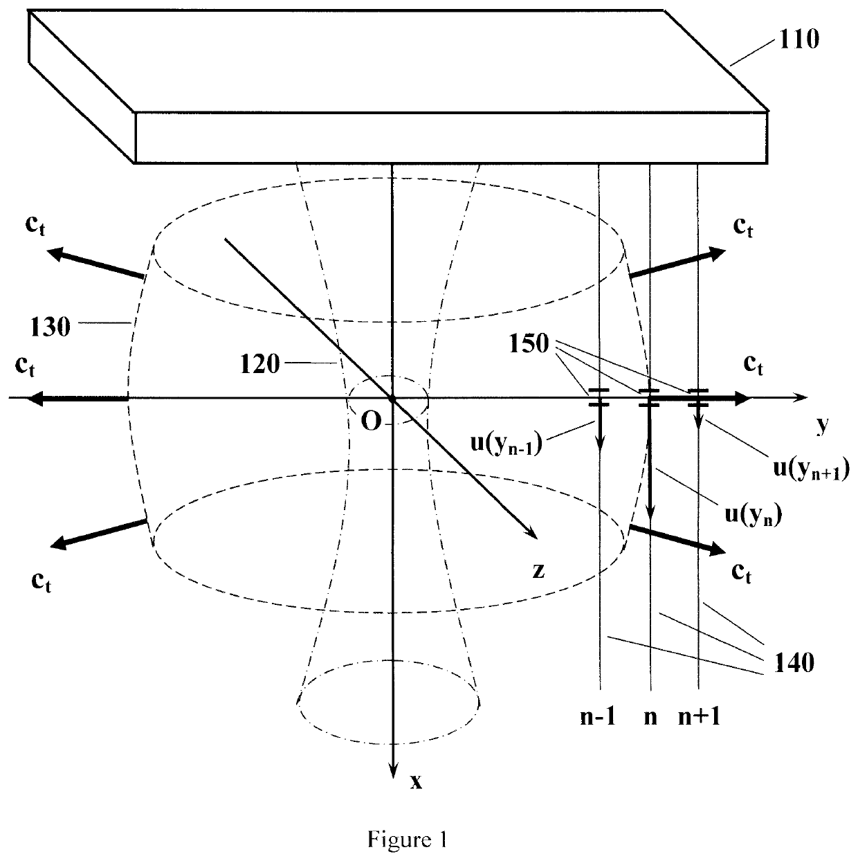
The invention relates to ultrasound methods and medical diagnostic apparatus using ultrasound probing for estimation of the biological tissue elastic properties by determining the propagation velocity of shear wave and imaging the elastic properties of tissue. The method is based on the determining the shear wave front propagation velocity along and perpendicular to the axis of excitation (Ox). In order to implement the method, a power ultrasound beam of waves is used for excitation of shear wave and a plurality of ultrasound pulses is sequentially transmitted to detect the tissue displacement by means of tissue response signals. The image of at least one parameter of tissue elasticity is acquired taking into account the noise level that occurred when determining the shear wave propagation velocity. The results are displayed in the form of images of the elasticity parameters and the noise level and indication of quantitative values of elasticity parameters and noise level. The technical result is to increase the accuracy and reliability of the measurements and increase the spatial resolution when imaging the biological tissue parameters in real time.

Description

FIELD OF THE INVENTION[0001]The invention relates to ultrasound methods and apparatus of medical diagnostic using ultrasound probing for estimation of the biological tissue elastic properties by determining the shear wave velocity and imaging the tissue elastic properties.BACKGROUND[0002]Pathological conditions can lead to a change of elasticity of soft biological tissue in comparison with the physiological conditions. As a result, physicians use palpation for estimation of the elasticity and identification of the pathological conditions of tissue. For example, in the case of such diffuse diseases of the liver as fibrosis, cirrhosis and others, the stiffness of the tissue increases and can be evaluated by palpation. However, such evaluation of elastic properties is not quantitative and this limits its diagnostic value.[0003]The method and apparatus for acquiring an image of the shear modulus or Young's modulus of biological tissue combining pulsed ultrasound probing with the simulta...

Claims


Application Information

Patent Timeline
IPC IPC(8): A61B8/08
CPCA61B8/485A61B8/5269A61B8/5223A61B8/085A61B8/08
Inventor BARANNYK, YEVGEN OLEKSANDROVYCHBOICHENKO, YURII PETROVYCHKNIAZEV, OLEKSII VALERIIOVYCHLINSKA, HANNA VOLODYMYRIVNAMARUSENKO, ANATOLII LLARIONOVYCHPUPCHENKO, VIKTOR IVANOVYCH
Owner AO NPF BIOSS
Learn More