Method and Apparatus for Treating Cranial Cruciate Ligament Disease in Canines

a cranial cruciate ligament and canine technology, applied in the field of surgery for the treatment of cranial cruciate ligament disease in canines, can solve the problems of limiting the extension further protecting of the canine knee, unable to combine hyperextension with high impact, and unable to achieve the common knee injury mode in humans, etc., to achieve balanced permanent stability

Active Publication Date: 2021-02-25
FITCH RANDALL +1
View PDF0 Cites 2 Cited by
  • Summary
  • Abstract
  • Description
  • Claims
  • Application Information

AI Technical Summary

Benefits of technology

[0019]The current invention provides balanced permanent stability for canines with cruciate ligament injuries combining the best corrective principles of mode

Problems solved by technology

In addition, the dog's knee geometry limits extension further protecting it (15 degrees of flexion is full extension with normal standing angles of 35 degrees of flexion).
Therefore coupling hyperextension with high impact, a common mode of knee injury in humans, is nearly impossible in the dog.
The cranial cruciate ligament has poor vascularity within this intra-articular environment with minimal healing capacity.
Repetitive strain therefore produces accumulating non-healing ligament fiber injuries increasing laxity resulting in even greater ligament strain cascading to complete ligament failure over time.
Traditionally, surgical repair techniques focused on functionally replacing the ruptured cranial cruciate ligament with limited success in dogs.
This is likely due in part to the unaccounted magnified continuous stress placed on the ligament repair during weight-bearing not present in humans.
However, all of these surgical techniques address only one component of t

Method used


Image

  • Method and Apparatus for Treating Cranial Cruciate Ligament Disease in Canines
  • Method and Apparatus for Treating Cranial Cruciate Ligament Disease in Canines
  • Method and Apparatus for Treating Cranial Cruciate Ligament Disease in Canines

Examples


Embodiment Construction

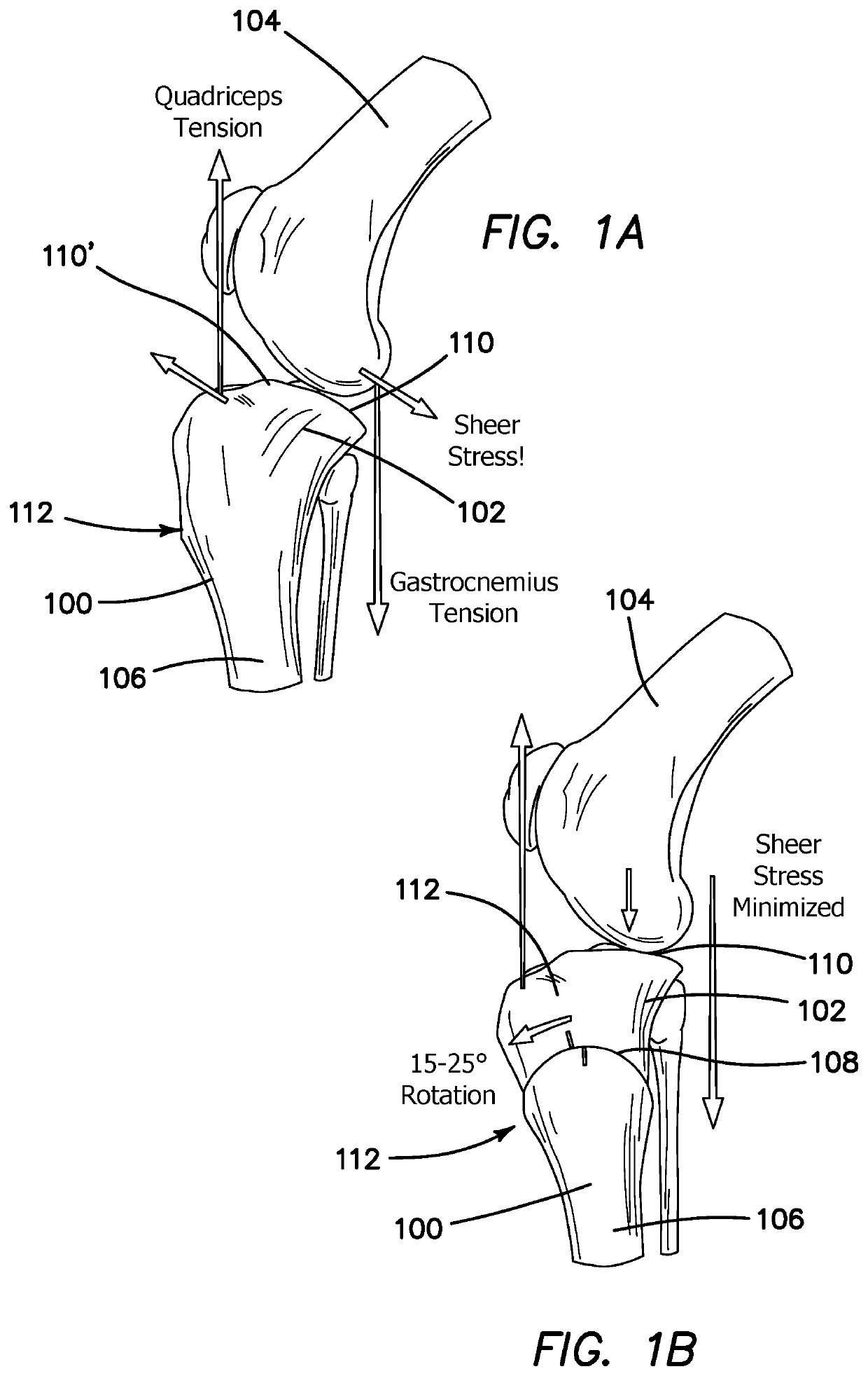
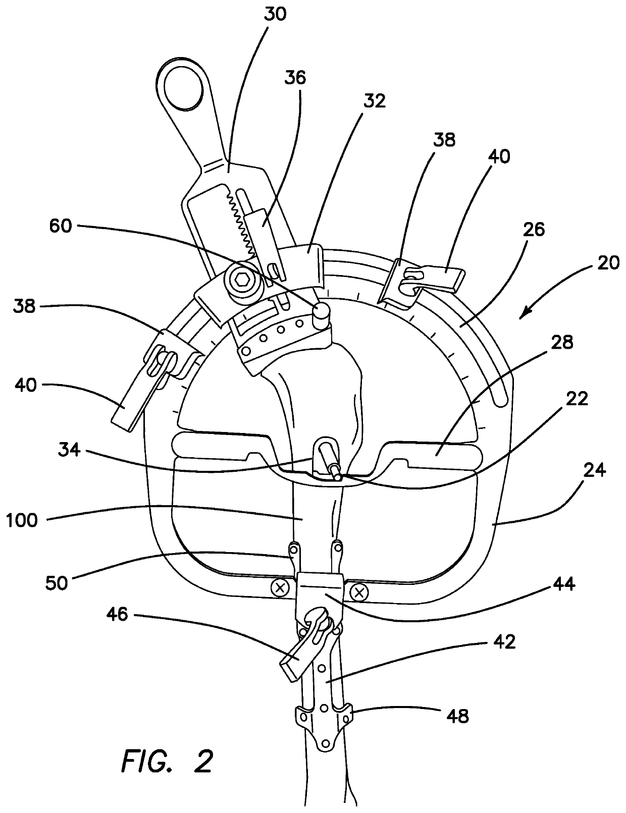
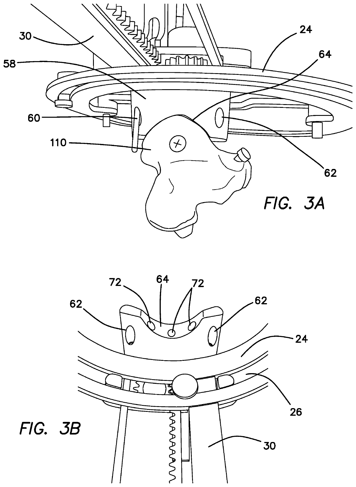
[0062]The current invention is a surgical guidance system (SGS) or kit 1 and a method for performing a cruciate pivot osteotomy, both of which are for surgically treating canine cranial cruciate ligament disease. FIGS. 1 and 1B are illustrations of a canine stifle comprising a tibia 100 and a femur 104 before and after performing a cruciate pivot osteotomy using the current SGS 1, respectively. The present invention provides a fast, accurate, and reliable means for performing an osteotomy 108 on the stifle of a canine, thereby allowing for a leveling of a proximal portion 102 of the tibia 100 relative to a distal portion 106 of the tibia 100. The current system and method further compresses the osteotomy 108 while providing an accurate template for a plate 80 which is then placed and affixed to the tibia 100 through a plurality of bone screws.

[0063]The current SGS 1 comprises a bi-planar geometry that will provide torsional stability when treating cranial cruciate ligament disease w...


PUM


Abstract

A surgical guidance system (SGS) for performing a cruciate pivot osteotomy in canines to treat cranial cruciate ligament disease. The SGS comprises a guide, a jig, and a plate. The guide is first placed over the tibia until it interacts with specific anatomical features of the tibia, thereby marking the proper position for the jig to be placed. After the jig has been secured, a blade defines an osteotomy within a proximal portion of the tibia. A portion of the jig is then cranially rotated providing a rotational correction of the proximal tibia. A compressive force is then applied to the osteotomy by the jig. Next the multiplane locking plate is placed over the osteotomy as dictated by the features of the jig. After initially securing the plate into its correct position, the jig is removed and the plate is then secured to the cranial surface of the tibia.

Description

BACKGROUNDField of the Technology[0001]The invention relates to the field of surgical techniques for the treatment of cranial cruciate ligament disease in canines, specifically to surgical methods based on CORA-based leveling osteotomy.Description of the Prior Art[0002]Cranial cruciate ligament injury is the most prevalent orthopedic injury in the dog requiring surgical repair with estimates of $1.32 billion spent in the United States on the treatment of this condition in 2003 (Wilke VL, JAVMA 2005). In that same year, the average spent per patient was $1840 with a range of 0 to $5000. Treatment costs have continued to escalate to the current $3500-6500. Implant costs likely do not substantially influence medical costs as the only make-up ˜3-5% of the total medical bill.[0003]Biomechanically, the canine stifle or knee has many advantages over the human knee that one would expect to protect the stifle from injury. The dog's stifle is spared from the type of destructive hyperextension...

Claims


Application Information

Patent Timeline
IPC IPC(8): A61B17/80A61F2/08A61B17/15A61B17/17A61D1/00
CPCA61B17/8095A61F2/0811A61B17/157A61F2/0805A61F2002/30703A61B17/151A61D1/00A61F2002/0882A61B17/1767A61B17/8061A61B17/1637A61B17/848A61B17/1764A61F2250/0081A61F2002/307
Inventor FITCH, RANDALLVON ZABERN, ROBERT
Owner FITCH RANDALL
Why Eureka
Try Eureka