Ultrasound Cardiac Processing

Active Publication Date: 2021-03-25
NORWEGIAN UNIVERSITY OF SCIENCE AND TECHNOLOGY (NTNU)
View PDF1 Cites 0 Cited by
  • Summary
  • Abstract
  • Description
  • Claims
  • Application Information

AI Technical Summary

Benefits of technology

[0038]In some embodiments, a spatiotemporal filter may be applied to the time series of three-dimensional Doppler data sets. This advantageously smooths the series of images, increasing the signal-to-noise ratio of the images.
[0039]In some embodiments, envelope detection may be carried out on the three-dimensional Doppler images. This may produce a 3D brightness-mode (B-mode) image with improved image quality.
[0040]In some embodiments, the method further comprises selecting the time series of frames in which a mechanical wave propagates through the region from a longer set of three-dimensional data frames, e.g. as a time window of interest. Data frames outside the period of interest may be discarded. This advantageously allows the particular time window of interest to be selected, and any data from outside of this time window to be removed, thus significantly reducing the quantity of data which is to be processed and thereby making the imaging method both faster and more efficient. In some embodiments, a period of interest, in which a mechanical wave propagates through the region, may be determined using electrocardiography (ECG). The ultrasound imaging apparatus may comprise an electrocardiogram device, which may be configured to be used to determine a period of interest in which a mechanical wave propagates through a region of the heart. This may allow the period of interest to be detected automatically, and the irrelevant d

Problems solved by technology

However, the information which is obtained using currently known

Method used


Image

  • Ultrasound Cardiac Processing
  • Ultrasound Cardiac Processing
  • Ultrasound Cardiac Processing

Examples


Example

[0060]Throughout the figures, like reference numerals have been used for like elements.

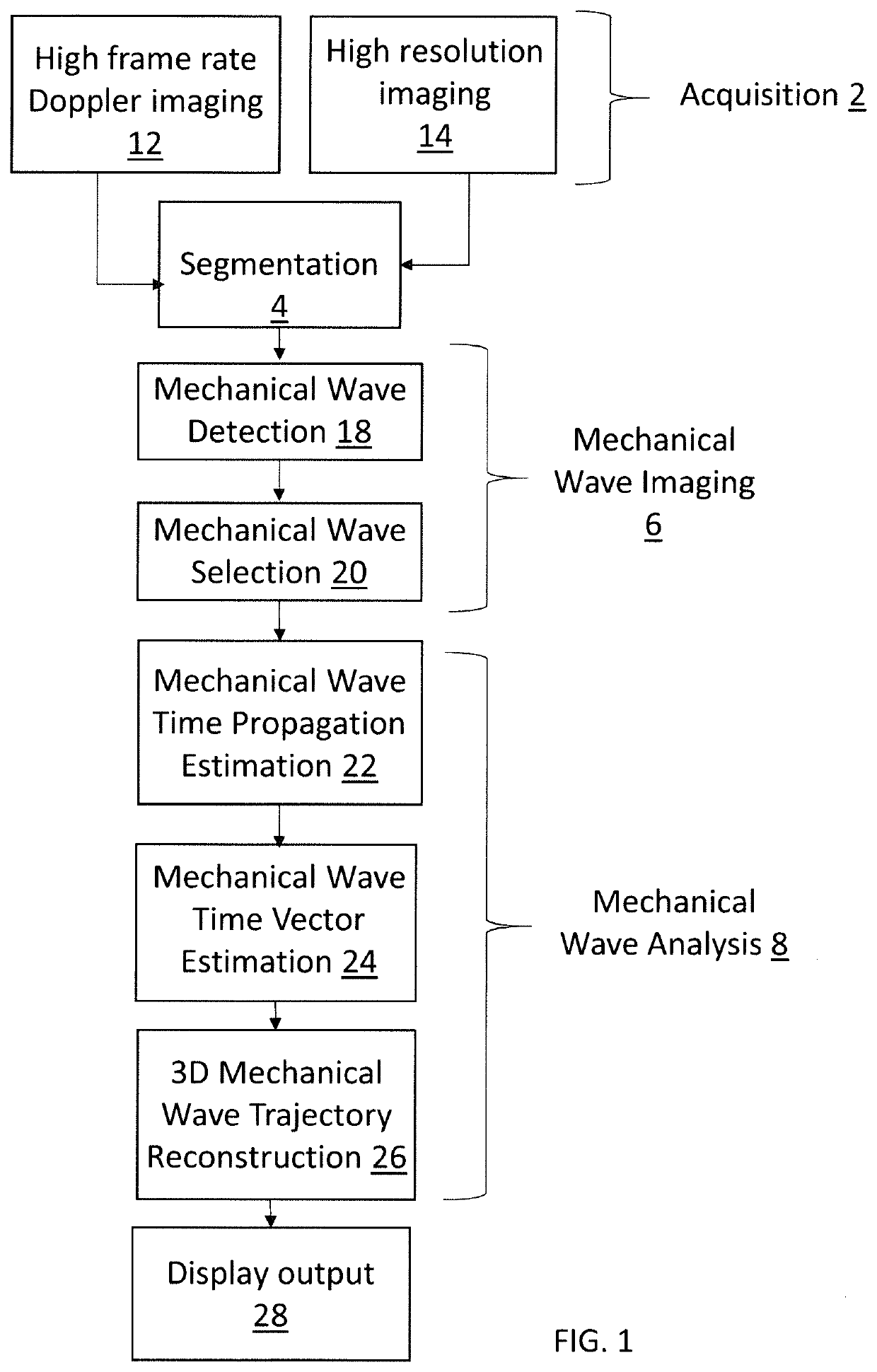
[0061]FIG. 1 is a flowchart showing an overview of the stages of a method of cardiac imaging embodying the present invention, in which the motion of natural mechanical waves in the heart are used to help visualise the heart tissue. Each of these stages will be described in detail with reference to the later Figures.

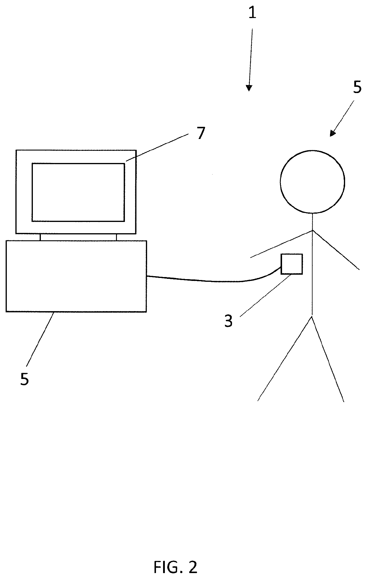
[0062]The ultrasound imaging process is carried out by an ultrasound imaging system 1, as shown in FIG. 2. The imaging system 1 includes a handheld ultrasound probe 3, a processing unit 5, and a display 7. The ultrasound probe 3 contains an array of ultrasound transducers for transmitting signals (e.g., a series of pulses) and for receiving reflections of the signals, under the control of the processing unit 5. The array of ultrasound transducers in the ultrasound probe 3 is a two-dimensional transducer array, which can capture three-dimensional data. This probe is used to generate imag...


PUM


Abstract

A method of processing cardiac ultrasound data for determining information about a mechanical wave in the heart. The method comprises receiving data representative of a time series of three-dimensional data frames, generated from ultrasound signals from a human or animal heart, each frame comprising a set of voxels, each voxel value representing an acceleration component of a respective location in the heart at a common time. The method also comprises identifying, for each voxel, a frame of the series in which the voxel value is at a maximum. A three-dimensional time-propagation data set is generated by assigning each voxel a value representative of the time of the respective frame in the time series for which the corresponding voxel is at a maximum. The method then comprises generating data representative of a three-dimensional velocity vector field by calculating time derivatives from the three-dimensional time-propagation data set.

Description

BACKGROUND OF THE INVENTION[0001]This invention relates to cardiac ultrasonography, e.g. for determining information about a mechanical wave in the heart.[0002]It has been known for many years to use ultrasound imaging techniques to image human and animal hearts, and to analyse the acquired image data in order to extract clinically useful information.[0003]Mechanical waves are oscillations of matter, which transfer energy through a medium. Mechanical waves are generated in the human body by the motion of the heart and blood passing through it—e.g. a shear wave is generated due to the closing of the aortic valve. These mechanical waves can be detected both in the heart and in the arteries closely surrounding the heart. Exactly how they travel through the heart can depend the health of the heart. Thus, it can be desirable to analyse the motion of such waves, e.g. in order to gain insights into patient health.[0004]It is known in the art to use ultrasound imaging to detect mechanical w...

Claims


Application Information

Patent Timeline
IPC IPC(8): A61B8/08G06T15/08A61B5/0402A61B8/00
CPCA61B8/485G06T15/08A61B8/0883G06T2210/41A61B8/488A61B5/0402A61B8/4488A61B8/483A61B8/466A61B8/5223A61B8/5269A61B8/543A61B8/5284A61B6/466A61B5/33G06T19/00A61B5/02108A61B8/00A61B8/02G06T7/0012G06T7/20G06T2200/04G06T2207/10136G06T2207/30048
Inventor SALLES, SEBASTIENLOVSTAKKEN, LASSETORP, HANS
Owner NORWEGIAN UNIVERSITY OF SCIENCE AND TECHNOLOGY (NTNU)
Why Eureka
Try Eureka