
Apparatuses and methods for digital pathology

a digital pathology and apparatus technology, applied in the field of apparatuses and methods for digital pathology, can solve problems such as inferior images that require significant user manipulation and processing

Pending Publication Date: 2021-07-08
PROTEIN METRICS LLC
View PDF16 Cites 0 Cited by
  • Summary
  • Abstract
  • Description
  • Claims
  • Application Information

AI Technical Summary

Benefits of technology

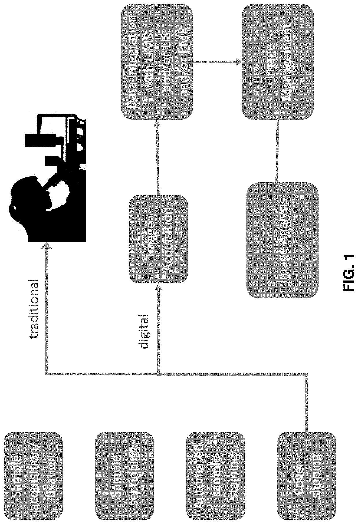
The present invention provides methods and software for improving the production and analysis of images in digital pathology. The invention addresses issues in current workflows by simplifying the process of analyzing large sets of images, reducing the need for manual intervention, and optimizing image data sets for easier handling and storage. The methods use a final mask to accurately and quickly identify cells and statistical information on positive cells. The invention also includes a ranking process that reduces the number of slide images needed for analysis. Overall, the invention provides improved efficiency and accuracy in digital pathology analysis.

Problems solved by technology

The difficulties include but are not limited to: laborious user involvement in the data acquisition software and image (data) analysis software; the absence of coordination between data acquisition and data analysis, leading to inferior images that require significant user manipulation and processing for each and every sample; the software and file incompatibility between different vendors of WSI instrumentation, between different implementations of image processing / analysis tools, and between combinations of the two; the need for a number of highly trained personnel in between the histology lab (where tissue samples are processed and stained), and the researcher's or pathologist's computer where digital images are analyzed (and in some cases paired with medical information, and decisions are made); and the data storage and handling requirements for large sets of image data.

Method used


Image

  • Apparatuses and methods for digital pathology
  • Apparatuses and methods for digital pathology
  • Apparatuses and methods for digital pathology

Examples


examples

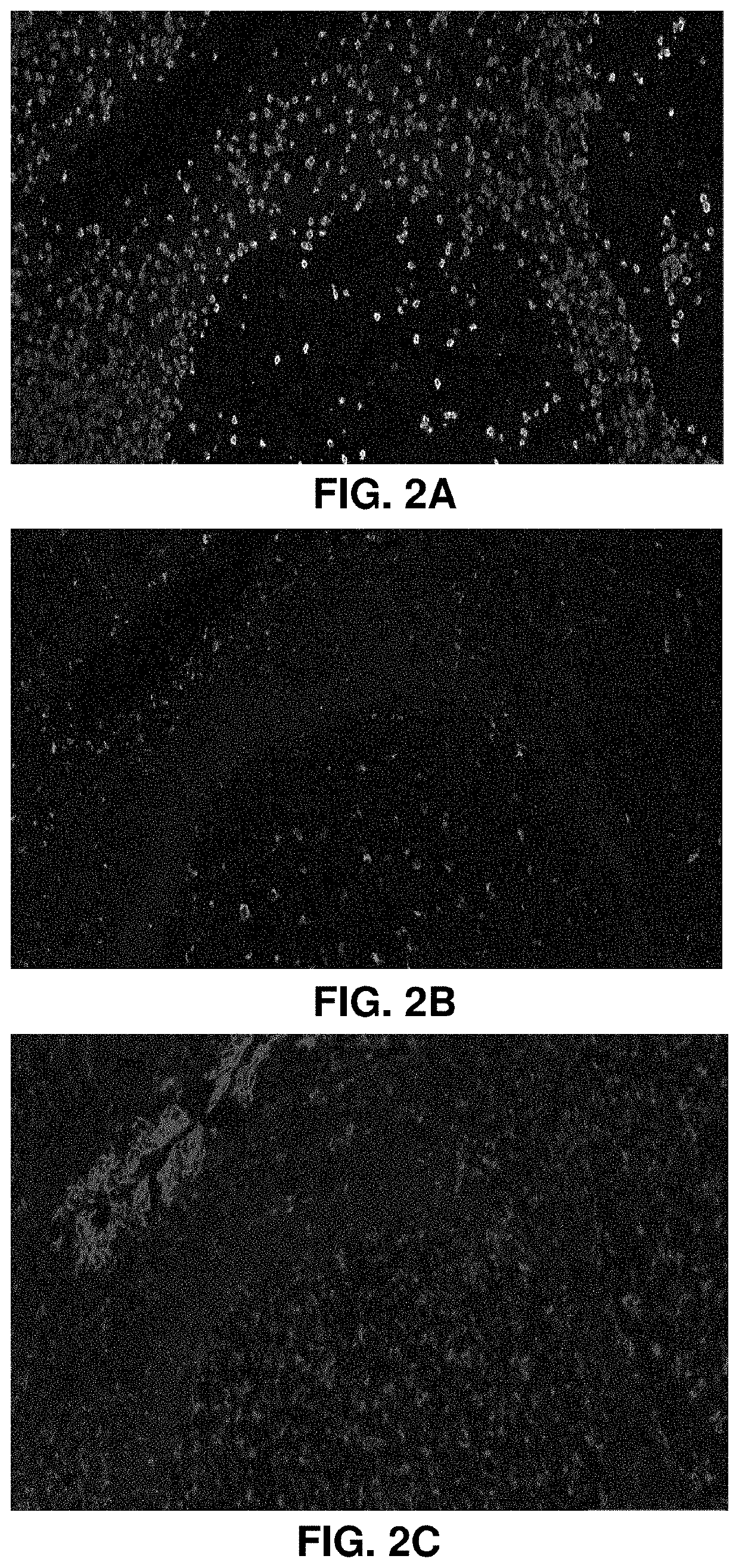
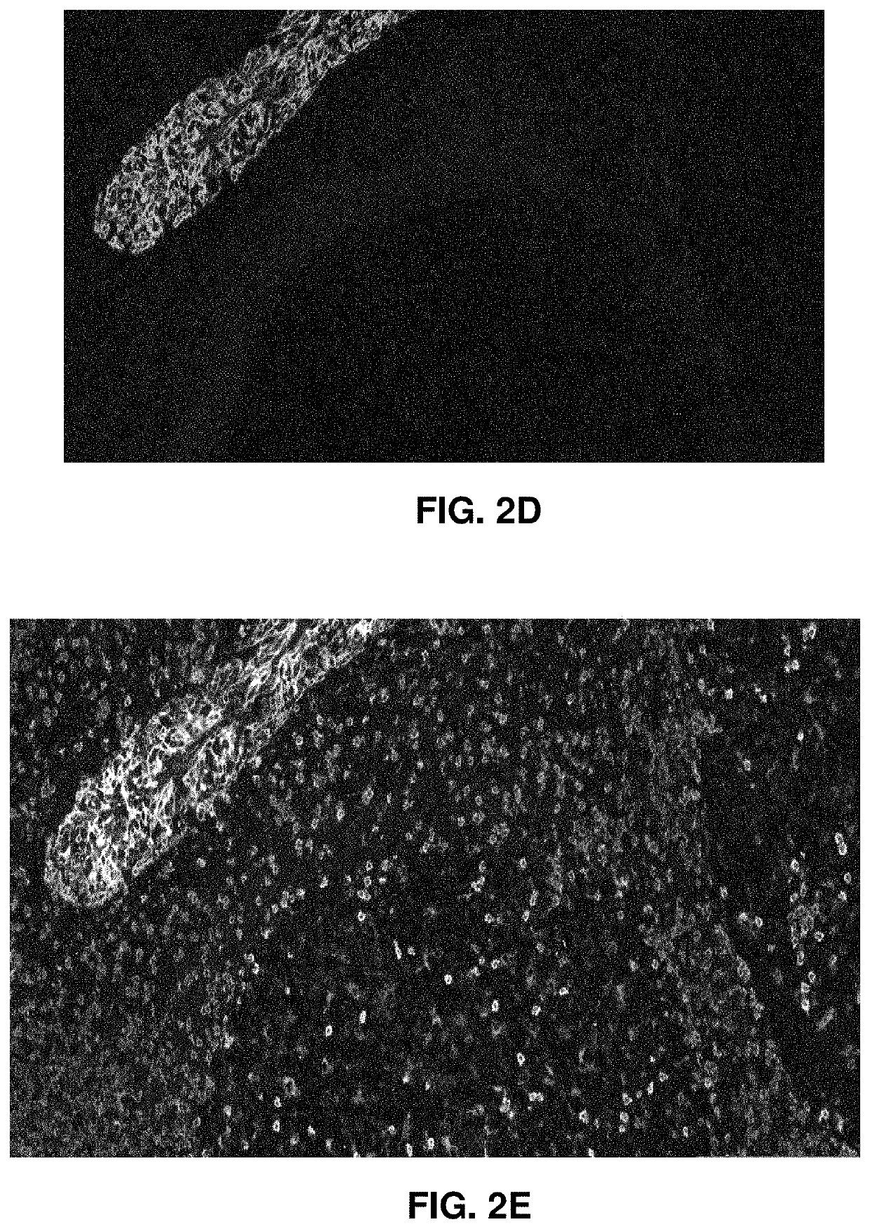
[0109]A series of 10 formalin-fixed, paraffin embedded (FFPE) tissue sections (approximately 4-5 μm thick) and two control slides were processed as described herein and compared with traditionally processing methods. The results show an improvement in the processing time as well as quality of the final data. The sections were obtained from 5 melanoma tumor blocks, with 2 serial sections per tumor. The control slides comprised tonsil and melanoma. Typical average sizes for the sections were 2.25 cm2±0.75 cm2. Each section had a complex shape (i.e. non-rectangular). An example is shown in FIG. 6. In FIG. 6 just one stained slide of a set of stained slides of the tissue sample is shown. In practice the set may include a plurality of (e.g., 5 or more, 10 or more, 15 or more, 20 or more 30 or more, 50 or more, etc.) stained slides.

[0110]The samples were stained using a commercially available Ultivue Ultimapper PD-L1 kit, using established protocols on a Leica Bond RX auto-stainer instrum...


PUM

PropertyMeasurementUnit
thicknessaaaaaaaaaa
thicknessaaaaaaaaaa
thickaaaaaaaaaa

Abstract

Digital pathology is a promising alternative to manual slide analysis, but improvements in imaging and analysis methods are needed to provide an analysis data set of images that can easily be handled, stored, and importantly, delivered from one data location to another. Methods and software are described herein to improve image collection and analysis.

Description

CROSS REFERENCE TO RELATED APPLICATIONS[0001]This patent application claims priority to U.S. Provisional Patent Application No. 62 / 957,095, filed on Jan. 3, 2020, titled “APPARATUSES AND METHODS FOR DIGITAL PATHOLOGY,” herein incorporated by reference in its entirety.INCORPORATION BY REFERENCE[0002]All publications and patent applications mentioned in this specification are herein incorporated by reference in their entirety to the same extent as if each individual publication or patent application was specifically and individually indicated to be incorporated by reference.FIELD[0003]Biopsy analysis by digital pathology for research, diagnosis and treatment monitoring requires development of instrumentation and methods for histologists, researchers, scientists, clinicians, and / or pathologists to obtain and identify multiple factors present in a single or a plurality of tissue sections on slides.BACKGROUND[0004]Increases in the number and complexity of tissue biopsy analyses during bi...

Claims


Application Information

Patent Timeline
Patent Type & Authority Applications(United States)
IPC IPC(8): G06T7/00G06T7/11G16H10/40G16H30/40G16H70/60G06N20/00G01N33/483
CPCG06T7/0012G06T7/11G16H10/40G16H30/40G06T2207/10056G06N20/00G01N33/4833G06T2207/30024G06T2207/30204G16H70/60G16H50/20G06N3/045
Inventor NATAN, MICHAEL J.BERN, MARSHALLCARLSON
Owner PROTEIN METRICS LLC
Learn More