
LED induced fluorescence detection system of epithelial tissue

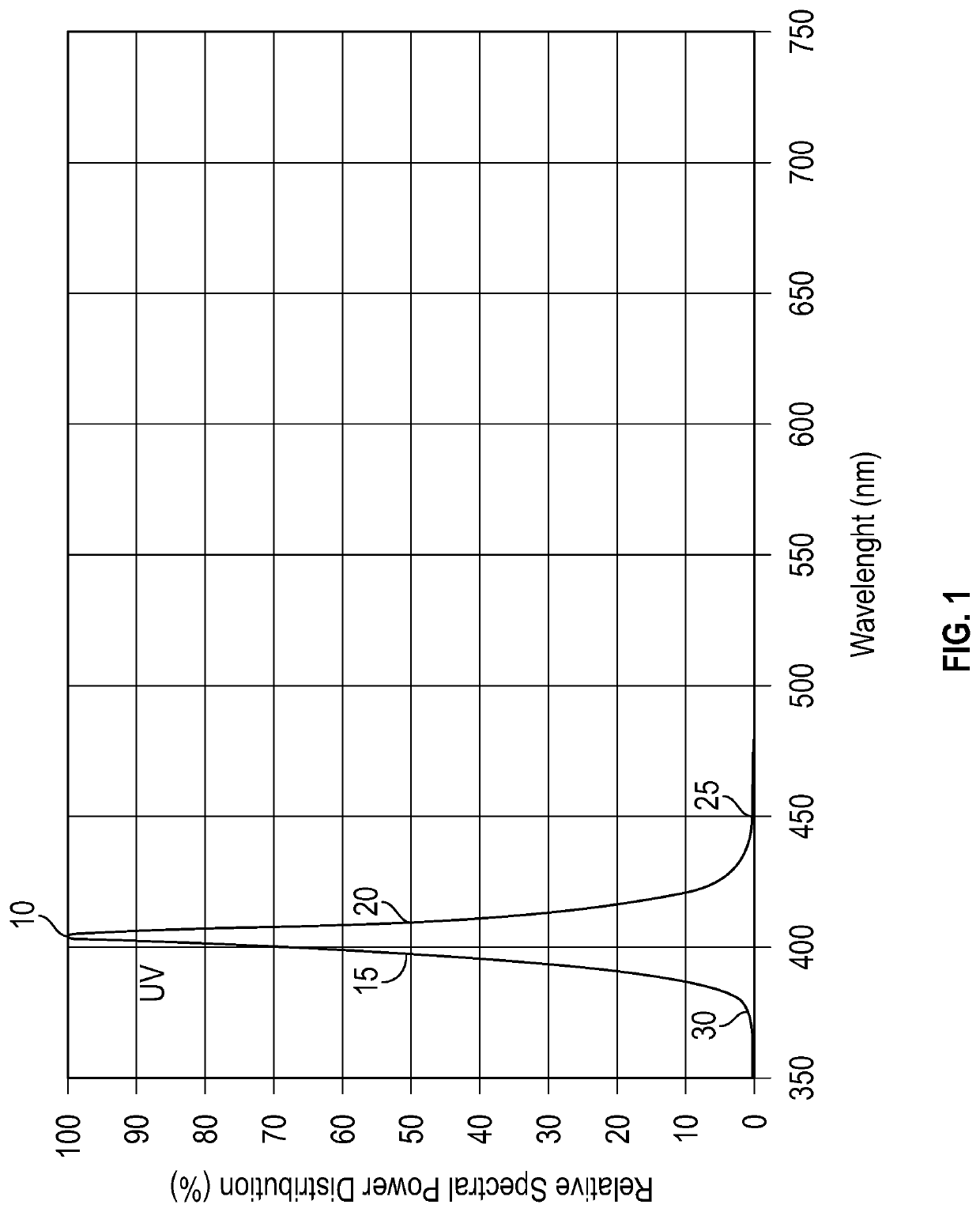
a fluorescence detection and epithelial tissue technology, applied in the field of epithelial tissue led induced fluorescence detection system, can solve the problems of insufficient sensitivity and specificity, detection techniques, tumors which may be benign or cancerous, etc., and achieve the effect of eliminating emissions and superior low-pass viewing filters

Inactive Publication Date: 2021-07-29
FORTIS BIOPHARMA LLC
View PDF0 Cites 1 Cited by
  • Summary
  • Abstract
  • Description
  • Claims
  • Application Information

AI Technical Summary

Benefits of technology

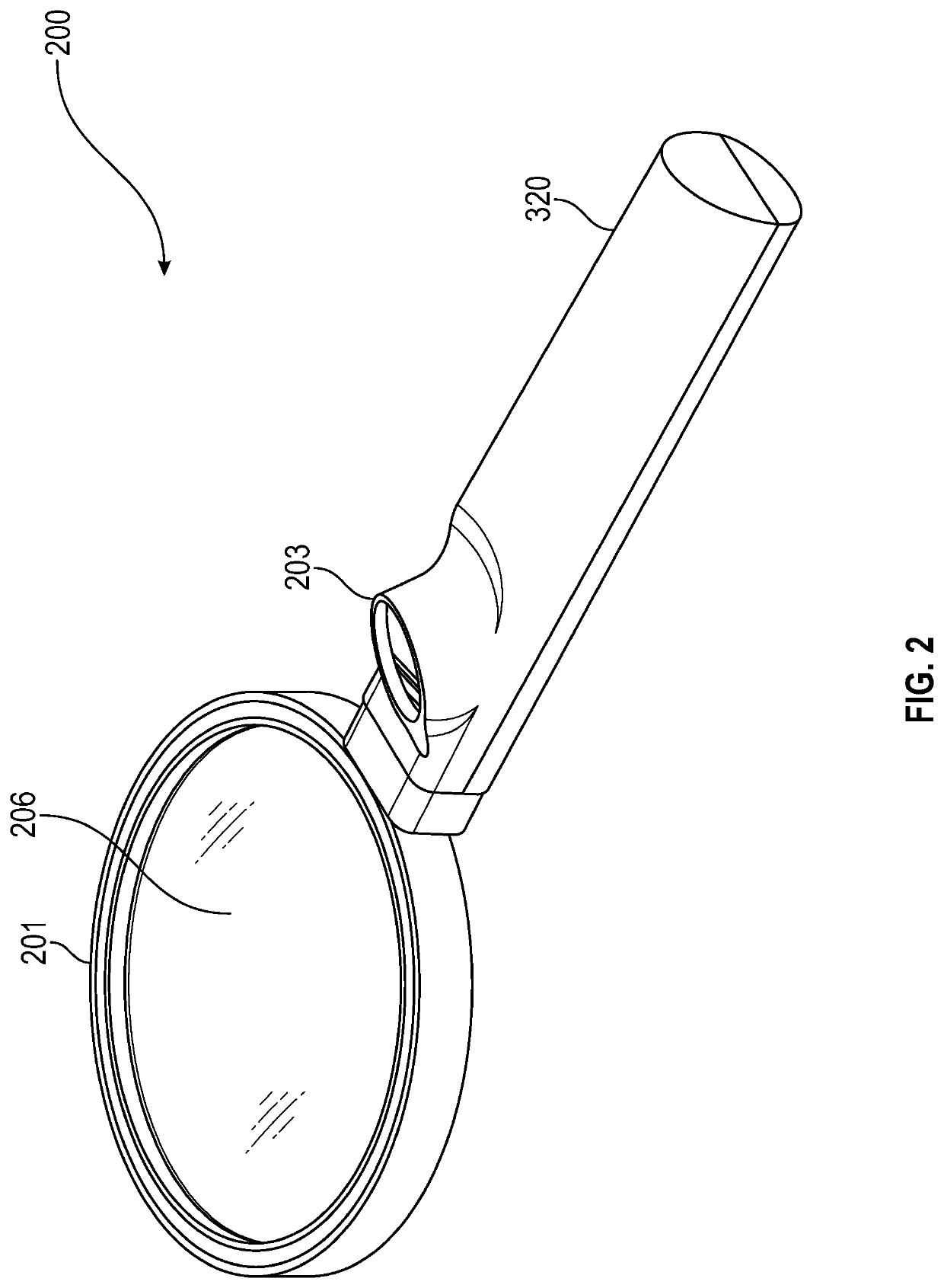
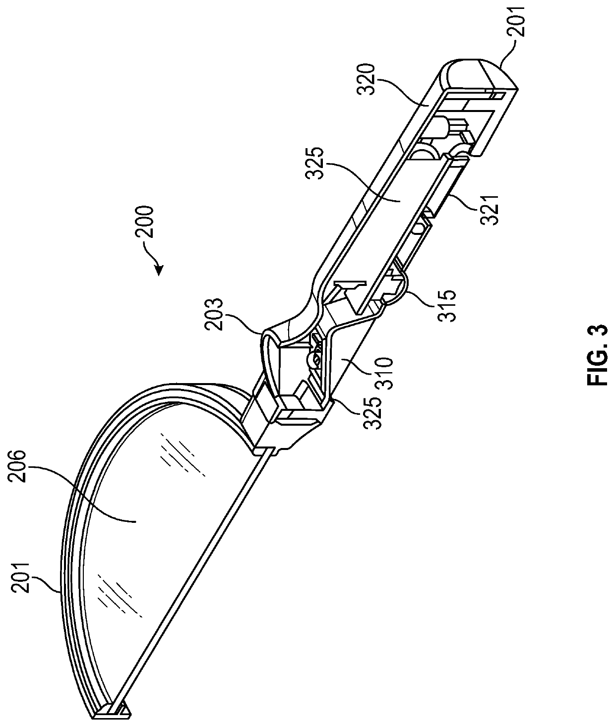
The patent is about a non-invasive tool for diagnosing pre-invasive epithelial neoplasia, such as oral cancer, skin cancer, cervical cancer, and colon cancer. The tool uses a simplified and low cost device that can examine skin tissue. The device uses the light-tissue interactions to visualize neoplastic lesions, specifically by exploiting tissue autofluorescence, which is the natural fluorescence of tissue. The device eliminates the need for a high pass filter at the light emission source and uses a better-matched LED emitter and superior low pass viewing filter to only illuminate the tissue surface with light having wavelengths less than approximately 460 nm. The device also includes a beam-shaping collimating optic and a charging port. The technical effects of this patent tool include increased patient survival, improved quality of life, and a better diagnosis for pre-invasive cancer.

Problems solved by technology

It can result in tumors which may be benign or cancerous.
Many of the currently available screening and detection techniques for epithelial pre-cancers do not provide adequate sensitivity and specificity; furthermore, many screening and detection methods require extensive training to yield adequate clinical results.
Consequently, practitioners often resort to the invasive and painful option of biopsies to confirm the presence of precancer or even early cancer.
Thus, despite the easy accessibility of the oral cavity for examination, current methods do not adequately screen and detect precancers in a non-invasive manner.

Method used


Image

  • LED induced fluorescence detection system of epithelial tissue
  • LED induced fluorescence detection system of epithelial tissue
  • LED induced fluorescence detection system of epithelial tissue

Examples


Embodiment Construction

[0025]Noninvasive and accurate techniques that facilitate the early detection of neoplastic changes improve cancer survival rates and lower treatment costs by reducing or eliminating other diagnostic procedures and allowing prompt commencement of treatment. Optical tools using knowledge of light and tissue interaction have been developed for noninvasive methods of cancer diagnosis. These tools have utilized broad ranges of light and required high frequency light band pass filter.

[0026]This disclosure utilizes a selected light emission source to induce fluorescence in living tissue, e.g., epithelial tissue. The selected light source features an output band whose higher frequency avoids overlap with the induced fluorescence output band of the target tissue. As will be explained in greater detail below, the collagen matrix present in healthy tissue fluoresces when exposed to a light source.

[0027]Normally, reflected white light on objects is observable because of a dominant light-tissue...


PUM

PropertyMeasurementUnit
wavelength peakaaaaaaaaaa
wavelength bandwidthaaaaaaaaaa

Abstract

A device for irradiating epithelial tissue with a light source between 410 nm and 420 nm and observing the presence of resultant fluorescence induced in the tissue. The device may include a long pass filter blocking light reflected from the tissue surface of 500 nm or shorter. A method for detecting epithelial neoplasia by irradiating tissue with a light source between 410 nm and 420 nm and observing the presence resultant fluorescence induced in the tissue. The method may include blocking reflected light having a wavelength of approximately 500 nm or shorter. The device may be a handheld device containing a light emission source and viewport with long pass filter. The light emission source can be positioned substantially normal to the tissue surface. The device may include a camera and a separately located display and recording capability. The light emission source can be positioned on a handle or upon the periphery of a viewport.

Description

RELATED APPLICATION[0001]This disclosure claims priority to provisional application Ser. No. 62 / 966,200 entitled “LED Induced Fluorescent Detection System of Epithelial Tissue” filed Jan. 27, 2020 and which is incorporated by reference herein in its entirety.FIELD OF USE[0002]This disclosure pertains to device using an LED light emission source emitting a specified narrow range of light wavelength. The center wavelength may preferably be between 410 nm and 420 nm and with an average of 415 nm. The light may be directed external to the skin or subcutaneously to living tissue. The disclosure detects the presence of fluorescence emanating from the tissue in response to the light emission source. This detected fluorescence may be indicative of the health of the tissue.PRIOR ART[0003]Devices utilizing light to induce fluorescence in tissue are known. However, such devices have utilized light sources whose output is poorly matched to the fluorescent light output band from the fluorescing ...

Claims


Application Information

Patent Timeline
Patent Type & Authority Applications(United States)
IPC IPC(8): A61B5/00G02B5/20
CPCA61B5/0071G02B5/208A61B5/444A61B5/742A61B2560/0214A61B2562/0233A61B2562/185A61B2560/0425A61B2560/0431A61B5/0077G02B25/02G01N21/6486G01N21/645
Inventor COLLINS, COOPER
Owner FORTIS BIOPHARMA LLC
Learn More