Mitochondrial augmentation therapy for primary mitochondrial diseases

a technology of mitochondrial disease and augmentation therapy, which is applied in the field of primary mitochondrial disease mitochondrial augmentation therapy, can solve the problems of inability to achieve the effect of successfully implementing human juvenile patients, long and costly process, and inability to achieve the effect of restoring the function of defective mitochondria, improving the deficits in various physiological parameters, and achieving long-lasting and highly beneficial improvement of patients' health

Pending Publication Date: 2021-08-26
MINOVIA THERAPEUTICS
View PDF0 Cites 0 Cited by
  • Summary
  • Abstract
  • Description
  • Claims
  • Application Information

AI Technical Summary

Benefits of technology

[0021]Mitochondrial augmentation therapy has for the first time been used to improve the deficits in various physiological parameters in children suffering from severe effects caused by defective mitochondria. While the positive effects of mitochondrial augmentation therapy have been postulated to be capable of restoring the function of defective

Problems solved by technology

While the positive effects of mitochondrial augmentation therapy have been postulated to be capable of restoring the function of defective mitochondria it was never successfully implemented in human juvenile patients.
For exa

Method used


Image

  • Mitochondrial augmentation therapy for primary mitochondrial diseases
  • Mitochondrial augmentation therapy for primary mitochondrial diseases
  • Mitochondrial augmentation therapy for primary mitochondrial diseases

Examples


example 2

rial Augmentation Therapy in Mice

[0244]Different mouse cells were incubated in DMEM containing 10% FCS for 24 hours at 37° C. and 5% CO2 atmosphere with isolated mitochondria in order to increase their mitochondrial content and activity. Table 1 describes representative results of the mitochondrial augmentation process, determined by the relative increase in CS activity of the cells after the process compared to the CS activity of the cells before the process.

TABLE 1RelativeCS activity ofincrease inOrigin ofmitochondria / CS activityOrigin of cellsmitochondrianumber of cellsof cellsICR Mouse -Human4.4 U CS / +41%Isolated from wholemitochondria1 × 10{circumflex over ( )}6bone marrowCellsFVB / N Mouse -C57 / BL placental4.4 U CS / +70%Isolated from wholemitochondria1 × 10{circumflex over ( )}6bone marrowCellsFVB / N Mouse -C57 / BL liver4.4 U CS / +25%Isolated from wholemitochondria1 × 10{circumflex over ( )}6bone marrowCells

[0245]FVB / N bone marrow cells (carrying a mutation in mtDNA ATP8) were incub...

example 3

ts in Co-Existence of Exogenous and Endogenous mtDNA Within Cells

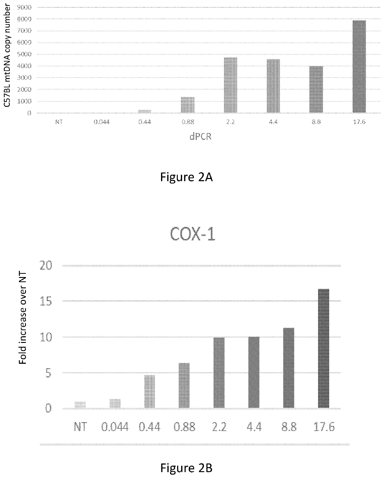
[0249]Mitochondrial augmentation of healthy donor CD34+ cells was performed with two different placenta-derived mitochondria batches, and cells were washed extensively after 24 h incubation. Illumina-based sequencing of the mtDNA show the presence of both transferred and endogenous mitochondria within the same cell.

[0250]As can be seen in FIG. 5 both MAT experiments from different placenta resulted in a similar augmentation percentage.

example 4

ria Can Enter Human Bone Marrow Cells

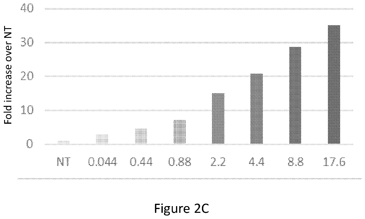
[0251]Human CD34+ cells (1.4*105, ATCC PCS-800-012) were untreated or incubated for 20 hours with GFP-labeled mitochondria isolated from human placental cells. Before plating the cells, mitochondria were mixed with the cells, centrifuged at 8000 g and re-suspended. After incubation, the cells were washed twice with PBS and CS activity was measured using the CS0720 Sigma kit (FIG. 6A). ATP content was measured using ATPlite (Perkin Elmer) (FIG. 6B), as previously described in WO 2016 / 135723.

[0252]The results demonstrated in FIG. 6 clearly indicate that the mitochondrial content of human bone-marrow cells may be increased many fold by interaction and co-incubation with isolated human mitochondria, to an extent beyond the capabilities of either human or murine fibroblasts or murine bone-marrow cells.


PUM


Abstract

The present invention provides human stem cells enriched with functional mitochondria, wherein the healthy functional exogenous mitochondria constitute at least 3% and less than 33% of the total mitochondria in the mitochondrially-enriched human stem cells. Further disclosed are methods for the production of such cells, and uses thereof for the treatment of primary mitochondrial diseases.

Description

FIELD OF THE INVENTION[0001]The present invention relates to mammalian stem cells, more specifically human stem cells, enriched with exogenous functional human mitochondria. The present invention further relates to methods for their production, and therapeutic methods utilizing such enriched cells.BACKGROUND OF THE INVENTION[0002]The mitochondrion is a membrane bound organelle found in most eukaryotic cells, ranging from 0.5 to 1.0 μm in diameter. Mitochondria are found in nearly all eukaryotic cells and vary in number and location depending on the cell type. Mitochondria contain their own DNA (mtDNA) and their own machinery for synthesizing RNA and proteins. The mtDNA contains only 37 genes, thus most of the gene products in the mammalian body are encoded by nuclear DNA.[0003]Mitochondria perform numerous essential tasks in the eukaryotic cell such as pyruvate oxidation, the Krebs cycle and metabolism of amino acids, fatty acids and steroids. However, the primary function of mitoch...

Claims


Application Information

Patent Timeline
IPC IPC(8): A61K35/545A61K35/28
CPCA61K35/545A61K35/28C12N5/0647A61P7/00
Inventor YIVGI OHANA, NATALIEHALAVEE, URIELBUKSHPAN, SHMUELSHER, NOABLUMKIN, MORIYA
Owner MINOVIA THERAPEUTICS
Why Eureka
Try Eureka