Therapeutic mitigation of epithelial infection
a technology for epithelial infection and therapy, applied in the field of epithelial infection therapy, can solve the problems of not addressing the comorbidities, no treatment that has been shown to reduce the risk of acquiring or containing these viral infections, etc., and achieve the effect of enhancing the safety of a vaccin
- Summary
- Abstract
- Description
- Claims
- Application Information
AI Technical Summary
Benefits of technology
Problems solved by technology
Method used
Image

Examples
example 1
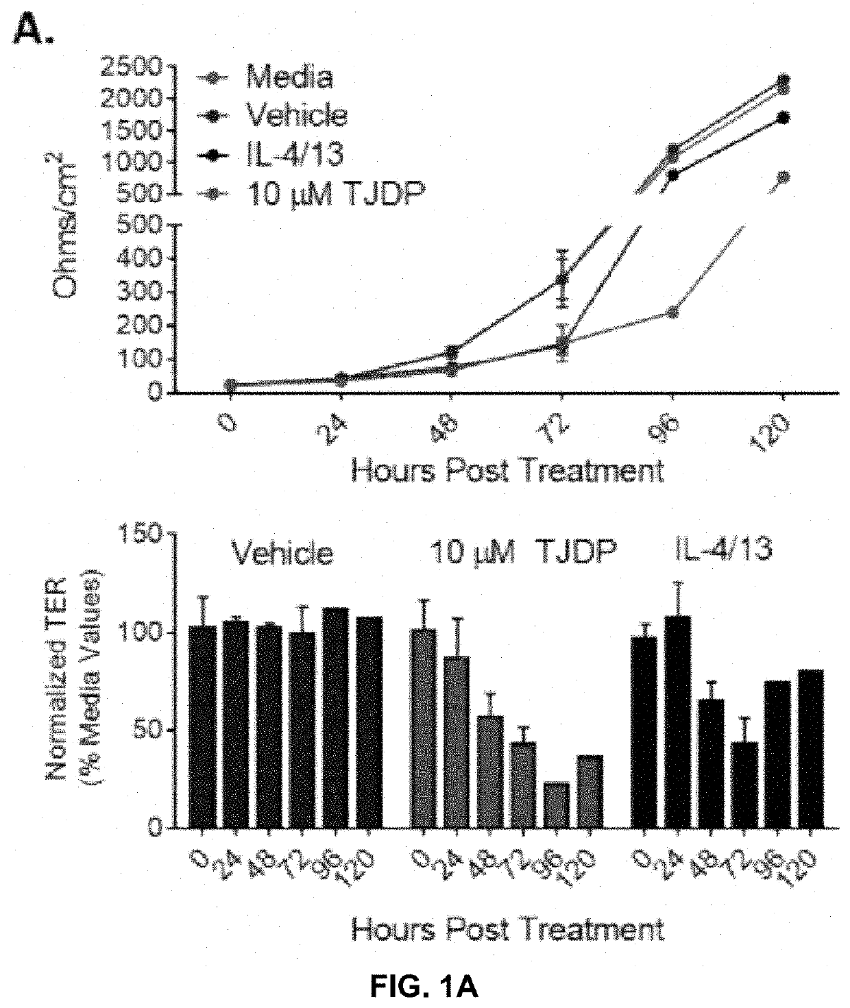
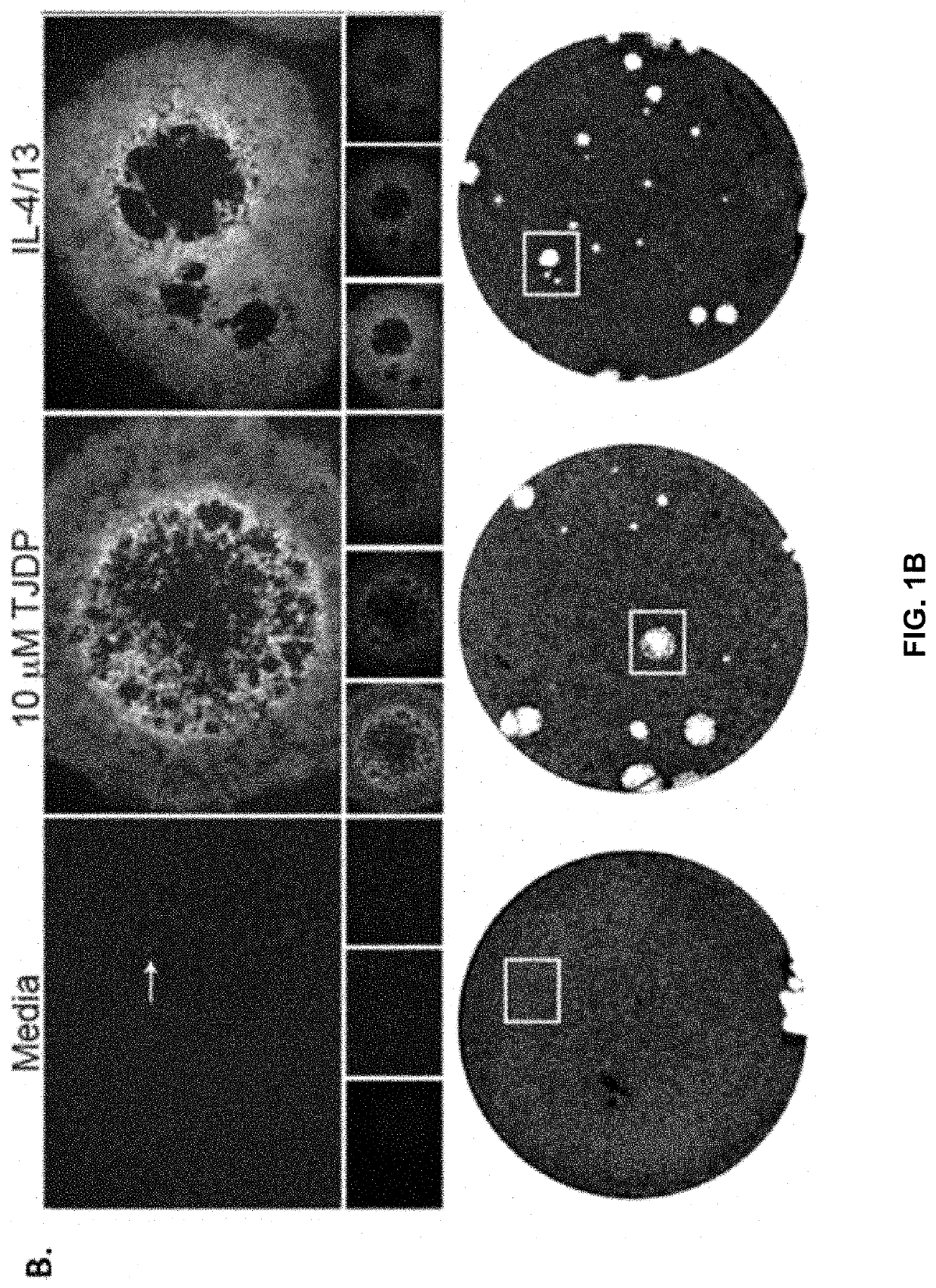
[0128]Patients with AD have increased Th2 cytokines and decreased barrier function, which is thought to make them more susceptible to infections of the skin. Taking this into account the inventors theorized that treatment of keratinocytes with Th2 cytokines or a tight junction disrupting peptide (TJDP) that reduces barrier function, should cause increased susceptibility to VV infection. To test this idea, primary human foreskin keratinocytes (PHFK) were seeded into multi-well plates and differentiated in high calcium media. Upon differentiation, cells were treated with IL-4 and IL-13, TJDP, or vehicle. Transepithelial electrical resistance (TER) was measured daily for 5 days to determine the establishment of barrier in the monolayer. It was found that skin barrier function was substantially delayed in the PHFK treated with TJDP and less so with Th2 cytokines, whereas cells treated with vehicle alone developed robust TJ within three days' post differentiation (FIG. 1A).
[0129]VV under...
example 2
[0131]In this example, assays are carried out to address the following questions: How does the transcriptome of differentiated PHFKs change in response to TJ disruption or Th2 cytokines? Can this transcriptional change be augmented to prevent enhanced viral infection? Do the same treatments used in vitro mitigate viral disease in a mouse model of AD?
[0132]Briefly, assays are carried out to characterize the transcriptional changes that occur as a consequence of exposure to presence of Th2 cytokines (IL-4 / 13) or the TJDP in differentiated PHFKs focusing on gene families implicated in barrier formation (SC, TJ and lipid biology), pathogen sensing and innate immunity. Previous studies have evaluated the transcriptome of keratinocytes during differentiation and by modeling an AD setting (IL-4 / 13 exposure), but these were with purchased keratinocytes and no study looked at differentiated PHFK exposed to Th2 cytokines or our TJDP [17-19].
[0133]To do this, keratinocytes (6-8 donors) during ...
example 3
[0134]In this example, assays are carried out to determine the potential for pan- and selective-JAK inhibitors to mitigate the enhanced viral susceptibility observed in PHFKs exposed to either Th2 cytokines (IL4 / IL13 alone or in combination) and / or TJDP.
[0135]To accomplish this goal, a minimum of eight PHFK donors are used to ensure that results are generalizable. Keratinocytes from these donors are used to form TJ in the presence of either Th2 cytokines or TJDP and paired cultures are exposed to JAK inhibitors, which are used at concentrations that are not cytotoxic but at doses that have been previously shown inhibit JAK signaling (tested by qPCR of relevant downstream genes). Cells are then infected with a low multiplicity of infection to observe viral dissemination (immunofluorescence) and pathogenicity (plaque formation) as described above. Additionally, JAK inhibitors are added at the time of VV infection as well as after infection (4-24 hours later) to test their therapeutic ...
PUM
| Property | Measurement | Unit |
|---|---|---|
| Electrical inductance | aaaaa | aaaaa |
| Magnetic field | aaaaa | aaaaa |
| Magnetic field | aaaaa | aaaaa |
Abstract
Description
Claims
Application Information