Systems and methods for imaging and manipulating tissue

a tissue imaging and tissue technology, applied in the field of systems and methods for imaging and manipulating tissue, can solve the problems of reducing the accuracy of procedures, increasing patient morbidity and mortality, and limited access to surgical sites for multiple systems, so as to reduce the risk of bacterial contamination, improve the prognosis of patients, and reduce the longevity of fertility.

Pending Publication Date: 2021-09-30
RES DEVMENT FOUND
View PDF11 Cites 4 Cited by
  • Summary
  • Abstract
  • Description
  • Claims
  • Application Information

AI Technical Summary

Benefits of technology

[0009]Exemplary embodiments of the present disclosure integrate three novel laser technologies for imaging, coagulation (e.g. blood flow interruption) and tissue removal (e.g. tissue “cutting”) and inspire a new surgical paradigm. Capability for image-guided high speed tissue removal with a cutting precision of a few cell layers is unprecedented in the surgical arts. Exemplary embodiments will allow access to remove previously inoperable tumors and other pathologic tissues in small confined spaces. Accordingly, surrounding nerves, specialized muscles and important glands can be spared, thereby substantially improving a patient's prognosis following surgery.
[0010]Exemplary embodiments may be utilized for surgical removal of many complex lesions in close proximity with specialized nerves, muscles, glands, and other normal organs and supporting structures. For example, endometriosis is frequently associated with the ovary, bowel and bladder, and other abdominal structures, and removal using conventional surgical procedures poses a risk of diminishing the longevity of fertility to the female patient due to a loss of ovum or eggs, or contamination of the abdomen with feces or urine. Exemplary embodiments may rapidly safely, and more precisely remove ovarian lesions sparing normal ovarian follicles, and remove endometrial tissue without the risk of bacterial contamination of the sterile abdominal cavity.

Problems solved by technology

The use of multiple independent systems can reduce the accuracy of the procedure, as the surgeon is not able to manipulate the tissue in spatial and / or temporal registration with preferred tissue visualization.
In addition, the use of separate systems for independent functions performed during surgical procedures can result in increased surgical times as each system must be introduced and withdrawn from the surgical site, increasing patient morbidity and mortality.
Other issues arising from the use of multiple surgical systems include limited access to surgical sites for multiple systems and increased costs.
In addition, the inability to concurrently image and cut has resulted in the inability to perform certain operations, or to perform operations inadequately requiring repeat procedures in the future.
For example, brain tumors are accessible via the ventricles but inoperable because the surgeon cannot concurrently image the boundary between tumor and healthy tissue and establish a safe margin.
Although prestaging imaging and surgical operating procedures have improved considerably over the last two decades, rate of successful outcomes (i.e., cancer-free patients) have not meaningfully improved.
A leading hypothesis for the existing high recurrence rate and corresponding poor outcomes is that cancer cells are being left within the patient due to improper selection of tumor margins.
Current methods utilized for tumor margin selection using traditional biopsy during surgery are limited and time-consuming.
Current practice requires the surgeon to physically label tissue regions for biopsy, perform the biopsy, manually transfer tissue to a pathology laboratory, creation of frozen sections and sectioning, staining the frozen section, diagnostic observation of frozen tissue sections under a microscope by a cancer pathologist and finally oral communication of the diagnostic results back to the surgeon—a process that requires about thirty minutes and is disruptive to the surgeon's preferred workflow.
For example, endometriosis is frequently associated with the ovary, bowel and bladder, and other abdominal structures, and removal using conventional surgical procedures poses a risk of diminishing the longevity of fertility to the female patient due to a loss of ovum or eggs, or contamination of the abdomen with feces or urine.

Method used


Image

  • Systems and methods for imaging and manipulating tissue
  • Systems and methods for imaging and manipulating tissue
  • Systems and methods for imaging and manipulating tissue

Examples


Embodiment Construction

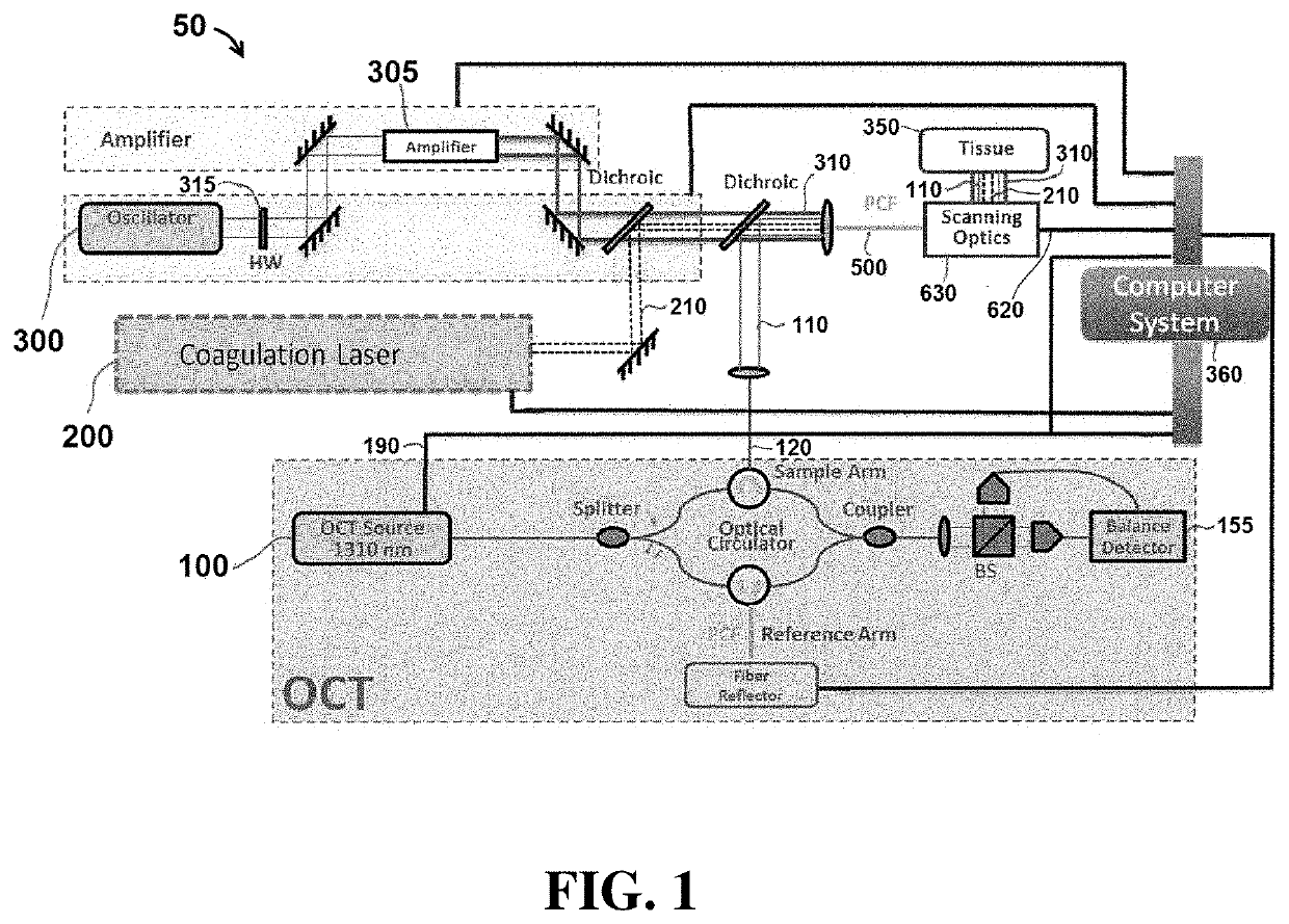
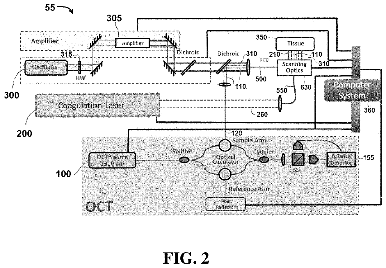
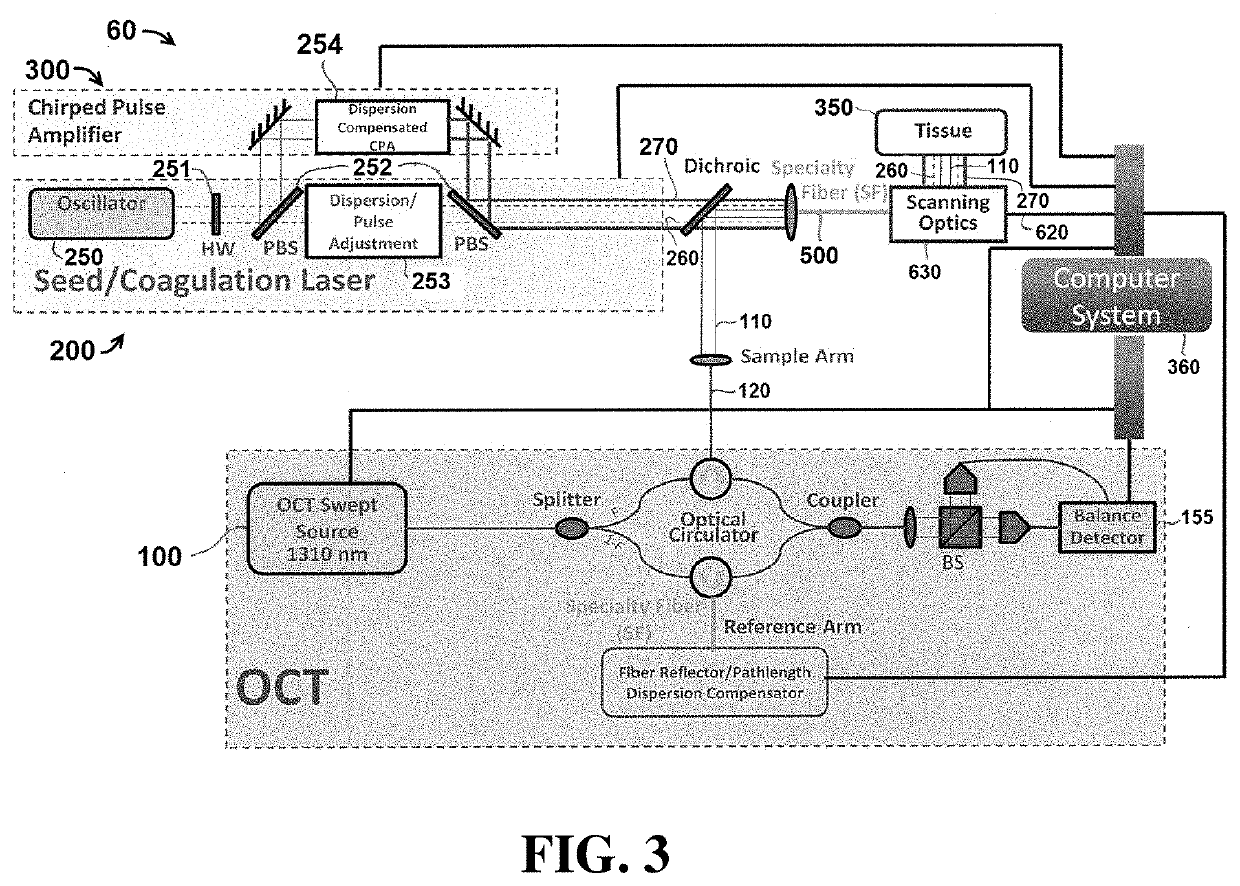
[0055]Exemplary embodiments of the present disclosure include systems and methods that utilize light to image tissue, coagulate tissue (e.g. blood vessels), and break molecular bonds of (e.g. cut) the tissue. In certain embodiments, the light used to perform each of these tasks may be produced by independent and separate components, including for example, lasers. In other embodiments, a single component (e.g. laser) may be configured to produce light energy that is used to perform multiple tasks, including coagulating the tissue and breaking molecular bonds of the tissue. It is understood that the embodiments described herein are merely exemplary, and that other embodiments are included within the scope of the invention.

[0056]As used herein, the term “coagulate” (and related terms, such as “coagulating”, “coagulation”, etc.) is used to refer to a process of interrupting blood flow (e.g. by damaging blood vessels so that when cut, blood does not flow outside the tissue) and rearrangi...


PUM


Abstract

Exemplary embodiments of the present disclosure include systems and methods capable of imaging, manipulating, and analyzing tissue using light, including for example, coagulating and breaking the molecular bonds (e.g. cutting) tissue.

Description

CROSS-REFERENCE TO RELATED APPLICATIONS[0001]This application is a divisional of U.S. application Ser. No. 14 / 852,460, filed Sep. 11, 2015, which claims priority to U.S. Provisional Patent Application Ser. No. 62 / 049,955 filed Sep. 12, 2014, the contents of each of which are incorporated by reference herein.BACKGROUND INFORMATION[0002]Traditional surgical techniques and apparatus have utilized independent systems and components for imaging and manipulating tissue. For example, a first system may be used to image the area in which the surgical technique is to be performed, while separate systems may be used to coagulate and cut the tissue.[0003]The use of multiple independent systems can reduce the accuracy of the procedure, as the surgeon is not able to manipulate the tissue in spatial and / or temporal registration with preferred tissue visualization. In addition, the use of separate systems for independent functions performed during surgical procedures can result in increased surgic...

Claims


Application Information

Patent Timeline
Patent Type & Authority Applications(United States)
IPC IPC(8): A61B5/00A61B18/20A61B18/22
CPCA61B5/0066A61B18/20A61B2018/00982A61B2018/2075A61B18/22A61B2018/00994A61B2018/00541A61B2018/00559A61B2018/00589A61B2018/00601A61B2018/207A61B2018/00607A61B2018/00636A61B2018/00702
Inventor FELDMAN, MARC D.MILNER, THOMAS E.
Owner RES DEVMENT FOUND
Why Eureka
Try Eureka